Translate this page into:
Caviar tongue
Correspondence Address:
Vishalakshi Viswanath
Department of Dermatology, Rajiv Gandhi Medical College and Chhatrapati Shivaji Maharaj Hospital, Kalwa, Thane - 400 605, Maharashtra
India
| How to cite this article: Viswanath V, Nair S, Chavan N, Torsekar R. Caviar tongue. Indian J Dermatol Venereol Leprol 2011;77:78-79 |
Sir,
A wide spectrum of disorders affects the tongue and clinical examination provides a clue to the diagnosis. Inspection of tongue helps in identifying systemic illnesses like anemia, cyanosis, nutritional deficiencies, and localized abnormalities like lichen planus, infections and tumors. However, the undersurface of the tongue is most often missed out on inspection. Lining the undersurface of the tongue are clusters of small dilated and tortuous veins that may attain a large size, appearing round and black akin to caviar. We present a case of this physiological change called caviar tongue. [1]
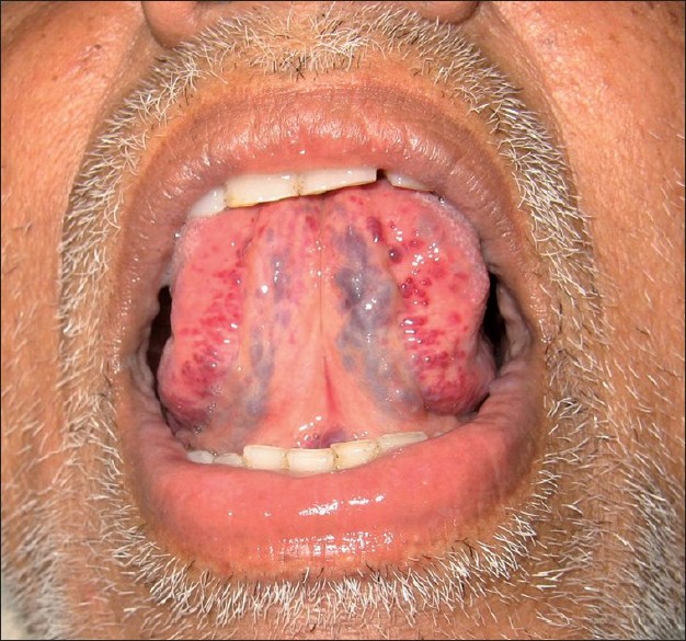
A 65-year-old man was referred for asymptomatic tortuous swellings on the undersurface of the tongue which he had noticed since 1 year [Figure - 1]. There was no history of bleeding from the site. On examination, dilated tortuous reddish vessels were seen along the lateral portions of undersurface of the tongue. No other mucosae and skin surface showed a similar change. He had no systemic complaints or past history of any major illnesses.
 |
| Figure 1 :Dilated tortuous vessels seen along the lateral portions of undersurface of the tongue |
The mucosal surface of the lower portion of the tongue is extremely thin and transluscent which permits ready inspection of submucosal vascular structures. Also, the supporting connective tissue framework is fragile and ill-developed so that even minor stimuli can cause dilatation and varicose changes to occur. Advancing age is a predisposing factor due to tissue loosening and elastic tissue degeneration in the vessels. The caviar lesion is thus commonly seen as a physiological change associated with senile elastotic degeneration of the sublingual veins. It occurs at three sites: under surface of tongue along the sublingual vein, floor of the mouth near ostia of the sublingual glands and along the lateral portions of the under surface of the tongue. [1] Uncommon sites are lips and buccal mucosae. It begins as a small outpouching of the veins, and the varicosities gradually elevate the overlying thin mucosa with color varying from red to purple, resembling buckshot or caviar with iridescent surface. Histologically, caviar lesion is a dilated vein with no inflammatory changes. The endothelium is hypoplastic but the wall is thick and cellular.
Sublingual varices are benign vascular dilatations, usually asymptomatic and affecting 10% of the population over the age of 40 years. [2] These are frequently observed by dentists and bleeding from these varices is uncommon. [3] In cases of bleeding, a detailed investigation should be undertaken for underlying associations. Caviar tongue can be found in association with conditions that result in increased venous pressure such as portal hypertension or superior venacava syndrome. [4] Phleboliths or thrombophlebitis may complicate this condition. An association with angiokeratoma of scrotum has also been reported. [5]
In a study by William Bean, caviar tongue was seen after fourth decade with increased frequency in sixth to seventh decade which is the same profile as that seen in cherry angiomas and venous stars. No evidence of any association with systemic diseases like pulmonary or vascular disease was found. [1] A recent study for prevalence of oral hemangioma, vascular malformation and varix in a Brazilian population revealed 65.6% prevalence of asymptomatic multiple sublingual varices with preponderance in females and Caucasians. [6]
Sublingual varices may be occasionally confused due to resemblance to tumors like hemangioma, lymphangioma, Kaposi′s sarcoma, melanoma or other conditions like Osler′s syndrome, blue rubber bleb nevus syndrome. [4] However, most of these conditions can be differentiated by a detailed history and thorough clinical evaluation. Caviar tongue on histology is just a dilated vein with no inflammatory changes. Vascular tumors like hemangioma and lymphangioma show endothelial cell proliferation and dilated lymphatics respectively, while Kaposi′s sarcoma shows tumor cells composed of vascular spaces and spindle cells on histology. Sublingual varices usually need no treatment but only reassurance regarding its benign nature. Sclerotherapy or surgery has been attempted in single lesions and unusual locations like lips or buccal mucosae. [6] This condition has been reported to highlight this rather interesting and benign physiological manifestation of aging process which is infrequently looked for by a dermatologist.
| 1. |
Bean WB. The caviar lesion under the tongue. Trans Am Clin Climatol Assoc 1952;64:40-9.
[Google Scholar]
|
| 2. |
Rogers RS, Bruce AJ. Tongue in clinical diagnosis. J Eur Acad Dermatol Venereol 2004;18:254-9.
[Google Scholar]
|
| 3. |
Pemberton MN. Sublingual varices are not unusual. BMJ 2006;333:202.
[Google Scholar]
|
| 4. |
Roy S, Rogers II, Mehregan DA. Disorders of oral cavity. In: Moschella SL, Hurley HJ, editors. Moschella's Dermatology. 3 rd ed. Philadelphia: WB Saunders Co; 1992. p. 2094.
rd ed. Philadelphia: WB Saunders Co; 1992. p. 2094. '>[Google Scholar]
|
| 5. |
James WD, Berger TG, Elston DM. Disorders of mucous membranes. In: James WD, Berger TG, Elston DM, editors. Andrews' Diseases of the Skin; Clinical Dermatology. 10 th ed. Philadelphia: W.B. Saunders Co; 2006. p. 803.
th ed. Philadelphia: W.B. Saunders Co; 2006. p. 803. '>[Google Scholar]
|
| 6. |
Corrκa PH, Nunes LC, Johann AC, Aguiar MC, Gomez RS, Mesquita RA. Prevalence of oral hemangioma, vascular malformation and varix in a Brazilian population. Braz Oral Res 2007;21:40-5
[Google Scholar]
|
Fulltext Views
29,075
PDF downloads
4,761


