Translate this page into:
Multiple brownish papules on the penile shaft
Correspondence Address:
Wu Bai-Yao
Department of Dermatology, Tri-Service General Hospital, No. 325, Sec. 2, Chenggong Road, Neihu Dist., Taipei City 114, Taiwan
China
| How to cite this article: Ching-Fu H, Wei-Ming W, Bai-Yao W. Multiple brownish papules on the penile shaft. Indian J Dermatol Venereol Leprol 2011;77:404 |
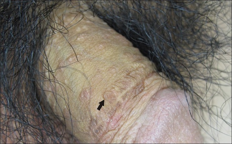
A 27-year-old man presented with a 2-month history of multiple asymptomatic skin lesions on the penile shaft. He was concerned about having a sexually transmitted disease. The general condition of the patient was good. He had no history of sexual exposure and no similar lesions were found in other family members. A physical examination revealed numerous, discrete, brownish papules with a smooth surface on the dorsal and lateral penile shaft [Figure - 1]. Ten papules were over the lesional site, and the diameter of these papules ranged from 1 mm to 3 mm. Examination of the rest of his body was unremarkable. The working diagnosis of bowenoid papulosis was considered, and a biopsy sample was obtained from one papule on the dorsal penile shaft [Figure - 1] for histological examination.
 |
| Figure 1: Multiple asymptomatic brown-yellow papules on the dorsal penile shaft |
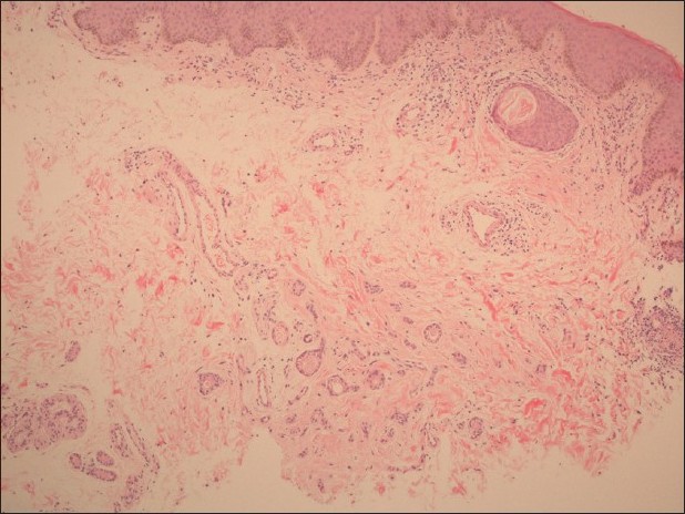
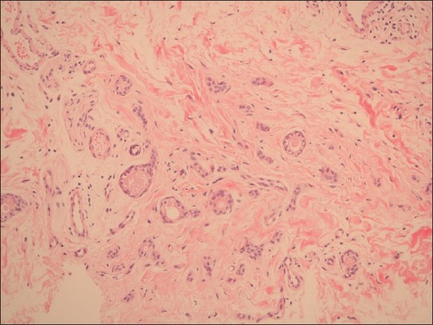
Histopathologically, the epidermis showed mild acanthosis with basal hyperpigmentation [Figure - 2] and comma-shaped (tadpole-shaped) structures originating from ducts dispersed within a sclerotic stroma. The ducts were lined by two or more epithelial cell layers [Figure - 3].
 |
| Figure 2: Epidermal basal hyperpigmentation. There are some cystic ducts with a tadpole-like appearance in a fibrotic stroma (H and E, ×100) |
 |
| Figure 3: The ductal structure is composed of two layers of epithelial cells (H and E, ×200) |
What is your Diagnosis?
Diagnosis
Eruptive penile syringoma.
Discussion
Syringoma is a common benign tumor of eccrine origin with a limited proliferative capacity. [1] Friedman et al.[2] classified four variants as a localized form, familial form, generalized form and a form associated with Down syndrome. Syringoma is more common in adolescent girls during puberty. The lesions usually localize on the lower eyelid, although the upper cheek, upper chest, axilla, abdomen and vulva are also common sites. Involvement of the penis is rare, and only a few cases have been reported.
Clinically, syringoma on the penis usually presents as numerous, asymptomatic flesh-colored or yellow-brown papules measuring 1-3 mm in diameter. [3] The involved area is usually located on the back and lateral surface of the shaft of the penis, and patients in early adult life or at puberty are affected most often. Because of the location, patients are usually distressed by the cosmetic effect and become concerned about its relationship with sexually transmitted disease. [4] The differential diagnosis of penile syringoma includes bowenoid papulosis, condyloma, lichen planus and genital warts. [3],[4] A skin biopsy to confirm the diagnosis is necessary when the lesions are resistant to treatment. Histopathologically, the characteristic finding is dermal proliferation of a double-layer cystic structure in a fibrous stroma. The structure presents as a comma- or tadpole-like appearance on sectioning.
In penile syringoma, treatment is usually unnecessary except for cosmetic reasons. Eliminating the lesions by surgical excision, cryotherapy with liquid nitrogen, electrodessication and curettage or carbon dioxide laser are the current treatment options. [4] However, recurrence and scarring can be bothersome after these treatments. Park et al.[5] tried a multiple-drilling method using a CO 2 laser to treat 11 patients with syringoma, and all patients had good clinical results without complications. Another report suggested that treatment with an intralesional insulated needle connected to an electrocoagulator may be a good therapeutic choice for a syringoma. [6] In conclusion, although eruptive penile syringoma is a rare condition, it should be added to the differential diagnosis of penile papules. Providing a definite diagnosis that is confirmed by histological examination may help patients avoid unnecessary aggressive treatment.
| 1. |
Hertl-Yazdi M, Niedermeier A, Hörster S, Krause W. Penile syringoma in a 14-year-old boy. Eur J Dermatol 2006;16:314-5.
[Google Scholar]
|
| 2. |
Friedman SJ, Butler DF. Syringoma presenting as milia. J Am Acad Dermatol 1987;16:310-4.
[Google Scholar]
|
| 3. |
Cassarino DS, Keahey TM, Stern JB. Puzzling penile papules. Int J Dermatol 2003;42:954-6.
[Google Scholar]
|
| 4. |
Olson JM, Robles DT, Argenyi ZB, Kirby P, Olerud JE. Multiple penile syringomas. J Am Acad Dermatol 2008;59:S46-7.
[Google Scholar]
|
| 5. |
Park HJ, Lee DY, Lee JH, Yang JM, Lee ES, Kim WS. The treatment of syrigomas by CO2 laser using a multiple drilling method. Dermatol Surg 2007;33:310-3.
[Google Scholar]
|
| 6. |
Hong SK, Lee HJ, Cho SH, Seo JK, Lee D, Sung HS. Syringomas treated by intralesional insulated needles without epidermal damage. Ann Dermatol 2010;22:367-9.
[Google Scholar]
|
Fulltext Views
20,316
PDF downloads
2,132


