Translate this page into:
Extrusion of pilosebaceous unit into blister cavity of pemphigus vulgaris: An artifact occurring as a natural phenomenon in pemphigus vulgaris
Correspondence Address:
B Vijaya
JSS Medical College, Mysore
India
| How to cite this article: Vijaya B, S, Manjunath G V, Priya L. Extrusion of pilosebaceous unit into blister cavity of pemphigus vulgaris: An artifact occurring as a natural phenomenon in pemphigus vulgaris. Indian J Dermatol Venereol Leprol 2010;76:575-576 |
Sir,
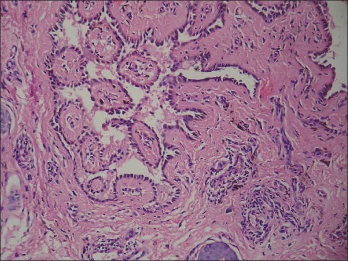
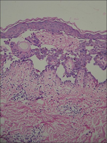
Extrusion of pilosebaceous unit into the blister cavity of pemhigus vulgaris is a peculiar phenomenon that has been reported. [1] We have observed four cases of pemphigus vulgaris (PV) over a period of 2 years that displayed this peculiar finding on histopathology. The biopsy specimen from all these cases showed typical microscopic features of PV, such as suprabasal bulla, acantholysis and inflammatory cells in the bulla cavity. An additional finding in two of these cases was extension of the acantholytic process to the adnexal epithelium, which is not always evident in histopathologic sections in all the cases of PV [Figure - 1]. Two cases showed extrusion of both hair follicle and sebaceous gland into the blister cavity, one case showed extrusion of sebaceous gland only and another case showed extrusion of the hair follicle only [Figure - 2]. Direct immunofluorescence studies in one case showed deposition of IgG in the squamous intercellular substance in a fishnet pattern. But, there were no adnexal structures in the tissue subjected for direct immunofluorescence studies to detect immunoreactants around the adnexal epithelium.
 |
| Figure 1 : Photomicrograph showing extension of the acantholytic process into the adnexal epithelium (H and E, ×200) |
 |
| Figure 2 : Photomicrograph showing hair follicle and sebaceous gland in the blister of pemphigus vulgaris (H and E, ×100) |
This unusual, interesting, histologic finding could possibly be a biopsy artifact occurring as a natural phenomenon in cases of PV. The possible explanation could be that there is loosening of the pilosebaceous unit at the suprabasal epithelial level of the hair follicle as a result of extension of the acantholytic process into the adnexal epithelium. The extrusion of the loosened pilosebaceous unit could have occurred during the process of section-cutting, when the microtome knife first hits the soft corium. In patients with PV, autoantibodies against desmoglein 3 (Dsg3) cause loss of cell-cell adhesion of keratinocytes in the basal and immediate suprabasal layers of the stratified squamous epithelia resulting in acantholysis. [2] Dsg3 also anchors the telogen hair to the outer root sheath of the follicle, and this observation underscores the importance of desmosomes in maintaining the normal structure and function of the hair. [3] The presence of Dsg3 in the hair follicle epithelium would explain the presence of acantholysis in the adnexal epithelium in PV. Follicular acantholysis may also be a subtle histologic manifestation of PV, as reported by Mahalingam. [4] This observation of pilosebaceous unit in the blister cavity of PV may suggest that even though this is considered an artifact, this artifactual change occurs in PV because of loosening of the pilosebaceous unit as there is acantholysis in the adnexal epithelium also. This process has also been observed in bullous pemphigoid. [5] This can be attributed to the same mechanism of extension/presence of the immunopathologic process around the basement membrane of the hair follicle resulting in loosening of the pilosebaceous unit.
We conclude that this displacement of pilosebaceous unit into the blister cavity in PV is because of the extension/presence of acantholysis in the adnexal epithelium, attributed to the antibodies against Dsg3 present in the suprabasal layers of the stratified squamous epithelial cells of the pilosebaceous unit.
| 1. |
Joshi R, Marwah HS. Extrusion of sebaceous gland into a blister of pemphigus vulgaris: an unusual processing artifact. Indian J Dermatol Venereol Leprol 2004;70:316-7.
[Google Scholar]
|
| 2. |
Koch PJ, Mahoney MG, Ishikawa H, Pulkkinen L, Uitto J, Shultz L, et al. Targeted disruption of the pemphigus vulgaris antigen (Desmoglein 3) gene in mice causes loss of keratinocyte cell adhesion with a phenotype similar to pemphigus vulgaris. J Cell Biol 1997;137:1091-102.
[Google Scholar]
|
| 3. |
Koch PJ, Mahoney MG, Cotsarelis G, Rothenberger K, Lavker RM, Stanley JR. Desmoglein 3 anchors telogen hair in the follicle. J Cell Sci 1998;111:2529-37.
[Google Scholar]
|
| 4. |
Mahalingam M. Follicular acantholysis: a subtle clue to the early diagnosis of pemphigus vulgaris. Am J Dermatopathol 2005;27:237-9.
[Google Scholar]
|
| 5. |
Veeranna S. Extrusion of sebaceous gland and hair follicle into a blister of bullous pemphigoid. Indian J Dermatol Venereol Leprol 2005;71:209-10.
[Google Scholar]
|
Fulltext Views
2,397
PDF downloads
2,105


