Generic selectors
Exact matches only
Search in title
Search in content
Post Type Selectors
Search in posts
Search in pages
Filter by Categories
15th National Conference of the IAOMFP, Chennai, 2006
Abstract
Abstracts from current literature
Acne in India: Guidelines for management - IAA Consensus Document
Addendum
Announcement
Art & Psychiatry
Article
Articles
Association Activities
Association Notes
Award Article
Book Review
Brief Report
Case Analysis
Case Letter
Case Letters
Case Notes
Case Report
Case Reports
Clinical and Laboratory Investigations
Clinical Article
Clinical Studies
Clinical Study
Commentary
Conference Oration
Conference Summary
Continuing Medical Education
Correspondence
Corrigendum
Cosmetic Dermatology
Cosmetology
Current Best Evidence
Current Issue
Current View
Derma Quest
Dermato Surgery
Dermatopathology
Dermatosurgery Specials
Dispensing Pearl
Do you know?
Drug Dialogues
e-IJDVL
Editor Speaks
Editorial
Editorial Remarks
Editorial Report
Editorial Report - 2007
Editorial report for 2004-2005
Errata
Erratum
Focus
Fourth All India Conference Programme
From Our Book Shelf
From the Desk of Chief Editor
General
Get Set for Net
Get set for the net
Guest Article
Guest Editorial
History
How I Manage?
IADVL Announcement
IADVL Announcements
IJDVL Awards
IJDVL AWARDS 2015
IJDVL Awards 2018
IJDVL Awards 2019
IJDVL Awards 2020
IJDVL International Awards 2018
Images in Clinical Practice
Images in Dermatology
In Memorium
Inaugural Address
Index
Knowledge From World Contemporaries
Leprosy Section
Letter in Response to Previous Publication
Letter to Editor
Letter to the Editor
Letter to the Editor - Case Letter
Letter to the Editor - Letter in Response to Published Article
LETTER TO THE EDITOR - LETTERS IN RESPONSE TO PUBLISHED ARTICLES
Letter to the Editor - Observation Letter
Letter to the Editor - Study Letter
Letter to the Editor - Therapy Letter
Letter to the Editor: Articles in Response to Previously Published Articles
Letters in Response to Previous Publication
Letters to the Editor
Letters to the Editor - Letter in Response to Previously Published Articles
Letters to the Editor: Case Letters
Letters to the Editor: Letters in Response to Previously Published Articles
Media and news
Medicolegal Window
Messages
Miscellaneous Letter
Musings
Net Case
Net case report
Net Image
Net Images
Net Letter
Net Quiz
Net Study
New Preparations
News
News & Views
Obituary
Observation Letter
Observation Letters
Oration
Original Article
ORIGINAL CONTRIBUTION
Original Contributions
Pattern of Skin Diseases
Pearls
Pediatric Dermatology
Pediatric Rounds
Perspective
Presedential Address
Presidential Address
Presidents Remarks
Quiz
Recommendations
Regret
Report
Report of chief editor
Report of Hon : Treasurer IADVL
Report of Hon. General Secretary IADVL
Research Methdology
Research Methodology
Resident page
Resident's Page
Resident’s Page
Residents' Corner
Residents' Corner
Residents' Page
Retraction
Review
Review Article
Review Articles
Reviewers 2022
Revision Corner
Self Assessment Programme
SEMINAR
Seminar: Chronic Arsenicosis in India
Seminar: HIV Infection
Short Communication
Short Communications
Short Report
Snippets
Special Article
Specialty Interface
Studies
Study Letter
Study Letters
Supplement-Photoprotection
Supplement-Psoriasis
Symposium - Contact Dermatitis
Symposium - Lasers
Symposium - Pediatric Dermatoses
Symposium - Psoriasis
Symposium - Vesicobullous Disorders
SYMPOSIUM - VITILIGO
Symposium Aesthetic Surgery
Symposium Dermatopathology
Symposium-Hair Disorders
Symposium-Nails Part I
Symposium-Nails-Part II
Systematic Review and Meta-Analysis
Systematic Reviews and Meta-analyses
Systematic Reviews and Meta-analysis
Tables
Technology
Therapeutic Guideline-IADVL
Therapeutic Guidelines
Therapeutic Guidelines - IADVL
Therapeutics
Therapy
Therapy Letter
Therapy Letters
View Point
Viewpoint
What’s new in Dermatology
View/Download PDF

Translate this page into:

Quiz
89 (
3
); 436-438
doi:
10.25259/IJDVL_618_20
pmid:
34379955

A patient with diffuse oedema of the eyelids, infraorbital area and indurated plaques over the scalp

Department of Dermatology, Apollo Hospitals, Hyderabad, Telangana, India
Department of Dermatology, Dermatrendz, Hyderabad, Telangana, India
Department of Pathology, Apollo Hospitals, Hyderabad, Telangana, India

Corresponding author: Dr. Indukooru Subrayalu Reddy, A-8 Anand Sheel Enclave, Nandi Nagar, Road Number 14 Banjara Hills, Hyderabad - 500 034, Telangana, India. drreddyis@gmail.com

Licence
This is an open-access article distributed under the terms of the Creative Commons Attribution-Non Commercial-Share Alike 4.0 License, which allows others to remix, transform, and build upon the work non-commercially, as long as the author is credited and the new creations are licensed under the identical terms.

How to cite this article: Reddy IS, Somani VK, de Padua M, Gowrishankar S. A patient with diffuse oedema of the eyelids, infraorbital area and indurated plaques over the scalp. Indian J Dermatol Venereol Leprol 2023;89:436-8.

A 65-year-old man presented with asymptomatic, indurated swelling of the eyelids and infraorbital area for eight months. Patient first noticed puffiness of eyelids and infraorbital areas, which progressively increased, followed by development of areas of induration over the scalp. Patient denied any preceding skin lesions over the face and scalp. He did not receive radiotherapy or chemotherapy in the past. The extent and degree of induration of eyelids progressively increased, resulting in impaired vision [Figure 1]. Patient first noticed puffiness of eyelids and infraorbital areas, which progressively increased, followed by development of areas of induration over the scalp. Patient denied any preceding skin lesions over the face and scalp. He did not receive radiotherapy or chemotherapy in the past. Two months later, the patient developed a well-delineated indurated plaque over the vertex of scalp with the surrounding area showing diffuse, brawny induration. There were discrete, hyperpigmented, follicular papules coalescing to form an indurated plaque over the left side of vertex [Figure 2]. Patient denied having any preceding skin lesions over the face and the scalp and not received radiotherapy nor chemotherapy in the past. No bleeding or ulceration was noted over the lesion. The posterior cervical and submental lymph nodes were enlarged, painless and firm to hard in consistency.

Skin biopsies were taken from the indurated plaque over the vertex and the left infraorbital area. Both the samples showed similar histopathology. The epidermis was unremarkable. The deep dermis showed prominent, dilated, irregular vascular channels of varying calibre [Figure 3]. The endothelial cells lining the vascular channels were atypical, plump and spindle shaped with hyperchromatic nuclei [Figure 4]. The high-power view showed neoplastic cells arranged in sheets exhibiting enlarged, pleomorphic and vesicular nuclei with scanty to moderate amount of cytoplasm and mitotic activity [Figure 5]. Immunohistochemistry with CD31 showed strong positivity of neoplastic cells through the entire thickness of the dermis [Figure 6].

Question

What is your diagnosis?
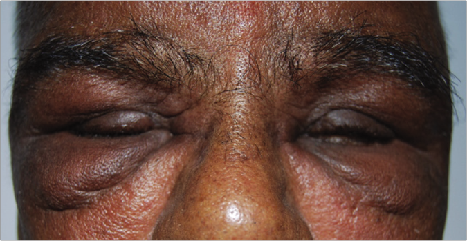
Oedema of eyelids and infraorbital area, more pronounced over the right side
Figure 1:
Oedema of eyelids and infraorbital area, more pronounced over the right side
Indurated plaque over the vertex, with surrounding area of brawny induration and hyperpigmented, indurated plaque over the left occipital area
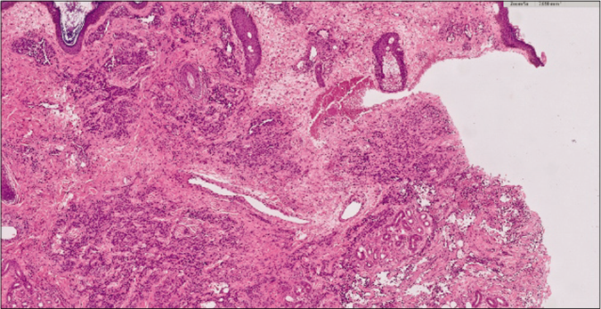
Figure 2:
Indurated plaque over the vertex, with surrounding area of brawny induration and hyperpigmented, indurated plaque over the left occipital area
Epidermis is normal. Deep dermis dermis shows prominent, dilated, irregular vascular channels of varying calibre (H&E ×5)
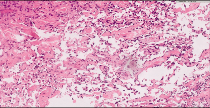
Figure 3:
Epidermis is normal. Deep dermis dermis shows prominent, dilated, irregular vascular channels of varying calibre (H&E ×5)
Dermis showing meshwork of anastomosing, dilated blood vessels. The blood vessels are lined by crowded, atypical endothelial cells with enlarged, hyperchromatic nuclei (H&E ×20)
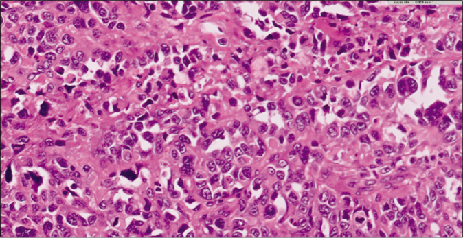
Figure 4:
Dermis showing meshwork of anastomosing, dilated blood vessels. The blood vessels are lined by crowded, atypical endothelial cells with enlarged, hyperchromatic nuclei (H&E ×20)
Neoplastic cells are arranged in sheets and exhibit enlarged, pleomorphic, vesicular nuclei with moderate to scanty cytoplasm and mitotic figures (H&E ×40)
Figure 5:
Neoplastic cells are arranged in sheets and exhibit enlarged, pleomorphic, vesicular nuclei with moderate to scanty cytoplasm and mitotic figures (H&E ×40)
Neoplastic cells are positive for CD31 (IHC ×10)
Figure 6:
Neoplastic cells are positive for CD31 (IHC ×10)

Answer

Idiopathic cutaneous angiosarcoma of the head and neck

Discussion

Angiosarcoma was first described by Wilson Jones in 1964 as a malignant angioendothelioma.1 It is a rare, malignant, mesenchymal tumour of vascular origin involving the face and scalp. There are three main clinicopathological subtypes of cutaneous angiosarcoma with an overall incidence of approximately 0.01/100,000 affecting primarily adult patients.2,3 The three clinicopathological subtypes are as follows: idiopathic cutaneous angiosarcoma of the head and neck, angiosarcoma complicating lymph edema (Stewart-Trevis syndrome) and post-irradiation angiosarcoma.

Rarely, angiosarcomas can occur sporadically in patients with xeroderma pigmentosum, epidermolysis bullosa, vinyl chloride disease, in gouty tophus, in renal transplant recipients and in patients with AIDS.

This patient falls in the category of the first type, namely, idiopathic cutaneous angiosarcoma of the head and neck. This typically affects patients in the seventh to ninth decades of life, more common in males with male-to-female ratio of 2.5:1. It commonly involves the upper part of the face and scalp.4 It usually presents as bruise-like patches, single or multiple reddish or purple papules, nodules and plaques. Ulceration and bleeding from the lesions is rarely observed. Exclusive periorbital involvement is occasionally observed.5 Very rarely, angiosarcoma can present as recurrent angioedema.6 The other rare presentations are rosacea-like eruption and rhinophyma-like lesions.7 Early lesions of angiosarcoma need to be differentiated from ecchymoses, port-wine stain and early Kaposi sarcoma.

The histopathology is similar in all three types of angiosarcoma.8 The tumour is poorly circumscribed, multifocal, involving the dermis and subcutaneous fat. The tumour shows an angiomatous and solid pattern. The angiomatous areas show a meshwork of anastomosing dilated blood vessels, permeating between the dermal collagen bundles (dissection of collagen). The vessels are lined by crowded endothelial cells with large, hyperchromatic nuclei. Solid areas show poorly differentiated tumor cells which are spindle shaped to polygonal. Focal epithelioid change is rarely observed.

All forms of angiosarcoma carry a poor prognosis. In one series, only 15% of patients survived for five years after the diagnosis.9 A retrospective study of angiosarcoma of the scalp and face showed an improved five-year survival rate of 43% by combined modality of therapy.10 Patients with lesions <5–10 cm in diameter were successfully treated with radical wide-field radiotherapy and surgery. Combination of surgery, radiotherapy and taxane (paclitaxel and docetaxel) has been shown to improve overall survival of the patients.

The present case is reported because of its rarity, extensive and multifocal involvement of the eyelids, periorbital area and the scalp.

Declaration of patient consent

The authors certify that they have obtained all appropriate patient consent.

Financial support and sponsorship

Nil.

Conflicts of interest

There are no conflicts of interest.

References

  1. . Malignant angioendothelioma of the skin. Br J Dermatol. 1964;76:21-39.
    [CrossRef] [PubMed] [Google Scholar]
  2. , . Cutaneous vascular proliferations. Part III Malignant neoplasms, other cutaneous neoplasms with significant vascular component, and disorders erroneously considered as vascular neoplasms. J Am Acad Dermatol. 1998;38:143-75.
    [CrossRef] [PubMed] [Google Scholar]
  3. , , , , . The spectrum of mesenchymal skin neoplasms reflected by the new WHO classification. Onkologie. 2004;27:401-6.
    [CrossRef] [PubMed] [Google Scholar]
  4. , , . Angiosarcoma of the scalp. Int J Dermatol. 1999;38:697-9.
    [CrossRef] [PubMed] [Google Scholar]
  5. , , , . Cutaneous angiosarcoma of the face. J Eur Acad Dermatol Venereol. 2003;17:594-5.
    [CrossRef] [PubMed] [Google Scholar]
  6. , . Cutaneous angiosarcoma presenting as recurrent angioedema of the face. Br J Dermatol. 2000;143:1346-8.
    [CrossRef] [PubMed] [Google Scholar]
  7. , , . Cutaneous angiosarcoma of the face: Clinicopathologic and immunohistochemical study of a case resembling rosacea clinically. J Am Acad Dermatol. 1998;38:837-40.
    [CrossRef] [PubMed] [Google Scholar]
  8. . Angiosarcomas of the skin. Semin Diagn Pathol. 1987;4:2-17.
    [PubMed] [Google Scholar]
  9. , , . Angiosarcoma of the face and scalp, prognosis and treatment. Cancer. 1987;59:1046-57.
    [CrossRef] [PubMed] [Google Scholar]
  10. , , . Outcomes after definitive treatment for cutaneous angiosarcoma of the face and scalp. Head Neck. 2010;33:661-71.
    [CrossRef] [PubMed] [Google Scholar]

Fulltext Views
5,257

PDF downloads
2,649
View/Download PDF
Download Citations
BibTeX
RIS
Show Sections