
Translate this page into:
Usefulness of ultrasound in diagnosis of plantar epidermal cyst
Corresponding author: Prof. Kapsok Li, Department of Dermatology, Chung-Ang University College of Medicine, Seoul, Korea. ksli0209@cau.ac.kr
-
Received: ,
Accepted: ,
How to cite this article: Park JW, Shin SH, Kim BJ, Li K. Usefulness of ultrasound in diagnosis of plantar epidermal cyst: A case series. Indian J Dermatol Venereol Leprol 2021;87:872-5.
Sir,
Epidermal cysts, also called epidermal inclusion cysts or epidermoid cysts, are common slow-growing dermal or subcutaneous epithelial cysts. It has been suggested that the cyst originates from the infundibular portion of the hair follicle.1 In rare cases, however, epidermal cysts occur in the palmoplantar area where there are no hair follicles.2 Herein, we aimed to evaluate the usefulness of ultrasonography in the diagnosis of plantar epidermal cysts by analyzing the sonographic features of nine cases of plantar epidermal cysts.
We retrospectively reviewed the medical records and pre-operative sonographic findings of patients with a confirmed histopathologic diagnosis (by punch biopsy or excisional biopsy) of epidermal cysts in the plantar area. Basic patient demographics, clinical manifestations and sonographic features were analyzed [Table 1]. All examinations were performed using ultrasonography equipment (Acuson P300; Siemens Medical Solutions, Mountain View, CA) using a high-resolution (up to 15 MHz) linear probe (LA435 producer). Sonographic features were evaluated using B-mode images (depth assessed up to four centimeters) and vascularity was analyzed by color Doppler mode. All patients provided written permission for the publication of their images. All patients had a slightly elevated subcutaneous firm mass on the sole. Furthermore, other clinical features including the texture of the surface, other associated symptoms, duration and whether the lesion is on pressure bearing site are described in Table 1.
| Case | Age/sex | Clinical presentation | Weight-bearing site (O, X) |
Duration (month) | Size on ultrasound (mm) | Shape | Margin | Homogenicity | Internal debris | Vascularity | Procedure | Follow-up after procedure (month) |
|---|---|---|---|---|---|---|---|---|---|---|---|---|
| 1 | F/25 | Non-symptomatic, skin colored, slightly elevated subcutaneous mass | O | 12 | 10.7 | Oval | Poorly circumscribed | Heterogeneous | Combination of hyperechoic strips and hypoechoic clefts | No increase | Punch biopsy → Excision | 2 |
| 2 | F/71 | Non-symptomatic, skin colored, hyperkeratotic, elevated subcutaneous mass | O | 4 | 8.6 | Oval | Poorly circumscribed | Heterogeneous | Hyperechoic strips | No increase | Punch biopsy → Excision | 1 |
| 3 | M/26 | Non-symptomatic, skin colored subcutaneous mass | O | 7 | 9.6 | Oval | Well-circumscribed | Homogeneous | Hyperechoic strips | Increase, periphery | Excisional biopsy | 1 |
| 4 | M/27 | Tender, skin colored, elevated subcutaneous mass | O | 3 | 6.6 | Round | Poorly circumscribed | Heterogeneous | Absent | Increase, periphery | Punch biopsy → Excision | 1 |
| 5 | F/27 | Tender, hyperkeratotic, black crusted, slightly elevated subcutaneous mass | O | 3 | 7.4 | Round | Well-circumscribed | Homogeneous | Hyperechoic strips | No increase | Excisional biopsy | 1 |
| 6 | M/12 | Tender, skin colored Subcutaneous mass | O | 5 | 7.6 | Oval | Well-circumscribed | Homogeneous | Absent | No increase | Excisional biopsy | 2 |
| 7 | F/13 | Tender, skin colored, hyperkeratotic subcutaneous mass | O | 1 | 9.7 | Oval | Well-circumscribed | Heterogeneous | Hyperechoic strips | No increase | Excisional biopsy | 1 |
| 8 | M/21 | Non symptomatic, skin colored hyperkeratotic, slightly elevated subcutaneous mass | O | 7 | 7.3 | Oval | Well-circumscribed | Heterogeneous | Hyperechoic strips | No increase | Excisional biopsy | 2 |
| 9 | F/77 | Non-symptomatic, skin colored, elevated subcutaneous mass | O | 12 | 12.8 | Irregular | Well-circumscribed | Heterogeneous | Hyperechoic strips | No increase | Punch biopsy → Excision | 1 |
On ultrasound imaging, plantar epidermal cyst appeared as a hypoechoic mass with posterior acoustic enhancement in all the cases (100%) [Figures 1-3]. The size of the epidermal cysts on ultrasound examination ranged from 6.6 to 12.8 mm (mean, 8.9 mm). Six (67%) cysts were oval shaped and two (22%) were round. Seven (78%) cysts exhibited inner hyperechoic or hypoechoic contents (debris). Two (22%) patients had signs of hypervascularization in the periphery of epidermal cysts.

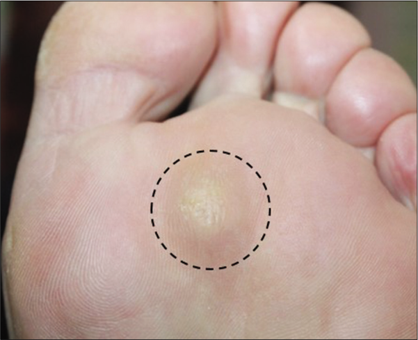
- Slightly elevated subcutaneous mass on the sole
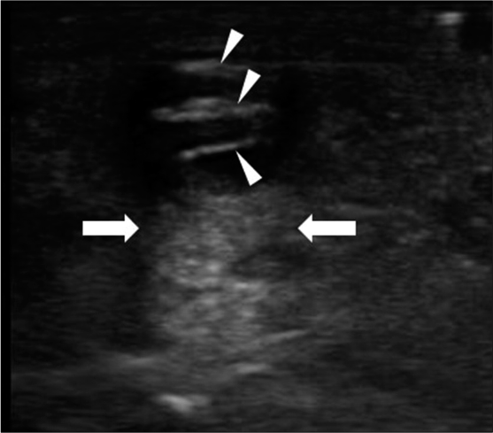
- Ultrasound image showing a well-circumscribed hypoechoic mass. Posterior acoustic enhancement (arrows) and hyperechoic strips (arrowheads) are seen
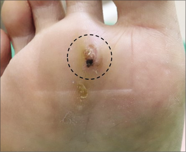
- Slightly elevated subcutaneous mass with hyperkeratotic surface on the sole
- Ultrasound image showing a well-circumscribed hypoechoic masswith posterior acoustic enhancement (arrows) and hyperechoic strips (arrowheads)
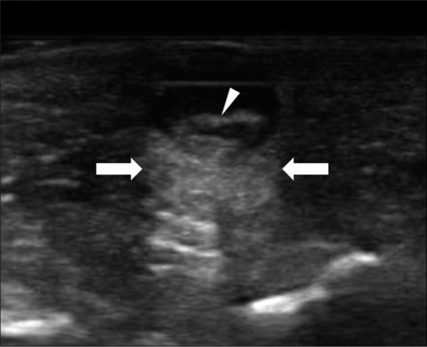
- Slightly elevated subcutaneous mass on the sole
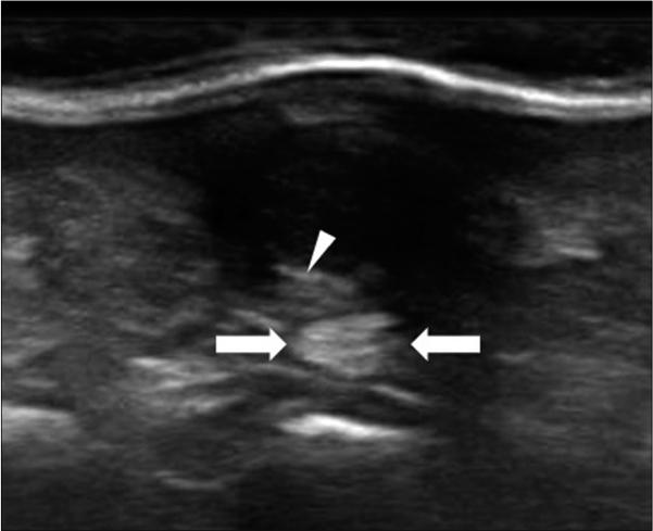
- Ultrasound image showing a poorly circumscribed hypoechoic mass with posterior acoustic enhancement (arrows) and hyperechoic strips (arrowheads)
Till now, studies analyzing the clinical and sonographic features of epidermal cysts at various sites have been conducted.1,3 However, there is a lack of studies evaluating sonographic features of plantar epidermal cysts. As the sole is a weight-bearing site, plantar epidermal cyst often presents as a slightly elevated firm nodule or mass. Thus, epidermal cysts of the sole are frequently misdiagnosed as a callus or viral wart which leads to inappropriate treatment.2 Therefore, the initial evaluation is important not only for diagnosis but also for proper management.
For the initial evaluation of soft-tissue masses, the ultrasound is increasingly being used in the field of dermatology. Although they sometimes have a non-specific appearance, epidermal cysts often show typical features on the ultrasound. They typically show well-circumscribed round-to-oval shaped cysts with posterior acoustic enhancement.4 In our study, it was remarkable that plantar epidermal cysts showed a mass with homogeneous or heterogeneous hypoechoic internal contents with posterior acoustic enhancement in all cases. Even though our study was conducted retrospectively, these findings agree with those of previous studies which indicate that the sonographic features of epidermal cysts of the plantar area are consistent with those of other sites.1,3
Furthermore, the differential diagnosis of epidermal cysts includes other soft-tissue tumors such as synovial cysts, ganglions, pilomatricomas, fibromas, lipomas and xanthomas.3 The typical sonographic features of these soft tissue tumors are summarized in Table 2. The diagnosis of epidermal cysts should be made based on sonographic features in correlation with clinical presentation, location and further histopathologic examination, if needed. Epidermal cysts contain variable amounts and arrangements of keratin debris.4 Scattered fragments of packed lamellae of keratin might appear as a hypoechoic or hyperechoic echotexture on the ultrasound. Hyperechoic strips might correlate with dense keratin debris, whereas hypoechoic debris might correlate with lipid fragments or keratin fragments containing additional water. In our case series, seven (78%) cysts had either hypoechoic or hyperechoic inner contents, and these two specific sonographic features suggest that the presence of keratin debris could be used to distinguish epidermal cysts from other soft-tissue tumors. Furthermore, Shimizu et al. demonstrated that an important pathological feature of plantar epidermal cysts is the predominance of compact orthokeratotic material as their content.5 The authors suggested that the pathological features of plantar epidermal cysts are due to invagination of keratin as a result of mechanical trauma or pressure on the sole. Thus, the anatomical site leads to an atypical clinical presentation; however, characteristic pathological features like more frequent inner echoes on the ultrasound could be identifying feature of plantar epidermal cysts.
| Shape | Margin | Homogenicity | Echogenicity | Internal debris | Posterior acoustic feature | Vascularity | |
|---|---|---|---|---|---|---|---|
| Pilomatricoma | Round to oval | Well-circumscribed | Heterogeneous | Variable | Echogenic foci | Shadowing | Variable |
| Xanthoma | Round to oval | Well-circumscribed | Heterogeneous | Hypoechoic | Absent | Absent | Absent |
| Ganglion cyst | Round to oval | Well-circumscribed | Homogeneous | Hypoechoic | Septations | Enhancement | Variable |
| Synovial cyst | Round to oval | Well-circumscribed | Homogeneous | Hypoechoic | Septations | Enhancement | Variable |
| Lipoma | Round to oval | Variable | Variable | Variable | Echogenic lines | Absent | Absent |
| Fibroma | Round to oval | Well-circumscribed | Heterogeneous | Hypoechoic to mixed | Alternating bands | Absent | Absent |
In conclusion, as the sonographic findings of plantar epidermal cysts are similar to those of epidermal cysts at other sites, initial evaluation by ultrasound would be helpful in the diagnosis and further proper management.
Declaration of patient consent
The authors certify that they have obtained all appropriate patient consent.
Financial support and sponsorship
Nil.
Conflicts of interest
There are no conflicts of interest.
References
- Epidermal cysts in the superficial soft tissue: Sonographic features with an emphasis on the pseudotestis pattern. J Ultrasound Med. 2011;30:11-7.
- [CrossRef] [Google Scholar]
- Differences in sonographic features of ruptured and unruptured epidermal cysts. J Ultrasound Med. 2012;31:265-72.
- [CrossRef] [Google Scholar]
- Relationship between sonographic and pathologic findings in epidermal inclusion cysts. J Clin Ultrasound. 2001;29:374-83.
- [CrossRef] [Google Scholar]
- Clinicopathologic features of epidermal cysts of the sole: Comparison with traditional epidermal cysts and trichilemmal cysts. J Cutan Pathol. 2005;32:280-5.
- [CrossRef] [Google Scholar]


