
Translate this page into:
Predictive equation to identify infection due to anthropophilic or zoophilic dermatophytes based on clinical features and risk factors: A ten-year retrospective study
Corresponding author: Assoc. Prof. Charussri Leeyaphan, Department of Dermatology, Faculty of Medicine Siriraj Hospital, Mahidol University, Bangkok, Thailand. charussrilee@gmail.com
-
Received: ,
Accepted: ,
How to cite this article: Bunyaratavej S, Kiratiwongwan R, Komoltri C, Lertrujiwanit K, Leeyaphan C. Predictive equation to identify infection due to anthropophilic or zoophilic dermatophytes based on clinical features and risk factors: A ten-year retrospective study. Indian J Dermatol Venereol Leprol 2022;88:416-9.
Sir,
Studies on the clinical features and risk factors differentiating anthropophilic cutaneous dermatophytosis and zoophilic cutaneous dermatophytosis are limited. Thus, we aimed to determine the correlation among the associated factors and the type of causative dermatophytes and further to develop an equation to predict the presence of zoophilic dermatophytes in patients with cutaneous dermatophytosis.
This ten-year, retrospective cross-sectional study was conducted at the Department of Dermatology, Faculty of Medicine Siriraj Hospital, Mahidol University, Bangkok, Thailand. We included patients diagnosed with either anthropophilic or zoophilic cutaneous dermatophytosis of glabrous skin (based on results of fungal culture) and whose clinical pictures were available. Patients with concomitant dermatologic conditions that may have interfered with the clinical evaluation, and cases without fungal culture results were excluded. The clinical findings were reviewed by dermatologists and the clinical features are illustrated in Figure 1.
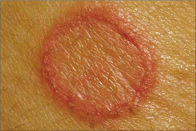
- A red-rubber-ring appearance
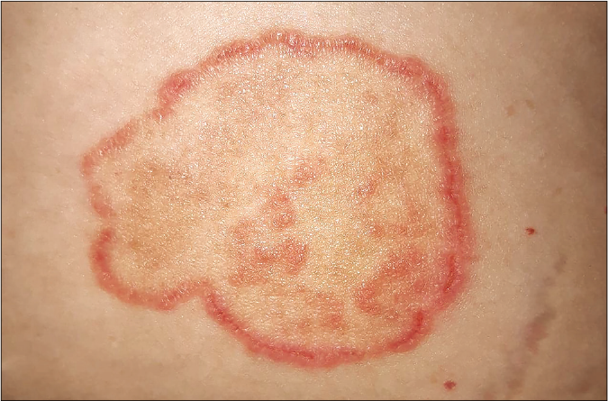
- A ring-within-a-ring appearance which is characterized by annular polycyclic erythematous rings with active border
The sample size was calculated using a Chi-square test. It was calculated based on prevalence of the causative organism. It was estimated that the error may be higher than 0.05, so it was recommended to use 0.07. However, this is a retrospective study and all data of patients were collected to do the analysis. A previous study reported the prevalence of Trichophyton mentagrophytes var. mentagrophytes to be 52%.1 Using a 2-sided Type I error of 0.07 and 95% confidence interval, a sample size of 196 patients was required. However, we only included patients who had complete data and pictures of the clinical findings. Therefore, 167 patients with complete data were included in the analysis.
Out of 167 patients (mean age, 44 years) included in the study, 108 (64.7%) patients had anthropophilic cutaneous dermatophytosis and the remainder (n = 59, 35.3%) had zoophilic cutaneous dermatophytosis. All patients were Asian. The patients with anthropophilic cutaneous dermatophytosis included Trichophyton rubrum (n = 95, 56.9%), T. tonsurans (n = 7, 4.2%), Epidermophyton floccosum (n = 3, 1.8%), Trichophyton interdigitale (n = 2, 1.2%) and Microsporum audouinii (n = 1, 0.6%). The zoophilic cutaneous dermatophytosis included T. mentagrophytes (n = 34, 20.4%), Microsporum canis (n = 23, 13.8%) and Trichophyton erinacei (n = 2, 1.2%). The baseline characteristics and morphological features are detailed in Table 1. Of the 51 patients who reported using topical corticosteroids, 26 (51%) had a ring-within-a-ring appearance. However, that appearance was not significantly associated with a history of previous topical corticosteroid usage (P = 0.307).
| Number (%) | P-value | ||
|---|---|---|---|
| Anthropophilic (n=108) | Zoophilic (n=59) | ||
| Female | 46 (42.6) | 39 (66.1) | 0.006* |
| Age (y), mean±SD | 44.9±18.5 | 42.9±17.8 | 0.497 |
| Median duration of symptoms (months) | 3.0 | 1.0 | 0.001* |
| Underlying diseases† | 44 (40.7) | 21 (35.6) | 0.619 |
| Dyslipidemia | 16 (14.8) | 8 (13.6) | 1.000 |
| Hypertension | 22 (20.4) | 13 (22.0) | 0.844 |
| Diabetes mellitus | 11 (10.2) | 6 (10.2) | 1.000 |
| Cardiovascular disease | 5 (4.6) | 1 (1.7) | 0.425 |
| Other underlying diseases | 22 (20.4) | 10 (16.9) | 0.683 |
| Previous topical medication usage‡ | 76 (70.4) | 40 (67.8) | 0.729 |
| Corticosteroids | 23 (21.3) | 28 (47.5) | 0.001* |
| Immunomodulators | 1 (0.9) | 0 (0) | 1.000 |
| Antifungals | 12 (11.1) | 4 (6.8) | 0.423 |
| Antibiotics | 1 (0.9) | 2 (3.4) | 0.285 |
| Local herb | 6 (5.6) | 1 (1.7) | 0.423 |
| Unidentified OTC medications | 39 (36.1) | 8 (13.6) | 0.002* |
| Contact with pets§ | 26 (37.1) | 38 (76.0) | <0.001* |
| Cat | 6 (8.6) | 29 (58.0) | <0.001* |
| Dog | 17 (24.3) | 9 (18.0) | 0.504 |
| Rabbit | 1 (1.4) | 3 (6.0) | 0.305 |
| Other | 4 (5.7) | 0 (0) | 0.141 |
| NA | 37 | 9 | |
| Affected area | |||
| Exposed area | 10 (9.3) | 30 (50.8) | <0.001* |
| Unexposed area | 59 (54.6) | 15 (25.4) | |
| Both exposed and unexposed areas | 39 (36.1) | 14 (23.7) | |
| Morphological feature | |||
| Redness | 83 (76.9) | 54 (91.5) | 0.020* |
| Induration | 81 (75.0) | 54 (91.5) | 0.013* |
| Vesicles/pustules | 14 (13.0) | 24 (40.7) | <0.001* |
| A red-rubber-ring appearance | 2 (1.9) | 14 (23.7) | <0.001* |
| Active border | 96 (88.9) | 48 (81.4) | 0.240 |
| Scale | 103 (95.4) | 54 (91.5) | 0.326 |
| Excoriation | 45 (41.7) | 23 (39.0) | 0.869 |
| PIH | 86 (79.6) | 26 (44.1) | <0.001* |
| A ring-within-a-ring appearance | 73 (67.6) | 22 (37.3) | <0.001* |
A logistic regression analysis was performed to obtain an equation to predict zoophilic cutaneous dermatophytosis. Variables in an equation predicting zoophilic dermatophytosis included contact with pets, vesicles/pustules, involving unexposed area, and a ring-within-a-ring appearance [Table 2]. The receiver operating characteristic curve of the simplified predictive equation for zoophilic cutaneous dermatophytosis had an area under the curve of 0.835. The scores ranged from –2 to +2 [Table 3]. With a score ≥ 0, the simplified equation showed the best sensitivity (80%), specificity (82%) and accuracy (81%) in the prediction of zoophilic cutaneous dermatophytosis. Thus, score ≥ 0 was used as the cut-off value.
| Model | Logistic regression equation | |||
|---|---|---|---|---|
| Final | ln odds | = | 0.158+ | 1.198X1+1.459X2–1.215X3–1.202X4 |
| Simplified | Score | = | X1+1.218X2–1.014X3–1.003X4 | |
| = | X1+X2–X3–X4 | |||
X1, contact with pets; X2, vesicles/pustules; X3, involving unexposed area; and X4, a ring-within-a-ring appearance, where 0=No and 1=Yes
| Number | Sensitivity (%) | Specificity (%) | Accuracy (%) | ||||
|---|---|---|---|---|---|---|---|
| Zoophilic dermatophytosis (n=50) | Anthropophilic dermatophytosis (n=71) | ||||||
| Score | –1, 0, 1, 2 | Indicate zoophilic dermatophytosis | 46 | 41 | 92 | 42 | 63 |
| –2 | Indicates anthropophilic dermatophytosis | 4 | 30 | ||||
| 0, 1, 2 | Indicate zoophilic dermatophytosis | 40 | 13 | 80 | 82 | 81 | |
| –2, –1 | Indicate anthropophilic dermatophytosis | 10 | 58 | ||||
| 1, 2 | Indicate zoophilic dermatophytosis | 24 | 5 | 48 | 93 | 74 | |
| –2, –1, 0 | Indicate anthropophilic dermatophytosis | 26 | 66 | ||||
As fungal culture is still limited to some hospitals and takes approximately one month to obtain results, this study created a new and simplified equation from clinical data to distinguish cases of zoophilic cutaneous dermatophytosis from those due to anthropophilic dermatophytes. The equation contained only four variables, showed high sensitivity and specificity, and made it easy for physicians to determine the type of causative dermatophytes resulting in appropriate treatment and disinfection methods to be implemented promptly well before the fungal culture results became available.
This study revealed that redness, induration, vesicles/ pustules and a red-rubber-ring appearance were significantly found in lesions from zoophilic cutaneous dermatophytosis, corresponding with the findings of earlier studies, which reported that zoophilic cutaneous dermatophytosis tend to form more inflammatory lesions.1-3 Moreover, a red-rubberring appearance, which is characterized by the presence of one or more bright erythematous and edematous rings with central clearing skin,1 was significantly reported in cases of zoophilic cutaneous dermatophytosis. As patients with zoophilic cutaneous dermatophytosis tend to have vesicles which resemble eczema, it follows that use of topical corticosteroids was found in a significantly higher proportion of cases of zoophilic cutaneous dermatophytosis.
A ring-within-a-ring appearance was seen predominantly in patients with anthropophilic cutaneous dermatophytosis. Specifically, it was significantly associated with Trichophyton rubrum infection (P = 0.016). Previous reports demonstrated a ring-within-a-ring appearance, or tinea pseudoimbricata, to be associated with repeated inflammatory responses and this may result from topical steroid abuse.4,5 However, a significant association between the use of topical corticosteroids and a ring-within-a-ring appearance was not found in our study, which comprised a larger number of patients than in the previous studies. Thus, a ring-within-a-ring appearance might be associated with anthropophilic cutaneous dermatophytosis rather than with a history of steroid use.
Differentiation between anthropophilic cutaneous dermatophytosis and zoophilic cutaneous dermatophytosis is important because it influences not only the appropriate treatment but also facilitates the elimination of the source of infection.3 In anthropophilic cutaneous dermatophytosis, apparels of the infected patients should not be shared with others and should be disinfected in order to prevent the spread of dermatophytes.2 As pets act as reservoirs for zoophilic cutaneous dermatophytosis,2,3 wearing of protective clothing before handling infected pets and timely and adequate treatment of those pets have been recommended.3
The main limitation of our study is that since it was a retrospective one, some data was unfortunately missing.
In conclusion, the presence of redness, inflammation, vesicles/ pustules, or a red-rubber-ring appearance may suggest zoophilic cutaneous dermatophytosis; in contrast, lesions with post-inflammatory hyperpigmentation or a ring-within-a-ring appearance may point towards a diagnosis of anthropophilic cutaneous dermatophytosis. Moreover, our new equation to differentiate anthropophilic cutaneous dermatophytosis and zoophilic cutaneous dermatophytosis is practically useful and can promptly guide treatment and disinfection methods.
Acknowledgments
We thank Dr. Supisara Wongdama for her kind support.
Declaration of patient consent
Institutional Review Board (IRB) permission obtained for the study.
Financial support and sponsorship
Nil.
Conflicts of interest
There are no conflicts of interest.
References
- Zoophilic dermatophytosis: A study of closed contact cases. Thai J Dermatol. 2008;24:194-207.
- [Google Scholar]
- Epidemiology of human dermatophytoses in Africa. Med Mycol. 2018;56:145-61.
- [CrossRef] [PubMed] [Google Scholar]
- 'Ring-within-a-ring' appearance: Morphological clue to topical steroid abuse in dermatophytosis. J Eur Acad Dermatol Venereol. 2017;31:e2-3.
- [CrossRef] [PubMed] [Google Scholar]
- Tinea pseudoimbricata as a unique manifestation of steroid abuse: A clinico-mycological and dermoscopic study from a tertiary care hospital. Indian Dermatol Online J. 2019;10:422-5.
- [CrossRef] [PubMed] [Google Scholar]


