
Translate this page into:
Current understanding of frictional dermatoses: A review
Corresponding author: Dr. Anuva Bansal, House No. 2, National Park, Lajpat Nagar-4, New Delhi, India. anuvabansal22@gmail.com
-
Received: ,
Accepted: ,
How to cite this article: Arora G, Khandpur S, Bansal A, Shetty B, Aggarwal S, Saha S, et al. Current understanding of frictional dermatoses: A review. Indian J Dermatol Venereol Leprol 2023;89:170-88.
Abstract
Human skin is continually exposed to internal and external forces, dynamic as well as static. The skin is normally flexible and can resist mechanical trauma due to friction, pressure, vibration, suction and laceration to a considerable degree. However, an excess of these forces can abnormally affect the structure and function of the skin, setting the stage for the development of a skin disorder. Repetitive trauma can cause lichenification, hyperpigmentation, erythema, scaling, fissuring, blisters, ulceration and chronic alterations. Frictional dermatoses is an under-recognised entity with no clear-cut definition and encompasses a variety of terms such as frictional dermatitis, frictional melanosis, frictional pigmentary dermatoses and certain other named entities, many of which are confusing. The authors propose to define frictional dermatoses as ‘a group of disorders caused by repetitive trauma to the skin as a result of friction of varied aetiology which can have a wide range of cutaneous manifestations depending on the type of insult.’ The exact prevalence of frictional dermatoses as a separate entity is unknown. Authors who conducted this review include a group of dermatologists and post graduate students from various institutions. Literature was reviewed through PubMed, Medscape, Medline, ResearchGate and Google Scholar using the terms ‘frictional dermatitis,’ ‘friction and skin,’ ‘dermatoses and culture,’ ‘clothing dermatitis,’ ‘friction melanosis,’ ‘PPE induced dermatoses in COVID-19 era,’ etc. A total of 122 articles were reviewed and 100 articles among them were shortlisted and included in the study, after removing duplications. The review was followed up with further deliberation which resulted in the formulation of a new definition and classification of frictional dermatoses taking into account the morphology, histopathological characteristics, anatomical region affected and the major predisposing factors. The rising incidence of mechanical dermatoses in the COVID-19 era was also emphasised.
Keywords
Cultural dermatoses
friction blisters
frictional dermatoses
occupational dermatoses
sports dermatoses

Introduction
Human skin is continually exposed to internal and external dynamic as well as static forces. The skin is normally flexible and can resist mechanical trauma to a considerable degree. However, an excess of these forces can abnormally affect the structure and function of the skin, setting the stage for the development of a skin disorder.
The term ‘frictional dermatoses’ encompasses a variety of entities such as frictional dermatitis, frictional melanosis, frictional pigmentary dermatoses and certain other named conditions, many of which are confusing. Friction is defined as the resistance experienced by a body when it comes in contact with another. Any imbalance in the frictional forces may lead to acute or chronic injury. Friction can also involve the mucosae1-4 hair5-8 and nails.9,10
Frictional dermatoses is an under-recognised entity with no clear-cut definition. The authors propose to define frictional dermatoses as ‘a group of disorders caused by repetitive trauma to the skin as a result of friction of varying aetiology which can have a wide range of cutaneous manifestations depending on the type of insult.’
The exact prevalence of frictional dermatoses as a separate entity is unknown. However, various studies have reported the prevalence of individual conditions. A study from Germany reported that 21.6% (89/412) musicians had musical instrument related frictional skin disorders.11,12 Friction accounted for 11.3% of occupational dermatoses due to non-glove personal protective equipment amongst healthcare workers in UK.1,11
Materials and Methods
Authors who conducted the review include a group of dermatologists and postgraduate students from various institutions. They are members of the Resident Connect Committee of the Indian Association of Dermatologists Venereologists and Leprologists of the Delhi State Branch. It was a voluntary exercise proposed by the Chairperson of the Resident Connect Committee, Delhi State Branch. After a framework was decided on by participation of all authors on a virtual platform, different sections of the ‘review of literature’ were assigned to each. Literature was reviewed through PubMed, Medscape, Medline, ResearchGate and Google Scholar using the terms ‘frictional dermatitis,’ ‘friction and skin,’ ‘dermatoses and culture,’ ‘clothing dermatitis,’ ‘friction melanosis,’ ‘PPE induced dermatoses in COVID-19 era,’ etc. A total of 122 articles were reviewed and 100 articles were shortlisted for inclusion in the study, after removing duplications. The selected articles included brief communications, letters to editor, case reports, case series, observational studies, review articles and randomised controlled trials. The review was followed up with further deliberation and a new definition and classification for frictional dermatoses was formulated. The rising incidence of mechanical dermatoses in the COVID-19 era was also emphasised upon.
Predisposing Factors
Friction not only causes new dermatoses in individuals but can also exacerbate existing ones.10 The susceptibility of an individual’s skin to friction differs and is influenced by factors both extraneous and inherent, such as the presence of pre-existing diseases and genetic or racial differences, respectively [Table 1].
| Predisposing factor | Pathogenesis |
|---|---|
| Inherent factors | |
Extremes of age13
|
|
| Gender10 |
|
| Body site14 |
|
| Sweating14 |
|
| Extraneous factors | |
| Temperature |
|
| Relative humidity14 |
|
| Clothing |
|
Occupation and habits16-18
|
|
COF: Coefficient of friction
It is important to identify the predisposing factors that may be largely apparent or sometimes inconspicuous, but which are critical in the management of frictional dermatoses.
Pathogenesis
In simple terms, friction is the rubbing of one surface over the other or the force that resists movement between two bodies [Figure 1].19 The manifestation of friction related skin injury depends on the following factors:

- Pathogenesis of frictional dermatoses. Friction is the rubbing of one surface over the other or the force that resists movement between two bodies. The manifestation of friction related skin injury depends on: type of friction which can be static or dynamic; intensity of the force (low or high); and, nature of the surface which includes body site, moisture content and the coefficient of friction
Type of friction
Static friction, which refers to the friction between two still surfaces, dynamic friction which prevents motion between two sliding surfaces and shear force causing shear strain on deeper layers.19
Intensity of the force
Low intensity stimulus, which causes rapid cellular turnover and hyperkeratosis, or a high intensity one leads increased hydrostatic pressure and intraepidermal bullae formation.20
Nature of the surface
The co-efficient of friction depends on the type of surface, moisture content, body site as well as type of fabric covering the skin.
Moisture content: The presence of moisture increases the co-efficient of friction making the well hydrated skin over palms and fingers amenable to frictional injuries. Conditions such as humidity, sweating and occlusion increase the moisture content of the skin, thereby increasing the Coefficient of friction14
Body site: There is increased moisture retention as well as constant rubbing between the skin surfaces of flexures and intertriginous areas. This further increases the co-efficient of friction14
Type of fabric: The type and structure of textiles can have a considerable impact on the type of friction forces as well as skin hydration. The fabrics most commonly associated with dermatoses include nylon and wool.15
Classification
As previously outlined, no single definition exists for frictional dermatoses and therefore currently there is no accepted classification encompassing the wide range of conditions that can be categorised under this entity. We propose a classification that takes into consideration the morphology, histopathological characteristics, anatomical region affected and the major predisposing factors such as the occupation of the individuals most commonly affected. This allows for a quick clinical diagnosis and treatment planning. However, it must be noted that the entities may not be specific to the occupation they are grouped under and may be found in patients with other occupations too.
Primary frictional dermatoses
Primary frictional dermatoses include entities where repeated friction is the major etiological factor responsible for development of the lesion. These conditions may be further classified based on the major predisposing factor such as the occupation, cultural or religious practices and the anatomical region affected [Figure 2], as outlined in Table 2.

- Important frictional dermatoses based on the location. Frictional dermatoses may be classified based on the major predisposing factor such as the occupation, cultural or religious practices and the anatomical region affected
| Entity | Primary Frictional Dermatoses | ||||||
|---|---|---|---|---|---|---|---|
| Anatomical Region | |||||||
| Generalised | Head and Neck | Trunk | Limbs | Hair | Mucosa | Nails | |
| 1. Occupational | |||||||
| A. Sports Related | - | - | Joggers nipples17 |
|
- | - | - |
| B. Soldiers | - | - | - | - | - | - | |
| C. Musicians | - | Cellists chest27 | - | - | - | ||
| D. Computer/Electronic device-related | - | - | - | - | - | - | |
| 2. Related to Cultural Practices | |||||||
| A. Frictional Melanosis | - | Facial frictional melanosis31 | - | - | - | ||
| B. Clothing Related | Nylon cloths friction dermatosis38 | - | Friction blisters due to socks15 | Friction alopecia due to socks7,8 | - | - | |
| AC. Religious Practices/ Praying related | - | Prayer’s Nodules18,41 | - | - | - | - | |
| 3. Miscellaneous | |||||||
| - | Acne mechanica43 Folliculitis mechanica43 Traumatic anserine folliculitis44 |
- | Amputation stump callosities45 | - | Frictional trauma to nails10 | ||
Secondary frictional dermatoses
This category includes entities where friction plays a contributory role in the pathogenesis; however, its presence alone is not sufficient for development of the lesion. Infection, sweat or other inciting agents and/or a pre-existing dermatoses are prerequisites for the development of this set of conditions.
Clinical Features [Table 3]
| Disease entity | Morphology | Location | Associated features |
|---|---|---|---|
| 1. Frictional Dermatoses in Sportspersons | |||
| Jogger’s nipple17 | Cracked & painful nipple Bleeding± |
Nipple | Long distance runners Continuous friction from cotton type T shirts |
| Pool palm21 | Recurrent painful, bilateral erythematous patches. | Finger pads, palms and soles | Swimmers |
| Rower’s hand20,48 | Erythematous patch or blisters | Fingers, back of hand | Rowers |
| Athlete’s nodule17,49 (Skater’s nodules, collagenomas, ‘Nike nodule’ in runners, ‘knuckle pad’ in boxers) |
Soft skin coloured painful/ painless keratinised nodule. | Lateral malleoli, lateral sides of feet, overlying the Achilles tendon | Skaters, runners, boxers |
| Talon noir17,50 Black heel, calcaneal bleed/petechiae |
Blue black, linear to oval macule | Posterior or posterolateral aspect of heel | Basketball & tennis players, gymnasts |
| Skater’s toe/ nail23 | Toe tip callus, nail thickening or SUH | Toe tip or toenail | Skaters, Runners, ice hockey players |
| Skater’s nodule23,49 (Double ankle bones) |
Collagenoma which presents with hyperkeratotic, smooth surface nodules | Lateral malleoli, lateral sides of feet or skin overlying Achilles tendon | Skaters |
| Skater's pad23 | Callus like hyperkeratosis. Superficial than skater’s nodule. | Heels | Skaters |
| Skater’s bite23,51,52 | Pain and swelling, radiating pain from lower leg towards toe | Dorsum of foot or near the tongue of the skate | Skaters, ice hockey players |
| Malleolar bursitis 23,53 | Intermittent painful S/C mass. Superimposed infection may occur | Tip of malleolus. Medial malleolus most common. | Skaters |
| Piezogenic pedal papule23,54,55 [Figure 3] |
Asymptomatic papule may be painful | Medial or lateral aspect of heels (visualised better in standing position) | Common in skaters, runners & weightlifters with underlying connective tissue disease, rheumatic heart diseases |
| Baseball pitcher’s friction dermatitis6 | Appears as discoid eczema. | Inner ankle, lower knee | Baseball players with endogenous eczema |
| Tyloma48 | Uniform thickening of skin. Pain & bleeding if fissures develop | Pressure bearing areas of foot, palms and fingers. | Athletes |
| Pump bump23 | Painful and inflamed bumps | insertion of Achilles tendon | Skaters with: High arched foot Tight Achilles tendons or Walking on outer aspects of heels |
| Hyperkeratosis and frictional dermatitis due to practicing kendo24 | Asymptomatic hyperkeratotic to eczematous lesion seen a kendo player (a Japanese sport) | Palmoplantar area | Kendo |
| 2. Frictional Dermatoses in Sportspersons | |||
| Corns and Calluses25 [Figure 4] | Yellow-white hyperkeratotic callosities | Pressure bearing areas | Protective response to constant mechanical stimuli |
| Friction Blisters63 | Blisters | Palms and soles | Constant stamping action of the feet; shearing force between skin and external surface; separation of epidermis at the level of the stratum corneum. |
| 3. Frictional Dermatoses in Musicians | |||
| a. Head and Neck | |||
| Fiddler’s neck56 | Eythematous, hyperpigmented, lichenified lesion | Angle of the mandible | Friction at the site where the violin contacts the chin |
| Clarinetists cheilitis57 | Presents with erythema, scaling and lichenification Fissuring, atrophy and depigmentation may also be seen. | Median part of the vermilion border of the lower lip, chin | Due to the continuous friction, pressure and saliva which collects under lower lip. It is an irritant dermatitis in the area where the wooden reed comes in contact with the skin |
| Lip callosity28 | Skin colored or hyperpigmented hyperkeratotic plaque | Mid portion of the lip | Repetitively irritated because of intense contact with parts of the instrument |
| Flautist’s Chin58 | Scaly, erythematous plaques | Chin | Friction, pressure and moisture Sweat and saliva at the site of contact ACD to nickel, chromium |
| b. Trunk | |||
| Cellist’s chest59 | Presents with xiphoid discomfort and hyperpigmented erythematous plaques | Lower end of the sternum at the xiphoid process | - |
| c. Limbs | |||
| Drummer’s digits12 | Yellow-white hyperkeratotic callosities | Lateral aspect of the left index finger | - |
| Guitarist’s fingers/harpists fingers59,60 [Figure 5] | Paronychia, blisters, calluses Onycholysis, SUH | Sides and tips of the fingers | Harpist who is just starting is most prone to injury as calluses have not yet developed |
| Garrod’s fingers/Violinist’s Pad12,59 | Thickening of the skin and underlying tissues | Backs of IP joints of the index, middle fingers of left hand | Force is applied to the strings while playing, calluses protective mechanism |
| Frictional Dermatoses in Writers and Computer users | |||
| Writer’s bump29 | Well circumscribed yellow-white plaque. Slight tenderness on pressure | Lateral aspect of DIP of the middle finger of dominant hand | Constant pressure of tightly gripped pencils and pens while writing |
| Cell thumb/ Playstation thumb29,61 | Well defined, tender calluses | Medial aspect of the thumb | Excess phone gripping, occasionally |
| Frictional dermatitis due to computer mouse/ mouse fingers62 | Sharply demarcated lesions with erythema and scaling. | Palmar aspect of first and fifth fingertips | Constant rubbing while using a mouse |
| Mousing callus30,63 | Painless, yellowish, thickened callus | Palmar aspect of the wrist | Friction and pressure, on the one hand, prolonged use of computer mouse |
| A. Frictional Melanosis | |||
| a. Face | |||
| Facial frictional melanosis31 | Deep dark brown pigmentation | Bony prominences of the face | Aggressive rubbing of the face with hand or handkerchief |
| b. Trunk | |||
| Lifa disease33 | Deep dark brown pigmentation | Clavicles, shins, upper back and lateral aspect of the arms | Repeated rubbing by lifa (brush for washing) |
| Orphan Rocker Tracks34 | Frictional dermatosis resembling train tracks | Bony prominences of the lumbosacral spine | Children with autistic behaviour, habitual of rocking movements |
| Frictional dermatoses due to car seat35 | Linear hyperpigmented patches | Bony prominences on the back | Prolonged periods of sitting and driving |
| c. Limbs | |||
| Frictional asymptomatic darkening of extensor surfaces36,64 | Asymptomatic darkening overlying sites of friction, ‘sign of dirty knees and elbows.’ | Extensor surfaces of elbows and knees | Increased frictional rubbing |
| Frictional melanosis of inner thighs37 | Asymptomatic darkening. | Inner thighs | Frictional stress -rubbing of thighs; obese females |
| B. Clothing related | |||
| a. Generalised | |||
| Nylon cloth frictional dermatoses38 | Dark brown pigmentation | Bony prominences: clavicle, back, shoulders, ribs, spinous processes, extensors (limbs) | Reported in Japan -practice of rubbing body with wet or dry nylon cloth or scrub brushes |
| b. Limbs | |||
| Frictional dermatoses due to socks7 | Dark brown pigmentation | Just below the knees or above the ankles up to mid-calf. | Repeated wearing of tight socks/knee length boots |
| c. Trunk | |||
| Dermatoses due to sari, petticoat, salwar drawstrings39 | Post inflammatory hyperpigmentation or hypopigmentation | Waist | Sari, petticoat/salwar tied to the waist via a drawstring, pressure and friction and formation of an artificial groove |
| C. Religious Practices/ Praying related | |||
| a. Face and Limbs | |||
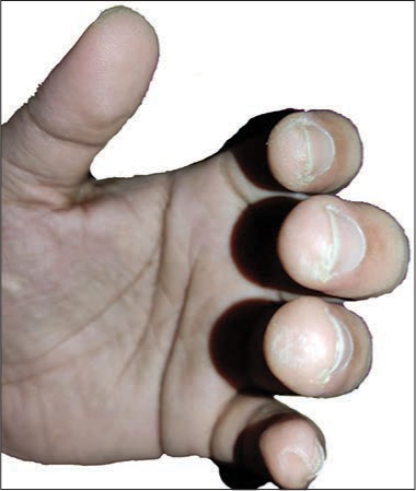
| Prayer Nodules18 [Figure 6] | Nodules or calluses | Forehead, Knees, ankles and dorsa of feet. | Squatting position -knees and ankles held against the floor, weight on the lower legs during prayers; touching forehead on the ground during prayer by Muslims. |
| Pew Blisters41 | Frictional blister | One or both knees | Repeated kneeling on pews in church |
| Davener’s dermatitis32 | Hyperpigmented ill-defined areas | Lower spinous processes | Exclusively in Jewish Israeli Yeshiva students, constant rocking of the upper body sitting on a firm wooden or metal chair with a rigid backrest during praying |
| Yoga sign42 | Hyperkeratotic, circumscribed, hyperpigmented plaques Patients suffering of neuropathy may ulceration of the callosities | Outer ankles and fifth toes | The characteristic Yoga sitting position on plain and hard floor exerts mechanical stress, that is, repeated and prolonged pressure and sheer forces Yoga sign is also seen in people who sit ‘cross legged’ |
SUH: Subungual hyperkeratoses, IP: Interphalangeal, DIP: Distal interphalangeal

- Piezogenic pedal papules-associated with figure skaters, runners and weightlifters, due to the high-impact surface collisions which cause herniation of subcutaneous fat into the dermis. Patients generally present with asymptomatic papules over the medial and lateral aspect of heels

- Callosities: Yellowish, hyperpigmented hyperkeratotic callosities present on the palm and proximal digits in a sportsperson
- Guitarist’s fingers: These present as calluses at the sides and tips of the fingers

- Prayer’s nodules: The religious practice of squatting and touching the forehead repeatedly on the ground during prayer by Muslims, leads to formation of nodules or calluses on the forehead
Primary
Occupational
Frictional dermatoses in sportspersons
Sports entail exposure to several factors such as trauma, prolonged sun exposure, infections and friction, thus increasing the propensity of a variety of skin problems amongst sportspersons. Frictional dermatoses are among the most common cutaneous manifestations in this group.6,17,2-24,50-56
Frictional dermatoses in soldiers
Friction-related skin injuries constitute one of the commonest dermatological conditions reported in military recruits and clinically present as corns, calluses or friction blisters localised to the pressure-bearing sites.25,26,50
Frictional dermatoses amongst musicians
Musicians are particularly predisposed to develop frictional dermatoses, which typically present at the site of contact with the musical instrument.16,27,58-60
Computer/electronic devices related frictional dermatoses
This category of frictional dermatoses has emerged in the past few decades due to the rampant use of computer electronics. The manifestations predominate on the users’ hands and fingers.29,30,61-63
Related to cultural practices
Indigenous cultural practices, religious customs and type of clothing also play a key role in causation of a friction related skin diseases and are especially relevant in the Indian setting.7,8,15,18,31-44,64
Miscellaneous
Certain clinically distinct frictional dermatoses are not specific to any category. Such entities, as well as those that manifest in the mucosa and nails, have been included here [Table 4].3,4,10,45-49,65,66
| Entity | Etiopathogenesis | Clinical feature | Diagnosis | Treatment |
|---|---|---|---|---|
| Face | ||||
| Acne mechanica43 |
|
|
|
|
| Traumatic anserine folliculosis44 [Figure 7] |
|
|
|
|
| Acanthoma fissuratum (misnomer: Granuloma fissuratum)65 [Figure 8] |
|
|
|
|
| Limbs | ||||
| Frictional dermatoses in amputees45 |
|
|
|
|
| Frictional dermatoses in oral mucosa | ||||
| Benign alveolar ridge keratosis3 |
|
|
|
|
| Morsicatio buccarum and linguarum4 |
|
|
|
|
| Frictional dermatoses of genitalia due to sexual practices | ||||
| Frictional trauma to nails10 |
|
|
|
|
| Frictional dermatoses of genitalia due to sexual practices | ||||
| Penile coital injury46 |
|
|
|
|
| Frictional dermatitis of Onan47 |
|
|
|
|
HPE: Histopathological evaluation, BCC: Basal cell carcinoma

- Traumatic anserine folliculosis: Characterised by multiple, closely-set, grouped follicular papules (‘anserine’ or goose-like appearance), usually on the chin, jaws or neck in children or adolescents, often with a history of resting or supporting the head in a particular position leading to repeated friction at the site

- Acanthoma fissuratum: Pruritic or asymptomatic unilateral; firm, folded coin-shaped lesion, flesh-coloured plaque with a central groove dividing the lesion into two halves (Coffee bean appearance) resulting from chronic persistent trauma due to ill-fitting spectacles
Secondary frictional dermatoses [Table 5]
| Secondary frictional dermatoses | Etiopathogenesis | Clinical features | Diagnosis | Treatment |
|---|---|---|---|---|
| Sweat dermatitis/Frictional sweat dermatitis5,67 |
|
|
|
|
| Diaper dermatitis68 |
|
|
|
|
| Juvenile plantar dermatoses/’sweating sock dermatitis’69 |
|
|
|
|
| Frictional lichenoid dermatitis/Dermatitis papulose adultorum/Sutton’s summertime prurigo70 [Figure 9] |
|
|
|
|
| Frictional dermatitis71 |
|
|
|
|
| Frictional amyloidosis/’Brush amyloidosis’72 |
|
|
|
|
DMSO: Dimethyl sulfoxide, HPE: Histopathological evaluation, KOH: Potassium hydroxide

- Frictional lichenoid eruption: Presents as grouped lichenoid papules over the elbows and knees in young children during spring and summer months
As we have already elucidated, entities traditionally classified as primary ‘frictional dermatoses’ are those caused due to the rubbing of one body against another, termed as dry friction. However, friction may be contributory to dermatoses wherein factors such as sweat, moisture or a pre-existing skin disease play a larger role. Friction also plays a pivotal role in many contact dermatoses, by disrupting the stratum corneum barrier.19,67-70,72-77
Differential Diagnosis
Careful history is of paramount importance in diagnosing frictional dermatoses and differentiating it from other clinically similar skin conditions. Differentials vary based on the location of the lesions, that is, skin, mucosa, hair and nail [Tables 6a and 6b].
| Entity | Differentials | ||||
|---|---|---|---|---|---|
| Differentials | Differentiating Features | Investigations | |||
| Head and neck | |||||
| Facial frictional melanosis30,73 |
|
|
Histopathology:
Dermoscopy:
|
||
| Facial macular amyloidosis |
|
Histopathology:
|
|||
|
|
Histopathology:
Dermoscopy: 30
|
|||
|
|
Histopathology:
|
|||
| Pigmented contact dermatitis |
|
Histopathology:
|
|||
|
|
Histopathology:
|
|||
|
|
- | |||
|
|
Dermoscopy:75,76
|
|||
| Fiddler’s neck16 |
|
|
Histopathology:
|
||
|
|
|
|||
| Traumatic anserine folliculosis44 |
|
|
Histopathology:
|
||
|
|
Histopathology:
|
|||
|
|
Histopathology:
|
|||
|
|
Histopathology:
|
|||
| Clarinetists cheilitis12 |
|
|
|
||
| Trunk | |||||
| Frictional melanosis77 |
|
|
Histopathology:
|
||
| Jogger’s nipple17 |
|
|
|
||
|
Long standing erythematous, scaly, or velvety patches or plaques over the nipple Usually unilateral
Serosanguinous discharge
|
Histopathology:
|
|||
|
|
- | |||
| Cellist’s chest12 |
|
Erythema, oedema, pruritus, oozing, vesiculation |
|
||
| Davener’s and lifa32,33 |
|
|
Histopathology:
Dermoscopy:31
|
||
OCPs: Oral Contraceptive Pills
| Entity | Differentials | Differentiating Features | Investigations | |||
|---|---|---|---|---|---|---|
| Extremities | ||||||
| Frictional melanosis of inner thigh37 Frictional asymptomatic darkening of extensor surfaces36 |
|
|
Histopathology:
Dermoscopy:31
|
|||
|
|
Histopathology:
|
||||
|
|
Histopathology:
|
||||
| Amputation stump callosities45 |
|
|
|
|||
|
|
Histopathology:
|
||||
| Socks alopecia8 |
|
|
|
|||
|
|
|
||||
|
|
Histopathology: Acute changes- sparse septal lymphohistiocytic infiltrates and foci of ischemic fat necrosis or hyalinisation of the lobules Late changes-
|
||||
|
|
- | ||||
| Mouse fingers62 |
|
|
Histopathology:
|
|||
|
|
|
||||
| Corns and callosities, tylomas22,23,56 |
|
|
Histopathology:
|
|||
| Black heel/hemorrhagic hyperkeratosis and calcaneal petechiae22,23,56 |
|
|
Histopathology:
|
|||
|
|
Histopathology:
Dermoscopy:91
|
||||
| Pool palms21,48 |
|
|
|
|||
| Skater’s nodules/Athlete’s nodules23 |
|
|
Histopathology:
|
|||
|
|
Histopathology:
|
||||
| Skater’s toenail23 |
|
|
|
|||
|
|
Histopathology:
|
||||
| Piezogenic pedal papules23 |
|
|
Histopathology:
Tumour cells are elongated with scant pink cytoplasm and vesicular nuclei and very rare mitotic figures27 |
|||
| Writer’s bump29 Prayers nodule18 Drummers digit12 Cell thumb29 Guitarists finger59,60 |
|
Exaggerated dermatoglyphics |
- | |||
| Mucosa | ||||||
| Frictional keratosis/ Benign alveolar ridge hyperkeratosis4 Morsicatio buccorum and linguarum/libea alba4 |
|
|
Histopathology:
|
|||
|
|
Histopathology:
|
||||
|
|
|
||||
|
|
Histopathology:
|
||||
| Frictional dermatitis of onan47 |
|
Erythema, oedema, pruritus, oozing, vesiculation | Patch test- positive | |||
|
|
- | ||||
|
|
Koh –pseudo hyphae Fungal culture- positive |
||||
|
|
Skin biopsy consistent with psoriasis | ||||
|
|
Histopathology:
|
||||
PPD: Paraphenylenediamine, USG: Ultrasonography, DVT: Deep vein thrombosis, KOH: Potassium hydroxide, EBV: Epstein-Barr virus
Diagnosis
The diagnosis of frictional dermatoses is based on a thorough history as well as complete physical examination of the patient. Examination must focus on the type, morphology, distribution and location of lesions. The pattern of involvement provides valuable clues to the probable sources of friction such as sports equipment, musical instruments and cultural practices. The morphology helps establish the cause as well as duration of the insult. Non-invasive modalities such as dermoscopy are a useful diagnostic aid [Figure 10a].5,7,31,78-81 In addition, patch and photo patch testing may be helpful to rule out contact dermatitis, an important differential. Diagnostic dilemma and differentiation from clinical mimics may warrant a skin biopsy [Figure 10b]. Most of the entities are characterised by lichenification and show hyperkeratosis, acanthosis and elongation of rete ridges. Hypergranulosis accompanying these features is seen in callus or nodule formation.22,23 Conditions presenting with folliculitis including traumatic anserine folliculitis and acne mechanica reveal dilated follicular infundibulum filled with keratin along with a mild perivascular infiltrate. Foreign-body reaction, with abscess, cyst and granuloma formation can be seen in Fiddler’s neck.16,41,43 Hyperpigmentation as seen in frictional melanosis, Lifa disease or Davener’s dermatitis correlates histologically with increased basal layer pigmentation and pigment incontinence.31-33 Friction blisters are characterised by an intraepidermal split without inflammation while eczematous lesions show oedema with intraepidermal neutrophilic and mononuclear infiltrate.25,57,71

- Important dermoscopic features of frictional dermatoses. Most of the entities are characterised by lichenification and show hyperkeratosis, acanthosis and elongation of rete ridges. Hypergranulosis accompanying these features is seen in callus or nodule formation. Conditions presenting with folliculitis including traumatic anserine folliculitis and acne mechanica reveal dilated follicular infundibulum filled with keratin along with a mild perivascular infiltrate. Foreign-body reaction, with abscess, cyst and granuloma formation can be seen in Fiddler’s neck. Hyperpigmentation as seen in frictional melanosis, Lifa disease or Davener's dermatitis correlates histologically with increased basal layer pigmentation and pigment incontinence. Friction blisters are characterised by an intraepidermal split without inflammation while eczematous lesions show oedema with intraepidermal neutrophilic and mononuclear infiltrate

- Important histopathological features of frictional dermatoses: Most of the entities are characterised by lichenification and show hyperkeratosis, acanthosis and elongation of rete ridges. Hypergranulosis accompanying these features is seen in callus or nodule formation. Conditions presenting with folliculitis including traumatic anserine folliculitis and acne mechanica reveal dilated follicular infundibulum filled with keratin along with a mild perivascular infiltrate. Foreign-body reaction, with abscess, cyst and granuloma formation can be seen in Fiddler’s neck. Hyperpigmentation as seen in frictional melanosis, Lifa disease or Davener's dermatitis correlates histologically with increased basal layer pigmentation and pigment incontinence. Friction blisters are characterised by an intraepidermal split without inflammation while eczematous lesions show oedema with intraepidermal neutrophilic and mononuclear infiltrate
Treatment
The principles of treatment include providing symptomatic relief, determining the mechanical aetiology and treating the lesion and preventing further recurrence. Various pressure relieving devices (e.g., footwear with cushioned insoles and silicon sheets lubricants and barrier creams) are effective tools to protect stratum corneum.82 The main treatment aims at the removal of triggering habits or contact with external causative agent. Topical agents such as keratolytics and humectants help in decreasing the hyperkeratosis and regulating epidermal proliferation. Various depigmenting agents such as retinoic acid analogues, kojic acid and arbutin help in decreasing hyperpigmentation. Q-switched Nd : YAG (1064 nm and 532 nm) laser has been found to be useful in frictional dermatoses with hyperpigmentation.27,83 Grenz rays help in decreasing the thickness of skin. A study reported remarkable improvement in patients with frictional hand dermatitis after six sessions, following a weekly dose of 400 rads (4Gy).84
Current Times: Mechanical Dermatoses in The Era of COVID-19
In the era of COVID-19 three major types of mechanical dermatoses have been reported, which include device-related pressure injury,85,86 moisture-associated skin damage and skin tears.87,88 According to a study done by Jiang et al. the prevalence of device-related pressure injury skin injuries amongst healthcare workers was 30% and this prevalence was reported to be higher as compared to patients using respiratory devices, tubes and splints (2.1–27.9%).89
This may be attributed to the prolonged duration of wearing personal protective equipment in combination with sweating and occlusion.
The common locations of device-related pressure injury among the medical personnel were bridge of the nose, ears, cheeks and forehead. Factors such as the pressure induced by the goggles on the bridge of nose and cheeks as well as the ear compression due to the mask strap, pressure on the forehead due to the surgical caps and face shield have been implicated [Figure 11].90

- Mask induced frictional dermatoses over the bridge of the nose
Sweating leads to moisture-associated skin damage, which causes skin maceration as well as redness, itching, pain and pricking. Sweat-induced skin soaking along with pressure can cause increased friction coefficient between the personal protective equipment and skin; thus, when the mask and goggles are removed quickly, it can lead to skin tears. The factors associated with increased skin tears on logistic regression analysis by Jiang et al., were grade of personal protective equipment, heavy sweating, daily wearing time and male gender.89 Bhoyrul et al., found a higher incidence of folliculitis/acne and occlusion and frictional dermatoses, when compared to glove related reactions.11 Rustemeyer et al., also found that tight fitting equipment induced pressure and friction could culminate in acne mechanica due to rupture of microcomedones.91 The authors suggested avoiding tight-fitting headgear and facemasks and recommended regular washing and cleansing to help improve this condition. In regard to dermatoses due to skin friction, mechanical factors including material and repetitive forces when wearing personal protective equipment can lead to occupational and frictional dermatoses.91 The recommendations are to use materials with less shearing forces which are more breathable and comfortable. Furthermore, non-glove personal protective equipment specially footwear provides ideal environment for the growth of human papilloma virus since the moist environment and frictional forces associated with it increase the risk of abrasions and thus entry of human papilloma virus, causing warts.11
Taking frequent breaks and removing excess sweat may facilitate alleviation of frictional dermatoses related to protective gear. Adequate hydration and regular use of emollients are essential to maintain barrier integrity and it is recommended that moisturisers should be used at least half an hour before wearing the mask. Low potency topical steroids or tacrolimus may be necessary in cases which fail to respond to conservative therapy.92
Conclusion
Frictional dermatoses may be defined as ‘a group of disorders caused by repetitive trauma to the skin as a result of friction of varying aetiology, which can have a wide range of cutaneous manifestations depending on the type of insult.’
The susceptibility of an individual’s skin to friction differs and is influenced by factors both extraneous and inherent. The manifestation of friction related skin injury depends on the type of friction, intensity of the force and the nature of the surface and can clinically present as lichenification, hyperpigmentation, erythema, scaling, fissuring, blisters, ulceration and chronic alterations. We have devised a simple classification based on morphology, histopathological characteristics, anatomical region affected and the major predisposing factors such as the occupation. In the current era of COVID-19, three major types of mechanical dermatoses including device-related pressure injury, moisture-associated skin damage and skin tears have been described.
The diagnosis of frictional dermatoses is based on a detailed history and examination where the pattern of involvement provides valuable clues to the probable sources of friction and morphology helps establish the cause as well duration of the insult. The principles of treatment include providing symptomatic relief, determining the mechanical aetiology, treating the lesion and preventing further recurrence.
Declaration of patient consent
Patients’ consent not required as there are no patients in this study.
Conflicts of interest
There are no conflicts of interest.
Financial support and sponsorship
Nil.
References
- Common cutaneous disorders in athletes. Sport Med. 1990;9:100-19.
- [CrossRef] [PubMed] [Google Scholar]
- Friction melanosis: A clinical, histologic, and ultrastructural study in Jordanian patients. Int J Dermatol. 2004;43:261-4.
- [CrossRef] [PubMed] [Google Scholar]
- Benign alveolar ridge keratosis (oral lichen simplex chronicus): A distinct clinicopathologic entity. J Am Acad Dermatol. 2008;58:151-7.
- [CrossRef] [PubMed] [Google Scholar]
- Frictional keratosis, contact keratosis and smokeless tobacco keratosis: Features of reactive white lesions of the oral mucosa. Head Neck Pathol. 2019;13:16-24.
- [CrossRef] [PubMed] [Google Scholar]
- Atypical presentation of sweat dermatitis with review of literature. Indian Dermatol Online J. 2019;10:698.
- [CrossRef] [PubMed] [Google Scholar]
- Baseball pitcher's friction dermatitis. Contact Dermatitis. 2002;47:176-7.
- [CrossRef] [PubMed] [Google Scholar]
- Frictional (Sock) alopecia of the legs: Trichoscopy as an aid. Int J Trichol. 2018;10:129-30.
- [CrossRef] [PubMed] [Google Scholar]
- Frictional alopecia of the distal legs: Case series and review. Dermatol Online J. 2016;22:129-30.
- [CrossRef] [Google Scholar]
- Decrease of superficial serine and lactate in the stratum corneum due to repetitive frictional trauma. Int J Dermatol. 2018;57:299-305.
- [CrossRef] [PubMed] [Google Scholar]
- Dermatitis from repeated trauma to the skin. Am J Ind Med. 1985;8:307-17.
- [CrossRef] [PubMed] [Google Scholar]
- A review of non-glove personal protective equipment-related occupational dermatoses reported to EPIDERM between 1993 and 2013. Contact Dermatitis. 2019;80:217-21.
- [CrossRef] [PubMed] [Google Scholar]
- Skin conditions in instrumental musicians: A self-reported survey. Contact Dermatitis. 2008;58:217-22.
- [CrossRef] [PubMed] [Google Scholar]
- Elastic properties of human skin: Relation to age, sex, and anatomical region. Arch Dermatol Res. 1990;282:283-8.
- [CrossRef] [PubMed] [Google Scholar]
- Friction of human skin against different fabrics for medical use. Lubricants. 2016;4:6.
- [CrossRef] [Google Scholar]
- Textiles and human skin, microclimate, cutaneous reactions: An overview. Cutan Ocul Toxicol. 2006;25:23-39.
- [CrossRef] [PubMed] [Google Scholar]
- Skin manifestations of athletes competing in the summer olympics: What a sports medicine physician should know. Sports Med. 2012;42:399-413.
- [CrossRef] [PubMed] [Google Scholar]
- Dermatoses due to Indian cultural practices. Indian J Dermatol. 2015;60:3-12.
- [CrossRef] [PubMed] [Google Scholar]
- Friction-induced skin injuries are they pressure ulcers? An updated NPUAP white paper. J Wound Ostomy Cont Nurs. 2015;42:62-4.
- [CrossRef] [PubMed] [Google Scholar]
- Occupational dermatitis from physical causes. Clin Dermatol. 1992;10:231-43.
- [CrossRef] [Google Scholar]
- Sports dermatology part 1: Common dermatoses. CMAJ. 2004;171:851-3.
- [CrossRef] [PubMed] [Google Scholar]
- Skin conditions in figure skaters, ice-hockey players and speed skaters: Part II cold-induced, infectious and inflammatory dermatoses. Sport Med. 2011;41:967-84.
- [CrossRef] [PubMed] [Google Scholar]
- Hyperkeratosis and frictional dermatitis from practicing Kendo. Case Rep Dermatol. 2010;2:65-8.
- [CrossRef] [PubMed] [Google Scholar]
- Friction blisters: Pathophysiology, prevention and treatment. Sport Med. 1995;20:136-47.
- [CrossRef] [PubMed] [Google Scholar]
- Rook's Textbook of Dermatology. (9th ed). Hoboken, New Jersey: Wiley-Blackwell; 2016. p. :4696.
- [CrossRef] [Google Scholar]
- Frequency and associated factors of instrument-specific dermatoses among musicians in a military band: A cross-sectional study. J Mar Med Soc. 2018;20:111-5.
- [CrossRef] [Google Scholar]
- Cell thumb replaces writer's bump: Changing times, changing callouses. Case Rep Dermatol. 2020;12:1-4.
- [CrossRef] [PubMed] [Google Scholar]
- Frictional lichenified dermatoses from prolonged use of a computer mouse: Case report and review of the literature of computer-related dermatoses. Dermatol Online J. 2010;16:12.
- [CrossRef] [Google Scholar]
- Facial frictional melanosis in Indian patients: Defining the entity. Clin Dermatol Rev. 2019;3:78.
- [CrossRef] [Google Scholar]
- Davener's dermatoses: A variant of friction hypermelanosis. J Am Acad Dermatol. 2000;42:442-5.
- [CrossRef] [Google Scholar]
- Frictional dermal melanosis (Lifa disease) over bony prominences. J Dermatol. 2001;28:12-5.
- [CrossRef] [PubMed] [Google Scholar]
- Orphan rocker tracks: A variant of friction melanosis in an institutionalized child. Pediatr Dermatol. 2013;30:198-9.
- [CrossRef] [PubMed] [Google Scholar]
- Frictional dermatoses in a courier driver. Open Access Maced J Med Sci. 2017;5:541-2.
- [CrossRef] [PubMed] [Google Scholar]
- Frictional melanosis of rubbing thighs in Iraqi patients. J Cosmet Dermatological Sci Appl. 2014;4:203-11.
- [CrossRef] [Google Scholar]
- Epidemiological survey of nylon cloths friction dermatoses in Japan. J Dermatol. 1985;12:498-501.
- [CrossRef] [PubMed] [Google Scholar]
- Dermatological signs in South Asian women induced by sari and petticoat drawstrings: Clinical dermatology. Clin Exp Dermatol. 2010;35:459-61.
- [CrossRef] [PubMed] [Google Scholar]
- Dermatoses secondary to Asian cultural practices. Int J Dermatol. 2012;51:372-82.
- [CrossRef] [PubMed] [Google Scholar]
- Religion and the skin: Devotional dermatoses. JAMA Dermatol. 2013;149:1322.
- [CrossRef] [PubMed] [Google Scholar]
- Callosities of cross-legged sitting: “Yoga sign”-an under-recognized cultural cutaneous presentation. Int J Dermatol. 2008;47:1212-4.
- [CrossRef] [PubMed] [Google Scholar]
- Inner thigh friction as a cause of acne mechanica. Pediatr Dermatol. 2019;36:546-7.
- [CrossRef] [PubMed] [Google Scholar]
- Traumatic anserine folliculosis. Indian Dermatol Online J. 2017;8:59-61.
- [CrossRef] [PubMed] [Google Scholar]
- Circumcision and reduced risk of self-reported penile coital injuries: Results from a randomized controlled trial in Kisumu, Kenya. J Urol. 2010;184:203-9.
- [CrossRef] [PubMed] [Google Scholar]
- Pulling boat hands: A unique dermatoses from coastal New England. J Am Acad Dermatol. 1985;12:649-55.
- [CrossRef] [Google Scholar]
- Athlete's nodule in a figure skater: An unusual presentation. Am J Dermatopathol. 2015;37:e21-5.
- [CrossRef] [PubMed] [Google Scholar]
- Black heel, talon noir or calcaneal petechiae? Australas J Dermatol. 2008;49:148-51.
- [CrossRef] [PubMed] [Google Scholar]
- On thin ice: Preparing and caring for the ice skater during competition. Curr Sports Med Rep. 2008;7:133-7.
- [CrossRef] [PubMed] [Google Scholar]
- How To Evaluate Figure Skating Injuries Podiatry Today. 2020. Podiatry Today. Available from: https://www.hmpgloballearningnetwork.com/site/podiatry/article/5374 [last accessed on 2022 Feb 01]
- [Google Scholar]
- Malleolar bursitis in figure skaters: Indications for operative and nonoperative treatment. Am J Sports Med. 2000;28:109-11.
- [CrossRef] [PubMed] [Google Scholar]
- Piezogenic pedal papules in a marathon runner. Clin J Sport Med. 2006;16:81-3.
- [CrossRef] [PubMed] [Google Scholar]
- Piezogenic pedal papules: Treated by resection and hernial closure. Foot. 1998;8:171-2.
- [CrossRef] [Google Scholar]
- Pressure and friction injuries in primary care. Prim Care. 2015;42:631-44.
- [CrossRef] [PubMed] [Google Scholar]
- Clarinetist's cheilitis caused by immediate-type allergy to cane reed. Contact Dermatitis. 2007;56:243-5.
- [CrossRef] [PubMed] [Google Scholar]
- Harpist's finger: Case report of a trauma-induced blister in a beginner harpist and review of string instrument-associated skin problems in musicians. Cutis. 2008;82:329-34.
- [Google Scholar]
- Modern electronic devices: An increasingly common cause of skin disorders in consumers. Dermatitis. 2016;27:82-9.
- [CrossRef] [PubMed] [Google Scholar]
- Mouse fingers, a new computer-related skin disorder (2) J Am Acad Dermatol. 2001;45:477.
- [CrossRef] [PubMed] [Google Scholar]
- A new computer-associated occupational skin disorder: Mousing callus. J Am Acad Dermatol. 2006;55:358-9.
- [CrossRef] [PubMed] [Google Scholar]
- The sign of dirty knees and elbows. Acta Endocrinol (Copenh). 1954;16:305-8.
- [CrossRef] [PubMed] [Google Scholar]
- Acanthoma fissuratum: Lest we forget. Indian Dermatol Online J. 2017;8:141.
- [CrossRef] [PubMed] [Google Scholar]
- Vulvar granuloma fissuratum: A description of fissuring of the posterior fourchette and the repair. Obstet Gynecol. 2005;105:1018-23.
- [CrossRef] [PubMed] [Google Scholar]
- Diaper dermatitis: Etiology, manifestations, prevention, and management. Pediatr Dermatol. 2014;31:1-7.
- [CrossRef] [PubMed] [Google Scholar]
- Juvenile plantar dermatoses in Singapore. Int J Dermatol. 1984;23:476-9.
- [CrossRef] [PubMed] [Google Scholar]
- Is frictional lichenoid dermatitis a minor variant of atopic dermatitis or a photodermatoses. Indian J Dermatol. 2015;60:66-73.
- [CrossRef] [PubMed] [Google Scholar]
- Irritant contact dermatitis ersus allergic contact dermatitis. Irritant Dermatitis. 2008;2008:11-8.
- [CrossRef] [Google Scholar]
- Frictional pigmentary dermatoses: A clinical and histopathological study of 27 cases. Indian J Dermatol Venereol Leprol. 1997;63:99-100.
- [Google Scholar]
- Clinical spectrum of facial hypermelanosis: A descriptive study from a tertiary care centre. Int J Res Dermatol. 2020;6:212.
- [CrossRef] [Google Scholar]
- Seborrheic melanosis: An entity worthy of mention in dermatological literature. Indian J Dermatol Venereol Leprol. 2017;83:285-9.
- [CrossRef] [PubMed] [Google Scholar]
- Pigmentary demarcation lines of Voigt-Futcher: Dermoscopic and reflectance confocal microscopy features In: Skin Research and Technology. Vol 26. Blackwell Publishing Ltd.; 2020. p. :440-2.
- [CrossRef] [PubMed] [Google Scholar]
- Dermatoscopic features of pigmentary diseases in ethnic skin. Indian Dermatol Online J. 2021;12:24-33.
- [CrossRef] [PubMed] [Google Scholar]
- A clinico-histopathological study of frictional melanosis. J Evid Based Med Healthc. 2016;3:2465-8.
- [CrossRef] [Google Scholar]
- Dermoscopy of general dermatological conditions in Indian population: A descriptive study. Clin Dermatol Rev. 2017;1:41-51.
- [CrossRef] [Google Scholar]
- Dermoscopy of frictional asymptomatic darkening of the extensor surfaces. Adv Dermatol Allergol. 2019;36:232-3.
- [CrossRef] [PubMed] [Google Scholar]
- Managing blisters in competitive athletes. Curr Sports Med Rep. 2002;1:319-22.
- [CrossRef] [PubMed] [Google Scholar]
- Efficacy and safety of 532-nm and 1,064-nm Q-switched Nd : YAG laser treatment of frictional dermal melanosis over bony prominences (lifa disease) Dermatol Surg. 2015;41:136-41.
- [CrossRef] [PubMed] [Google Scholar]
- Frictional hyperkeratotic hand dermatitis responding to Grenz ray therapy. Contact Dermatitis. 2008;58:49-51.
- [CrossRef] [PubMed] [Google Scholar]
- The incidence and prevalence of medical device-related pressure ulcers in intensive care: A systematic review. J Wound Care. 2019;28:512-21.
- [CrossRef] [PubMed] [Google Scholar]
- Prevalence and analysis of medical device-related pressure injuries: Results from the international pressure ulcer prevalence survey. Adv Ski Wound Care. 2018;31:276-85.
- [CrossRef] [PubMed] [Google Scholar]
- CMS MDS 3.0 section M skin conditions in long-term care: Pressure ulcers, skin tears, and moisture-associated skin damage data update. Adv Ski Wound Care. 2017;30:415-29.
- [CrossRef] [PubMed] [Google Scholar]
- Understanding moisture-associated skin damage, medical adhesive-related skin injuries, and skin tears. Adv Ski Wound Care. 2017;30:372-81.
- [CrossRef] [PubMed] [Google Scholar]
- The prevalence, characteristics, and prevention status of skin injury caused by personal protective equipment among medical staff in fighting COVID-19: A multicenter, cross-sectional study. Adv Wound Care. 2020;9:357-64.
- [CrossRef] [PubMed] [Google Scholar]
- The Management of COVID-19 Prevention and Control. (5th ed). http://www.nhc.gov.cn/jkj/s3577/202002/a5d6f7b8c48c451c87dba14889b30147/files/3514cb996ae24e2faf65953b4ecd0df4.pdf [Last accessed on 2020 Feb 21]
- [Google Scholar]
- Jogger's facial dermatoses: An emerging entity in COVID-19 pandemic. Dermatol Ther. 2020;33:e14293.
- [CrossRef] [Google Scholar]


