Translate this page into:
Recurrent targetoid hemosiderotic hemangioma with spontaneous resolution in a male
Corresponding author: Prof. Bhushan Madke, Department of Dermatology, Venereology and Leprosy, Jawaharlal Nehru Medical College, Datta Meghe Institute of Higher Education & Research (DMIHER) (Deemed to be University), Wardha, Maharashtra, India. drbhushan81@gmail.com
-
Received: ,
Accepted: ,
How to cite this article: Chopra SP, Madke B, Bhardwaj M. Recurrent targetoid hemosiderotic hemangioma with spontaneous resolution in a male. Indian J Dermatol Venereol Leprol. 2024;90:94. 10.25259/IJDVL_985_2022
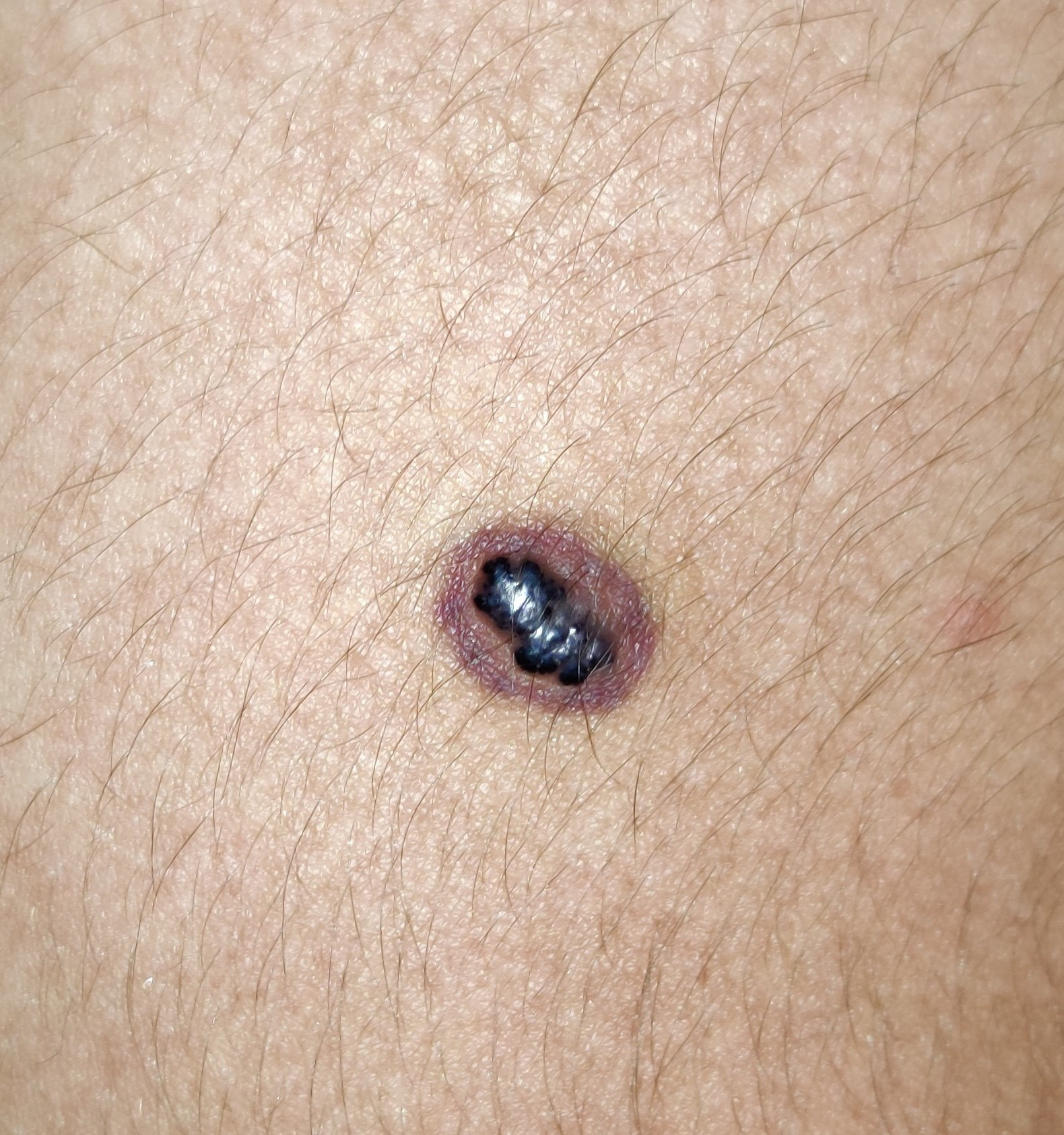
A 21-year-old healthy medical student presented with an asymptomatic bluish-purple vascular lesion surrounded by a purple-red ring on his left arm for 3 years. There was history of multiple spontaneous resolution and reappearances of this lesion. It measured 3 × 1.5 cm and was located on the extensor aspect of the left arm [Figure 1].

- A single violaceus purple nodule with surrounding ecchymotic ring on left arm.
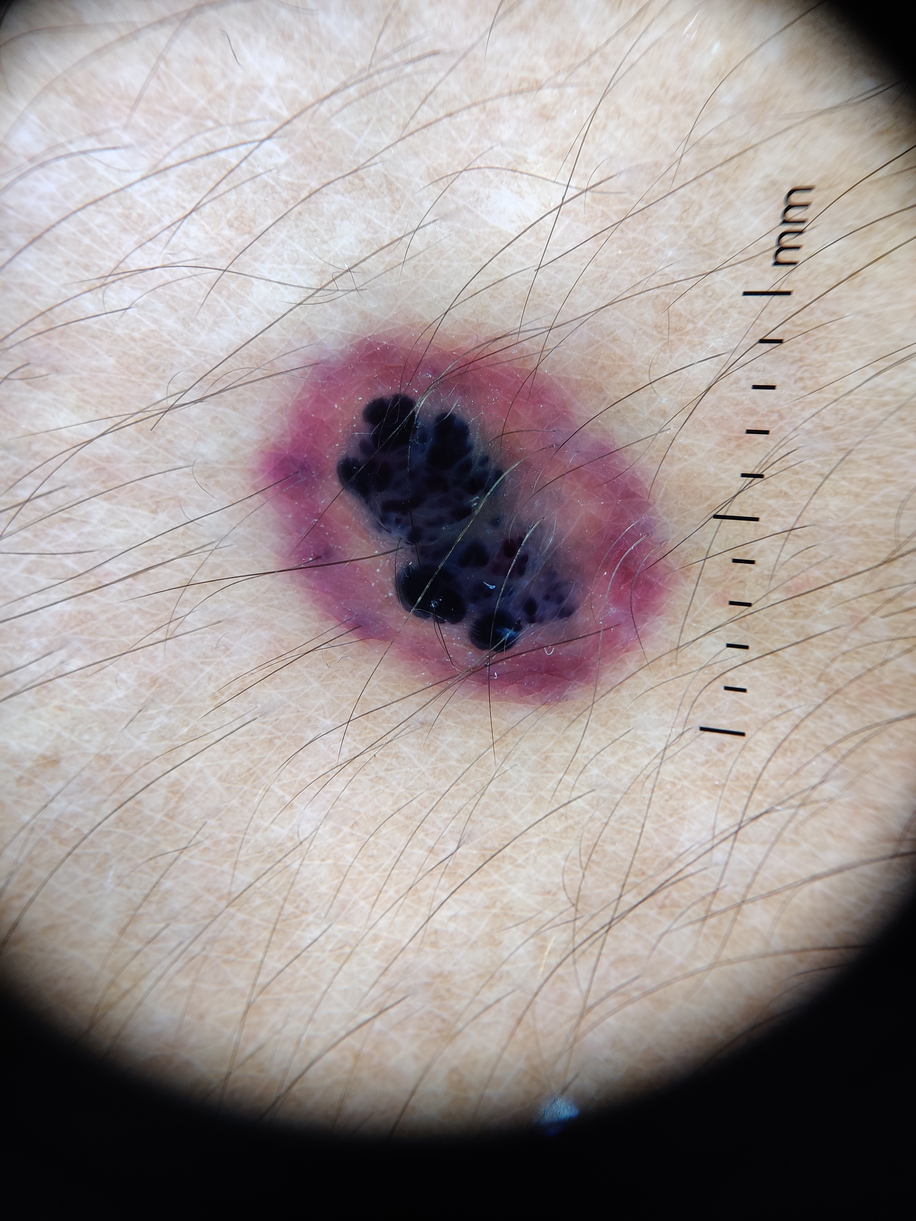
Contact dermoscopy (Dermlite, DL4, 3Gen Inc) showed a central dark bluish-black area surrounded by reddish-purple homogeneous area with vascular structures. The margin of the central purple area showed whitish-blue feathery borders [Figure 2].

- Contact dermoscopy (Dermlite, DL4) showing a bluish black well-defined vascular lesion at centre with a red violaceus peripheral ring.
Excisional biopsy and histopathology showed features consistent with a targetoid hemosiderotic hemangioma.
Declaration of patient consent
Patient’s consent not required as patients identity is not disclosed or compromised.
Financial support and sponsorship
Nil.
Conflicts of interest
There are no conflicts of interest.


