Translate this page into:
Rupioid psoriasis with inverse psoriasis
Corresponding author: Dr. Vichitra Sharma, Department of Dermatology, Venereology & Leprosy, Era’s Lucknow Medical College and Hospital, Lucknow, Uttar Pradesh, India. vichitrasharma26@gmail.com
-
Received: ,
Accepted: ,
How to cite this article: Sharma V, Mohanty S, Chaudhary G. Rupioid psoriasis coexisting with inverse psoriasis. Indian J Dermatol Venereol Leprol. 2024;90:390-1. doi: 10.25259/IJDVL_1000_2022
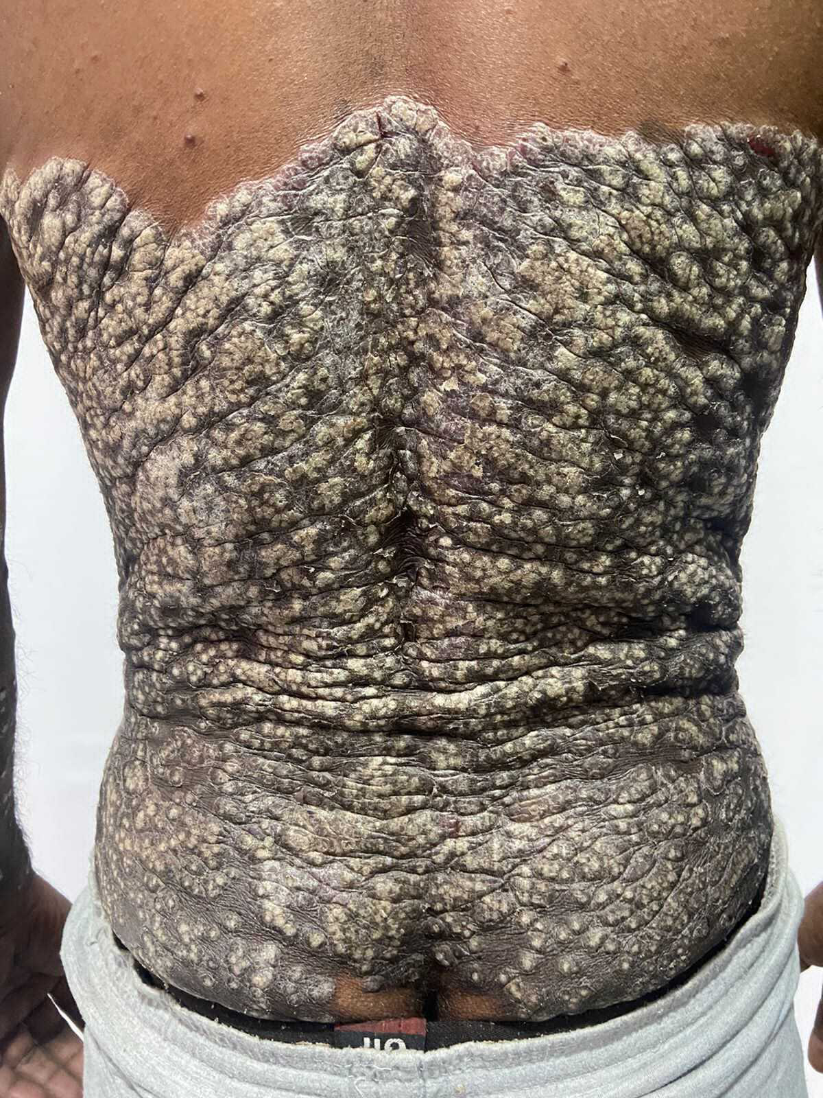
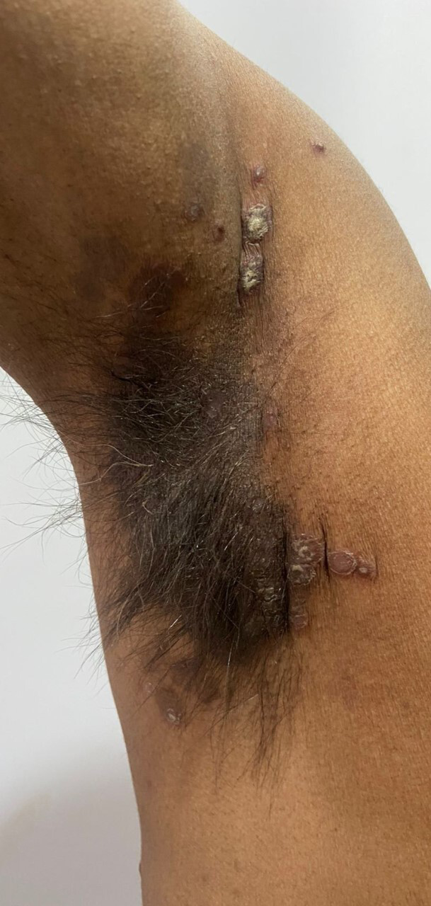
A 50-year-old male presented with thick, scaly lesions over the body for 15 years. On examination, he had multiple, well-defined, hyperpigmented discrete plaques studded with thick white limpet-like scales over the trunk [Figure 1], bilateral upper and lower limbs. Bilateral axillae showed well-defined hyperpigmented to erythematous plaques with minimal scaling [Figure 2]. Pitting was seen in the finger and toenails. The Psoriasis Area and Severity Index score was 16.9. Human immunodeficiency virus serology was negative. Skin biopsy showed hyperkeratosis with confluent parakeratosis, neutrophils within the stratum corneum, diminished granular layer, regular acanthosis and dilated vessels within the dermis. Diagnosis of rupioid psoriasis with inverse psoriasis was made based on clinico-histopathological features.

- Well-defined, thick hyperpigmented plaque studded with limpet-like scales on the back

- Well-defined hyperpigmented to erythematous plaques on axilla
Declaration of patient consent
Patient’s consent not required as patients identity is not disclosed or compromised.
Financial support and sponsorship
Nil.
Conflicts of interest
There are no conflicts of interest.


