Translate this page into:
Bilateral congenital cartilaginous rest of the neck: A rare presentation of accessory tragus
Corresponding author: Dr. Geeti Khullar, Department of Dermatology & STD Vardhman Mahavir Medical College & Safdarjung Hospital, New Delhi, India. geetikhullar@yahoo.com
-
Received: ,
Accepted: ,
How to cite this article: Dev PP, Khullar G, Sharma S, Alex P. Bilateral congenital cartilaginous rest of the neck: A rare presentation of accessory tragus. Indian J Dermatol Venereol Leprol. 2025;91:377-8. doi: 10.25259/IJDVL_517_2023
Dear Editor,
Congenital cartilaginous rest of the neck (CCRN) is a rare benign congenital anomaly which arises due to failure of fusion of the first and second branchial arches.1 It is a variant of accessory tragus presenting on the neck and can be associated with various syndromes. We could find only one previously reported case of bilateral CCRN in the literature.2 It is crucial for clinicians to be aware of this condition and the importance of performing a thorough clinical examination for any associated anomalies.
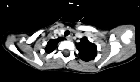
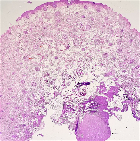
A 5-year-old boy presented with asymptomatic swellings bilaterally on the lower part of his neck since birth. The prenatal and natal histories were unremarkable. The papule on the left side was pedunculated and measured 10 × 5 mm, while the papule on the right side was 4 × 4 mm in size [Figure 1]. The lesions were skin-colored, non-tender, smooth, and had a hard consistency, with no movement on the tongue protrusion. No regional lymphadenopathy was detected. A systemic examination did not reveal any abnormalities. An ultrasonographic study of the neck showed hypoechoic areas in the subcutaneous plane of the lower neck with no deep communication. A computerized tomography scan of the neck showed two ill-defined, hyper- to isodense, mildly enhancing lesions bilaterally in the subcutaneous plane of the lower neck, anterior to the sternal part of the sternocleidomastoid [Figure 2]. Histopathological examination of the pedunculated lesion showed vellus hair follicles in the entire dermis, interspersed with collagen fibres and a focus of cartilage tissue [Figure 3]. After clinicopathological correlation, a diagnosis of CCRN was made.

- Pedunculated papules of size 10 × 5 mm on the left side of the neck and measuring 4 × 4 mm on the right side of the neck.

- Computed tomography scan of the neck at the axial plane above the sternoclavicular joint shows bilateral, mildly enhancing lesions in the subcutaneous plane anterior to the sternal part of the sternocleidomastoid (white arrows). The lesions are ill-defined and hyper to isodense.

- Numerous tiny vellus hair follicles (red arrows) in a prominent connective tissue framework and central cartilage (black arrow) in the subcutaneous fat (Haematoxylin & eosin, 40x).
The accessory tragus is a subcutaneous, firm papule or nodule that develops due to dysplasia or failure in the fusion of auricular hillocks of the first (mandibular) and second (hyoid) branchial arches. It is estimated that accessory tragus can occur as frequently as in 1 to 2 births per 1000. By the fifth and sixth weeks, these arches produce six mesenchymal tubercles, referred to as “hillocks of His.” Each arch forms three hillocks that gradually merge and transform into the recognizable structures of the auricle, which gradually ascends from the neck to the side of the head. The accessory tragus is found near the tragus or along an imaginary line from the tragus to the angle of the mouth but can also be located on the lateral neck along the anterior margin of the sternocleidomastoid.1 The accessory tragus located near the tragus originates from the first arch, while the cervical tragus is believed to originate from the second arch.1 In our case, accessory tragi were present on the anterior aspect of the lower part of the sternocleidomastoids in the cervical region, a rare variant known as CCRN.3 CCRN is usually present at birth and can be unilateral or rarely bilateral, like in our patient.
CCRN may be accompanied by syndromes like Goldenhar’s and Townes-Brocks syndrome.2 Goldenhar syndrome (oculo-auriculo-vertebral syndrome) includes a triad of mandibular hypoplasia resulting in facial asymmetry, ear and eye malformations, and vertebral anomalies.2 Townes-Brocks syndrome is characterised by a triad of imperforate anus, overfolded superior helices, and accessory tragus. Individuals with Townes-Brocks syndrome may also experience hearing impairments, thumb malformations, renal impairment, congenital heart disease, foot malformations, and genitourinary malformations.2 Other rare associations are Wolf-Hirschhorn syndrome and VACTERL syndrome. Thus, ophthalmological, otorhinolaryngological, oral, and spinal examinations should be performed to rule out associations with congenital syndromes. Histopathological examination of CCRN reveals the presence of elastic cartilage surrounded by various skin structures, including eccrine glands, adipose tissue, and pilosebaceous units.4 Ultrasonography and computed tomography scans can be utilised to determine the extent of the lesion and to look for any underlying sinus tracts.3
Differential diagnoses for CCRN include thyroglossal duct cyst, hair follicle naevus, fibroepithelial polyp, and branchial cleft cyst.5 Thyroglossal duct cysts are most prevalent in the neck’s midline near the hyoid bone, moving on tongue protrusion or swallowing. Hair follicle naevus presents as a solitary, skin-coloured papule and is sometimes accompanied by hypertrichosis.6 Fibroepithelial polyps are benign, soft, fleshy growths hanging off the skin of collagen fibres and blood vessels.7 Branchial cleft cysts can appear as cysts, fistulas, sinus tracts, or cartilaginous remnants, typically found on the front of the neck and upper chest.
While CCRN is benign and does not require treatment, it can also be managed with surgical excision or laser treatment. Care should be taken to ensure that the deep cartilage component is also removed. In our case, the parents chose conservative management after receiving adequate counselling.
Declaration of patient consent
The authors certify that they have obtained all appropriate patient consent.
Financial support and sponsorship
Nil.
Conflict of interest
There is no conflict of interest.
Use of artificial intelligence (AI)-assisted technology for manuscript preparation
The author(s) confirms that there was no use of artificial intelligence (AI)-assisted technology for assisting in the writing or editing of the manuscript and no images were manipulated using the AI.
References
- Accessory auricles affecting the tragus and cheek occurring with cervical chondrocutaneous branchial remnants: A case report. JPRAS Open. 2015;6:20-4.
- [Google Scholar]
- Bilateral cervical accessory tragus: a rare pediatric neck mass. Int J Otorhinolaryngol Head Neck Surg. 2018;4:579-81.
- [Google Scholar]
- Congenital cartilaginous rest of the neck in a boy. Dermatol Online J. 2016;22:1-4.
- [Google Scholar]
- Pathologic Quiz Case- An Anterior Neck Mass in a 5-Month-Old Female Infant. Arch Pathol Lab Med. 2004;128:1453-4.
- [CrossRef] [PubMed] [Google Scholar]
- The accessory tragus—no ordinary skin tag. J Dermatol Surg Oncol. 1989;15:304-7.
- [CrossRef] [PubMed] [Google Scholar]
- Hair follicle nevus: case report and review of the literature. Pediatr Dermatol.. 1996;13:135-8.
- [CrossRef] [PubMed] [Google Scholar]
- Skin tags: localization and frequencies according to sex and age. Dermatologica. 1987;174:180-3.
- [CrossRef] [PubMed] [Google Scholar]


