Translate this page into:
Scratch amyloidosis over nose: A rare site of cutaneous amyloidosis
Corresponding author: Dr. Preema Sinha, Department of Dermatology, Base hospital Lucknow, Lucknow, India. drpreemasinha@gmail.com
-
Received: ,
Accepted: ,
How to cite this article: Sinha P, Tripathi A, Madakshira MG, Prashantha GB, Raj CS. Scratch amyloidosis over nose: A rare site of cutaneous amyloidosis. Indian J Dermatol Venereol Leprol. 2025;91:379-81. doi: 10.25259/IJDVL_448_2023
Dear Editor,
Primary localised cutaneous amyloidosis is a rare group of skin disorders characterised by the deposition of extracellular homogenous hyaline material (amyloid) in the dermis without systemic involvement.1 The different subtypes of primary localised, cutaneous amyloidosis include lichen amyloidosis, macular amyloidosis and nodular amyloidosis. A joint manifestation of lichen and macular amyloidosis is called biphasic amyloidosis.2,3 The disease is more common in females with macular amyloidosis being the commoner variant. The disease manifests as hyperpigmented macules and patches in a rippled pattern on the extensor aspect of forearms and upper back.
Frictional melanosis is a close differential diagnosis with a clinical and histopathological overlap. Demonstration of amyloid deposits is diagnostic of cutaneous amyloidosis.3 Here, we discuss an uncommon presentation of cutaneous amyloidosis on the nose.
A 52-year-old woman, with no known comorbidities, presented with a dark discolouration of the nose with itching of 9 months duration. The patient gave a history of frequent scratching of the nose with no oozing or pain. Dermatological examination revealed a well-defined hyperpigmented plaque over the nose [Figure 1].

- Well-defined hyperpigmented plaque over the nose.
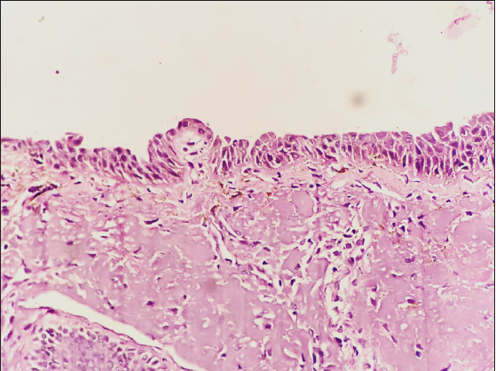
Dermoscopy showed a hyperpigmented reticular pigment network over a dark background [Figure 2]. Histopathological examination showed pigment incontinence, and the papillary dermis had nodular acellular deposits that were pale eosinophilic in character [Figure 3a]. High power highlighted the focal basal cell vacuolar degeneration with melanophages in the upper dermis [Figure 3b], while the Congo red stain showed birefringence of nodular material in the papillary dermis [Figure 3c]. Immunohistochemistry (IHC) (400x magnification) with serum-associated amyloid (SAA) stained the deposits in the papillary dermis [Figure 4]. All routine investigations to rule out systemic causes of amyloidosis were normal. A final diagnosis of cutaneous amyloidosis was made, and the patient was advised not to scratch the nose and was treated with mid-potent topical corticosteroid cream (0.1% mometasone). After 2 weeks, her lesions showed improvement with remarkable regression of the hyperpigmentation and reduced pruritus [Figure 5].
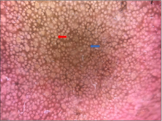
- Dermoscopy revealed a reticular pigmented network over a brown background. Red arrow indicates accentuation of reticular pigment network and blue arrow indicates sparing of hair follicles. (Dermlite dl4, polarised mode with 10x magnification)
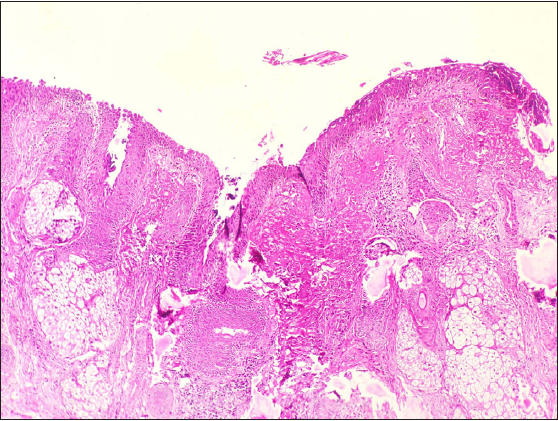
- Pale eosinophilic nodular acellular deposits in the papillary dermis (Haematoxylin & eosin, 100x).
-
Focal basal cell vacuolar degeneration with melanophages in the upper dermis (Haematoxylin & eosin, 400x).
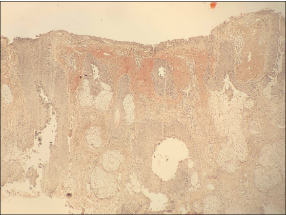
- Polarised microscopy showed congophilic nodular acellular material in the papillary dermis (Congo red stain, 100x).
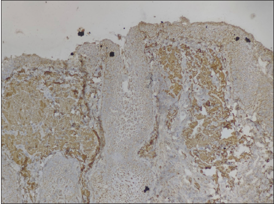
-
Serum-associated amyloid (SAA) highlights the acellular deposits in the papillary dermis (100x).
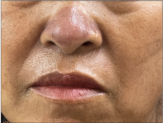
- Regression of the hyperpigmentation with reduced pruritus after 2 weeks.
Primary localised cutaneous amyloidosis is a disorder where amyloid deposits are seen in previously normal skin, with no evidence of deposits in internal organs. Macular amyloidosis, a common variant, has a female preponderance with age of onset ranging between 21 and 50 years. Clinically, macular amyloidosis presents as poorly delineated hyperpigmented lesions of greyish-brown macules with a rippled pattern, associated with deposition of amyloid material in the papillary dermis on histopathology.4 The sites most commonly involved are the interscapular area and extremities (shins and forearms), while clavicles, breast, face, neck and axillae are rarely involved.
Friction or often repeated trauma by towels, nylon scrubbers and clothes has been implicated in the causation of macular amyloidosis, and is described as friction amyloidosis or nylon friction dermatitis.5,6 Though the rippled and reticulate pattern is the most common presentation, many unusual forms, like poikilodermatous, diffuse, bullous, nevoid, linear, amyloidosis cutis dyschromica and incontinentia pigmenti-like have been reported.6
We report this case for its unusual appearance as well as rare site of presentation.
Declaration of patient consent
The authors certify that they have obtained all appropriate patient consent.
Financial support and sponsorship
Nil.
Conflicts of interest
There are no conflicts of interest.
Use of artificial intelligence (AI)-assisted technology for manuscript preparation
The authors confirm that there was no use of artificial intelligence (AI)-assisted technology for assisting in the writing or editing of the manuscript and no images were manipulated using AI.
References
- Primary cutaneous amyloidosis: A clinical, histopathological and immunofluorescence study. J Clin Diagn Res. 2017;11:WC01-5.
- [Google Scholar]
- Primary localized cutaneous amyloidosis: A retrospective study of an uncommon skin disease in the largest tertiary care center in Switzerland. Dermatol. 2022;238:579-86.
- [Google Scholar]
- Frictional melanosis and macular amyloidosis-exploring the link. Pigment Int. 2022;9:166-75.
- [Google Scholar]
- A clinico-epidemiological study of macular amyloidosis from North India. Indian J Dermatol. 2012;57:269.
- [CrossRef] [PubMed] [PubMed Central] [Google Scholar]
- Frictional melanosis and its clinical and histopathological features. Iran J Dermatol. 2018;21:124-7.
- [Google Scholar]


