Translate this page into:
Vermilion: An underemphasised anatomical area in dermatology
Corresponding author: Dr. Biswanath Behera, Department of Dermatology, All India Institute of Medical Sciences, Sijua, Patrapada, Bhubaneswar, India. biswanathbehera61@gmail.com
-
Received: ,
Accepted: ,
How to cite this article: Behera B. Vermilion: An underemphasised anatomical area in dermatology. Indian J Dermatol Venereol Leprol. 2024;90:562-3. doi: 10.25259/IJDVL_1413_2023
The anatomy of vermilion
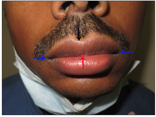
Lips comprise of vermilion, lip skin and inner lip mucosa. Vermilion is the area between the vermilion border and the wet line which meets at the lip angle [Figure 1]. Many times, the area of vermilion is inaccurately used as synonymous with lip. However, vermilion is a unique lip component distinct from lip skin and lip mucosa.

- Vermilion is the area bound by lip angles (blue arrow), vermilion border (black arrow) and wet line (red arrow).
The vermilion is lined by stratified squamous epithelium and is the transition zone between the skin and oral mucosa. The following are the unique features of vermilion: it is partially keratinised; stratum corneum is thinner than lip skin; concentration of rete pegs is maximum; rete pegs are narrow, long and slender; depth of the dermis is minimum; and the reflection of the blood vessels imparts a red colour. The colour of the vermilion can vary from pink, red and pinkish-brown to reddish-brown. The differential melanisation can explain the colour variability despite having the same melanocyte concentration as in the skin.1 The other characteristics that make the vermilion unique from the skin are the presence of moisture, the lack of pilosebaceous units and rapid regenerative power.
Applied aspect
Compared to the lip skin and lip mucosa, vermilion, especially the lower one, is vulnerable to various inflammatory and neoplastic conditions due to its continuous exposure to various extrinsic and intrinsic harmful factors, such as saliva, physical trauma, ultraviolet light, wind, air pollution, smoking, contact allergens and irritants. The dermatoses on the vermilion require their own depiction, as these are transitions between the oral and cutaneous variants. Discoid lupus erythematosus is difficult to recognise on vermilion due to the absence of two essential clues commonly seen in cutaneous counterparts: follicular plugging and epidermal atrophy. In addition, clinically, the blurring of the vermilion border is considered a typical feature of lip discoid lupus erythematosus (DLE).2 From a management point of view, the orabase formulations should be avoided for the treatment of cheilitis localised to vermilion due to poor absorption and the treatment should be similar to the skin using cream or ointment.3 Furthermore, vermilion should be protected from ultraviolet light in disorders like lip DLE and lip lichen planus to prevent the development of squamous cell carcinoma.4
In conclusion, it is of utmost importance to familiarise ourselves with the normal anatomy, dermoscopic features and histopathological characteristics of the vermilion to facilitate the understanding of the changes occurring due to various dermatoses.
Declaration of patient consent
The authors certify that they have obtained all appropriate patient consent.
Financial support and sponsorship
Nil.
Conflicts of interest
There are no conflicts of interest.
Use of artificial intelligence (AI)-assisted technology for manuscript preparation
The authors confirm that there was no use of artificial intelligence (AI)-assisted technology for assisting in the writing or editing of the manuscript and no images were manipulated using AI.
References
- Melanin: The biophysiology of oral melanocytes and physiological oral pigmentation. Head Face Med. 2014;10:8.
- [CrossRef] [PubMed] [PubMed Central] [Google Scholar]
- The lip in lupus erythematosus. Clin Exp Dermatol. 2014;39:563-9.
- [CrossRef] [PubMed] [Google Scholar]
- Isolated lichen planus of the lips: Cases reports and literature review. J Oral Med Oral Surg. 2020;26:14.
- [Google Scholar]
- Discoid lupus erythematosus leads to squamous cell carcinoma. Med J Armed Forces India. 2007;63:184-5.
- [CrossRef] [PubMed] [Google Scholar]


