
Translate this page into:
Dermoscopic cues of Paget disease: A series of three cases
Corresponding author: Dr. Eeshaan Ranjan, Department of Dermatology, Base Hospital, Delhi Cantt, India. eeshaan.ranjan@gmail.com
-
Received: ,
Accepted: ,
How to cite this article: Oberoi B, Tripathy DM, Neema S, Ranjan E, Chand S, Goel S. Dermoscopic cues of Paget disease: A series of three cases. Indian J Dermatol Venereol Leprol. doi: 10.25259/IJDVL_199_2024
Dear Editor,
Paget disease (PD) is an intraepithelial adenocarcinoma, which develops over the nipple-areola complex (NAC), and has a high association with underlying breast cancer.1,2 PD can have classical and pigmented variants. The available dermoscopic literature has laid more emphasis on the pigmented variant and very few authors have described the dermoscopy of the classical variant.1–3 We hereby describe the dermoscopic features of three cases of classical Paget disease.
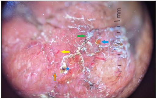
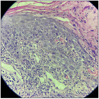
The 1st patient, a 75-year-old-woman presented with a six-month history of a slow-growing erythematous proliferative plaque over the right NAC associated with mild pain and discharge. Examination revealed a 1 cm × 0.5 cm well-defined firm erythematous moist plaque with surface crusting [Figure 1a]. There were no palpable lymph nodes. Dermoscopy of the lesion revealed pink background, yellow and white scales, dotted vessels, linear vessels, blood spots and chrysalis-like structures, brown peppering [Figure 1b]. Histopathology showed intraepidermal collection of malignant cells with large pale cells with eosinophilic cytoplasm and prominent nuclei (Paget cells), which formed duct-like nests in the basal and supra-basal layers [Figure 1c]. Immunohistochemistry (IHC) was positive for CK7 (cytokeratin 7) and epithelial membrane antigen (EMA) and negative for CK20. A diagnosis of Paget disease of the nipple was made, and subsequent work up for underlying malignancy, including mammography and MRI (magnetic resonance imaging) of the breast was performed and was normal.
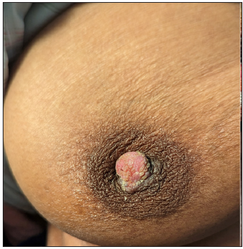
- Clinical image showing a well-defined firm erythematous plaque with surface crusting over right NAC.
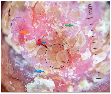
- Dermoscopy (DermLite DL4, polarised, 10x) showing a pinkish-orange background, yellow scales (blue arrow) and white scales (green arrow), dotted vessels (blue circle), blood spots (blue star), brown peppering (black circle), chrysalis like structure (orange arrow).
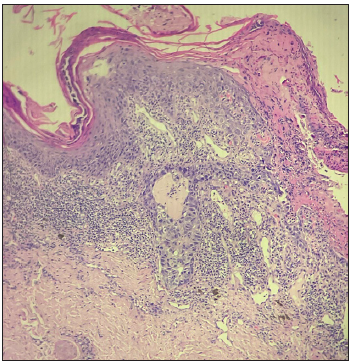
- Histopathological examination (Haematoxylin & eosin, 10x) showing intraepidermal collection of malignant cells.
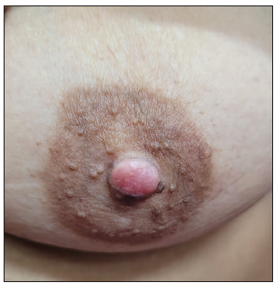
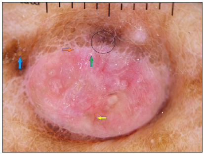
The 2nd patient, a 67-year-old woman presented with a nine-month history of proliferative plaque over the right NAC associated with pain and tightness over the area. Examination revealed a 2 cm × 3 cm well-defined firm brown plaque tethered to the nipple distorting its morphology [Figure 2a]. No other skin lesions or palpable lymph nodes were noted elsewhere on the body. Dermoscopy showed yellowish-white scales, a pink-orange background, large polymorphous vessels, yellow and white scales, blood spots, chrysalis-like structure and diffuse brown pigmentation in the periphery [Figure 2b]. Histopathology was suggestive of Paget disease with large pale cells with eosinophilic cytoplasm and prominent nuclei (Paget cells) forming duct-like nests in the basal and supra-basal layers. Prominent collagen bundles could be seen in the dermis [Figure 2c] The patient was diagnosed with Paget disease of the nipple without any evidence of underlying breast malignancy.
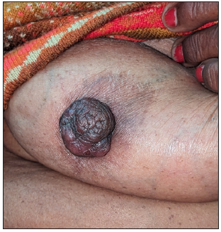
- Clinical image showing a well-defined firm brown plaque tethered to the nipple distorting the morphology.
- Dermoscopy (DermLite DL4, polarised, 10x) showing pinkish-orange background, yellow scale (blue arrow) and white scale (green arrow), linear vessels (yellow arrow), blood spots (blue star), chrysalis-like structure (orange arrow).
- Histopathological examination (Haematoxylin & eosin, 40x) showing large pale cells with eosinophilic cytoplasm and prominent nuclei (Paget cells) forming duct-like nests in the basal and supra-basal layers. Prominent collagen bundles could be seen in the dermis.
Our 3rd patient was a 54-year-old woman with mildly itchy lesions on the right nipple [Figure 3a], Dermoscopy of the lesion showed a pink-orange background, yellow and white scales, linear vessels, brown peppering and chrysalis-like structures [Figure 3b]. Patient was later diagnosed as Paget disease with histopathology showing paget’s cells with abundant pale cytoplasm and phagocytosed melanin seen inside and around the cells [Figure 3c]. There was no underlying malignancy in the breast.
- Clinical image showing an erythematous scaly plaque over NAC.
- Dermoscopy (DermLite DL4, polarised, 10x) showing pinkish-orange background, yellow scales (blue arrow), white scales (green arrow), linear vessels (yellow arrow), brown peppering (black circle) chrysalis-like structure (orange arrow).
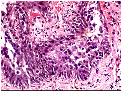
- Histopathological examination (Haematoxylin & eosin, 40x) confirming Paget’s cells with abundant pale cytoplasm and phagocytosed melanin seen inside and around the cells. The cells have large irregular nuclei with prominent nucleoli and vague gland formation is noted. Here, too, the prominent collagen bundles can be seen.
Paget disease of the nipple is a relatively rare malignancy originating from the apocrine duct-derived epithelial cells. Any dermoscopic features which can help clinch the diagnosis need to be reported. Dermoscopy of the lesion was found to show irregular linear vessels in the early stage and large polymorphous vessels in a later stage (as per our cases); however, a study with a larger sample size needs to be carried out to confirm this finding. Multiple blue-grey dots (peppering) corresponding to melanophages in the papillary dermis were seen in our cases, which were previously reported as well.2 Shiny white streaks or chrysalis-like structures were seen, which have been described as common features of Paget disease4 and represent new or remodelled collagen bundles.2 Similar chrysalis-like structures can be seen in dermoscopy of dermatofibroma, scars, basal cell carcinoma, pyogenic granuloma, spitz naevus and melanoma.4 A case-control study comprising of 22 cases and 43 controls concluded that amongst non-pigmented lesions, pink structureless areas, white lines, erosion/ulceration and white scales served as predictors of PD.5 The NAC can develop a multitude of lesions which need to be differentiated from Paget disease. Dermoscopic findings of various conditions have been summarised in Table 1.4,6
| Nipple lesions | Epithelial/follicular pattern | Pigment pattern | Vascular pattern | Lesion-specific features |
|---|---|---|---|---|
| Eczema | Yellowish-white crust | Faint pink background | Dotted/patchy/comma-like/linear vessels | |
| Bowen’s disease | Surface scales/hyperkeratosis | - | Glomerular vessels/clustered vascular pattern | |
| Pigmented Bowen’s | Scales | Patchy distribution of regularly packed small brown globules | Glomerular vessels | |
| Pink or skin-coloured eccentric structureless areas | ||||
| Nevoid hyperkeratosis | Papillomatous surface, scales, small erosions | Pink homogenous areas, blue-grey globules | Red dots | |
| Erosive adenomatosis of the nipple | Whitish/yellowish hyperkeratosis a | Reddish-whitish background | Sparse dotted vessels | Increased red serpiginous and annular structures |
| Pinkish background | ||||
| lichenoid keratosis | Scalloped border and scale | Pinkish background, annular granular structures, gray pseudonetwork and diffuse blue-gray dots | Dotted/short thin vessels | Shiny white structures/crystalline structures (appear as strands or bloches) |
| Pagets disease (non-pigmented) | Whitish scales/yellow scales/reddish-brown crust | Pinkish background, brown peppering | Polymorphous vessels/dotted/glomerular vessels | Chrysalis-like (whitish streak-like) structures) |
| Pagets disease (pigmented) | Whitish scales | Diffuse brown structureless areas, reticular pigmentation, irregular black dots, blue-grey dots, scar-like depigmentation | Polymorphous vessels/dotted/glomerular vessels | Chrysalis-like (whitish streak-like) structures) |
| BCC | Small erosions | Blue-grey globules | Arborising vessels | Maple leaf appearance |
| Spoke wheel areas | ||||
| Malignant melanoma | Ulceration | Atypical pigment network, multicomponent pattern | Atypical vascular pattern | Blue-white wheel |
| Regression structures |
BCC: Basal cell carcinoma
Paget disease of the nipple has varied presentations and can be difficult to diagnose in early stages. Dermoscopy can aid histopathology and immunohistochemistry in early diagnosis and prompt treatment of the potentially malignant entity.
Declaration of patient consent
The authors certify that they have obtained all appropriate patient consent.
Financial support and sponsorship
Nil.
Conflicts of interest
There are no conflicts of interest.
Use of artificial intelligence (AI)-assisted technology for manuscript preparation
The authors confirm that there was no use of AI-assisted technology for assisting in the writing or editing of the manuscript and no images were manipulated using AI.
References
- Polarized dermoscopy of mammary Paget disease. An Bras Dermatol. 2013;88:290-2.
- [CrossRef] [PubMed] [PubMed Central] [Google Scholar]
- Dermoscopy of Paget’s disease. Indian Dermatol Online J. 2020;11:674-5.
- [CrossRef] [PubMed] [PubMed Central] [Google Scholar]
- Dermoscopy as a supportive instrument in the early recognition of erosive adenomatosis of the nipple and mammary Paget’s disease. Ann Dermatol. 2017;29:365-7.
- [CrossRef] [PubMed] [PubMed Central] [Google Scholar]
- Dermoscopic features of mammary Paget’s disease: A retrospective case-control study by the International Dermoscopy Society. J Eur Acad Dermatol Venereol. 2019;33:1892-8.
- [CrossRef] [PubMed] [Google Scholar]
- Nipple and areola lesions: Review of dermoscopy and reflectance confocal microscopy features. J Eur Acad Dermatol Venereol. 2019;33:1837-46.
- [CrossRef] [PubMed] [Google Scholar]


