
Translate this page into:
Beyond the surface: Link between Demodex mite and frictional facial melanosis
Corresponding author: Dr. Nayan Sevantilal Prajapati, Department of Dermatology, Govt. Medical College Baroda, Deesa, Gujarat, India. nayanprajapati4863@gmail.com
-
Received: ,
Accepted: ,
How to cite this article: Patel KS, Parekh AR, Prajapati NS, Shah HA. Beyond the surface: Link between Demodex mite and frictional facial melanosis. Indian J Dermatol Venereol Leprol. doi: 10.25259/IJDVL_1383_2023
Dear Editor,
Frictional facial melanosis is not uncommon but an under-recognised entity with no clear-cut definition and unknown prevalence. It has a significant impact on the quality of life and self-esteem of those who are affected1. The present case illustrates the pathogenic potential of Demodex in frictional facial melanosis.
A 52-year-old married woman presented to us with a complaint of facial hyperpigmentation associated with itching for two years. The patient had no history of photosensitivity or atopy and denied the use of any topical medication. The patient was a known case of diabetes mellitus for five years. The patient complained of itching over the face followed by the development of red-coloured skin lesions that eventually turned brownish.
On clinical examination, the patient had ill-defined diffuse hyperpigmented patches present on bilateral cheeks and chin with sparing of forehead, nose, and sides of the face [Figure 1]. The skin surface showed increased skin markings with the rough texture. The rest of the clinical and systemic examinations were non-contributory.
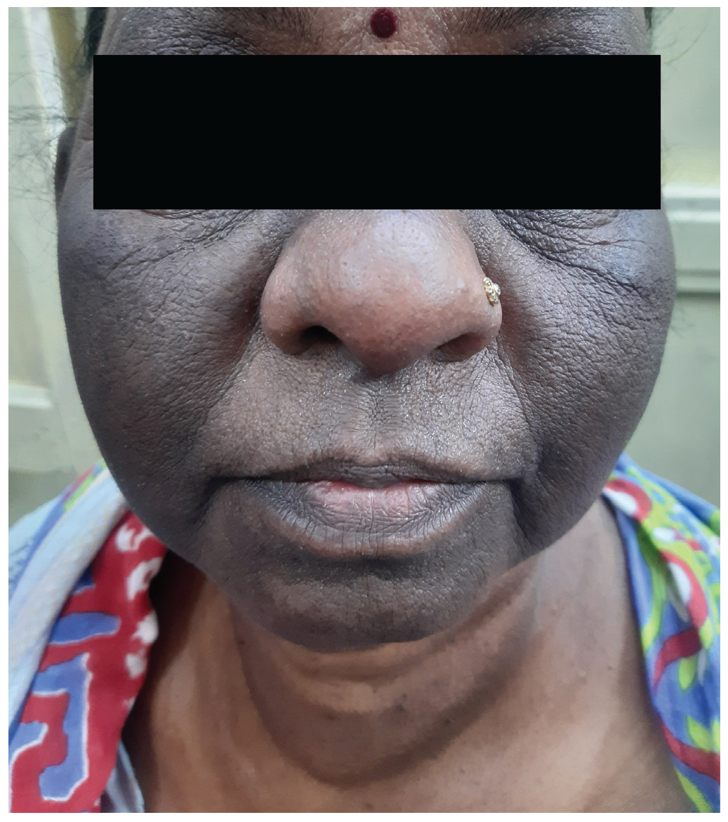
- Pre-treatment diffuse ill-defined hyperpigmentation with sparing of the nose.
On Wood’s lamp examination, no enhancement of skin pigmentation was observed. Polarised-light dermoscopy showed dark blotches of pigmentation in perifollicular areas with a background pseudo-reticular pigment network In total, three demodex mites were appreciated on dermoscopic examination of the various affected regions on the face [Figure 2a]. Based on clinical and dermoscopic findings, pigmented demodicosis, pigmented contact dermatitis, and acanthosis nigricans were considered as our differential diagnoses.

- Dermoscopic image (10x magnification polarised mode) of demodex tail appearing as perifollicular gelatinous, filament-like structure, with background pseudo-reticular pigment network. (yellow circle).
Histology showed a compact stratum corneum, irregular epidermal acanthosis, and increased pigmentation in the basal layer. Most interestingly, a well-preserved demodex mite was seen in the dermis, surrounded by lymphohistiocytic cell infiltrate, in the vicinity of the hair follicle [Figure 2b].

- Histopathological image (Haematoxylin & eosin,100x and inset 400x) – The image shows a demodex mite in the dermis surrounded by lymphohistiocytic infiltrate (red circle), The inset figure shows a magnified image of the demodex mite surrounded by lymphohistiocytic infiltrate.
Based on specific dermoscopic and histopathologic findings attributable to demodex infestation, we started anti-demodectic treatment. The patient was treated with oral ivermectin 12mg (200μg/kg/dose), three doses a week apart, topical ivermectin (1%), twice a day, along with emollients for three weeks. Within a span of three weeks, the patient observed a significant reduction in itching and improvement in facial hyperpigmentation [Figure 3].

- Improvement after anti-demodectic treatment.
Facial melanosis is multifactorial in aetiology. Melasma, lichen planus pigmentosus, acanthosis nigricans, Riehl’s melanosis, naevus of Ota, and post-inflammatory hyperpigmentation are among the common causes of facial melanosis.
Facial hyperpigmentation attributed to Demodex mites is also referred to as pigmented demodicosis.2 Demodex folliculorum and Demodex brevis are 8-legged microscopic ectoparasites that inhabit exclusively, at lower densities, in human pilosebaceous follicles, especially over the scalp and face, as part of the normal human microbiota.3 As Demodex is a commensal organism, it may via unknown mechanisms alter the host immunity, thus permitting its own survival. In addition, Demodex mites are known to exacerbate various chronic inflammatory skin diseases. Recent studies show the effect of Demodex mite on the host’s immunity to be conflicting, one being immunosuppressive and so promoting its proliferation, whereas the other being defensive to eliminate the mite. Various factors, including immunosuppression, diabetes, or sebaceous hyperplasia, can displace the subtle host/Demodex stability in favour of Demodex proliferation.
The density of mites or their extra-follicular location is more relevant in identifying demodicosis.2 The presence of >5 Demodex mites per cm2 area in standardised skin surface biopsy (SSB) specimens is required for the diagnosis of demodicosis.4
With dermoscopic examination, the Demodex tail appears as a gelatinous, filament-like structure in the peri-follicular area. Histopathology is diagnostic, but dermoscopy is recommended as a non-invasive diagnostic method with an efficacy of 88%.5
As seen in the histology of the present case, possible routes for the presence of the Demodex mite within the dermis include either active penetration or passive transmission via a damaged pilosebaceous follicle. No findings favouring damaged follicles like foreign body granulomatous inflammation were seen in multiple sections studied. So, in the present case, Demodex might have actively crossed the follicular wall, penetrated the dermis, and incited inflammation. One report claims to have documented a mite penetrating an intact follicle wall.6 Inflammatory reaction to the intradermal mite leads to pruritus and chronic rubbing. The resultant pigmentation causes a paradoxical increase in forceful rubbing, exacerbating the pigmentation and building up a vicious cycle of friction and reactive hypermelanosis. The treatment is aimed towards reduction of Demodex mite density, and hence intensity of pruritus, rubbing, and resulting hyperpigmentation. Topical agents like permethrin, ivermectin, sulphur, selenium sulphide, lindane, benzyl benzoate, crotamiton, malathion, metronidazole, and systemic agents like ivermectin and metronidazole have shown satisfactory results.7
Declaration of patient consent
The authors certify that they have obtained all appropriate patient consent.
Financial support and sponsorship
Nil.
Conflicts of interest
There are no conflicts of interest.
Use of artificial intelligence (AI)-assisted technology for manuscript preparation
The authors confirm that there was no use of artificial intelligence (AI)-assisted technology for assisting in the writing or editing of the manuscript and no images were manipulated using AI.
References
- Current understanding of frictional dermatoses: A review. Indian J Dermatol Venereol Leprol.. 2023;89:170-188.
- [CrossRef] [PubMed] [Google Scholar]
- Friction melanosis: A clinical, histologic, and ultrastructural study in Jordanian patients. Int J Dermatology. 2004;43:261-4.
- [Google Scholar]
- Human demodex mite: The versatile mite of dermatological importance. Indian J Dermatol. 2014;59:60-6.
- [CrossRef] [PubMed] [PubMed Central] [Google Scholar]
- Pigmented demodicidosis - an under-recognized cause of facial hyperpigmentation. Int J Dermatol. 2022;61:564-9.
- [CrossRef] [PubMed] [Google Scholar]
- Rosacea, an infectious disease: Why rosacea with papulopustules should be considered a demodicosis A narrative review. Acad Dermatol Venereol. 2022;36:987-1002.
- [Google Scholar]
- High accuracy of recognition of common forms of folliculitis by dermoscopy: An observational study. J Am Acad Dermatol. 2019;81:463-71.
- [CrossRef] [PubMed] [Google Scholar]
- Demodex and perifollicular inflammation in man: review and report of 69 biopsies. Ann Dermatol Venereol. 1986;113:1047-1058.
- [PubMed] [Google Scholar]


