Translate this page into:
Thermocol-based spacer for interventional dermoscopy
2 Department of Dermatology, Medical Trust Hospital, Cochin, Kerala, India
Correspondence Address:
Feroze Kaliyadan
Department of Dermatology, College of Medicine, King Faisal University, Al-Ahsa, 31982
Saudi Arabia
| How to cite this article: Kaliyadan F, Puravoor J. Thermocol-based spacer for interventional dermoscopy. Indian J Dermatol Venereol Leprol 2020;86:218-219 |
Problem
Recently, there have been reports dealing with the modification of standard dermoscopes for dermoscopy-guided procedures.[1],[2] While these modifications are simple and effective, the limited field of work is one of the limitations. We suggest a simple modification for hand-held dermoscopes which will enable dermoscopy-guided procedures with a larger field available for the same.
Solution
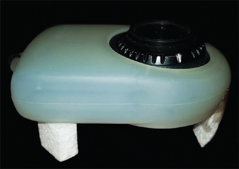
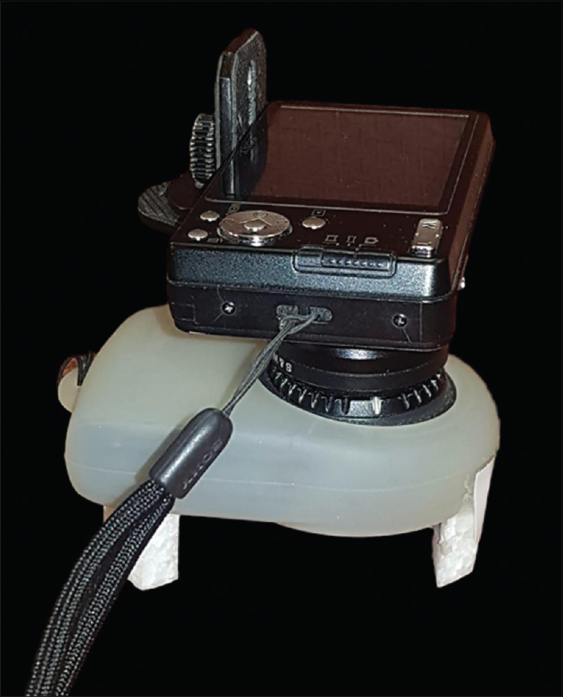
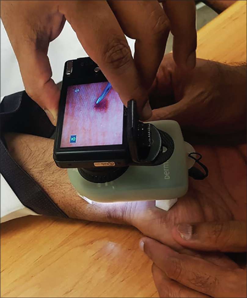
We attached two thermocol pieces with height adjusted according to focusing distance of the dermoscope, with double-sided adhesive tape [Figure - 1]. The apparatus thus assembled can be used as such or with a camera attached to it [Figure - 2], [Figure - 3]. The arrangement leaves plenty of free space for marking biopsy areas or for procedures such as needling, comedone extraction and radiofrequency ablation. For radiofrequency ablation, it is important to ensure that the gaps are large enough so as to prevent contact of the cautery tip with the thermocol. Cutting the thermocol pieces and attaching them is also much easier as compared to cutting a syringe or custom-made acrylic spacers.
 |
| Figure 1: Thermocol spacers attached to the dermoscope |
 |
| Figure 2: Assembly with camera attached |
 |
| Figure 3: Use of the apparatus for dermoscopy-guided procedures |
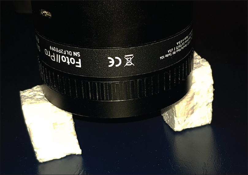
The advantage of using the thermocol spacer is that it can be used for most dermoscopes. We have tried the same for a larger one also—the Dermlite foto ii pro [Figure - 4]. However, because of the smaller diameter of the tip, moulding a thermocol spacer would be difficult for a USB dermoscope. The height of the spacer is equivalent to the original spacer that comes with the dermoscope and this ensures that there are no changes in magnification of field of vision, which can be a problem when distance of viewing is adjusted.[3]
 |
| Figure 4: Spacer attached to Dermlite Foto ii pro |
The main limitation at this stage would continue to be the limited vertical height, corresponding to the optimal focusing distance of the dermoscope. In addition, it is important not to apply too much pressure on the thermocol spacer, especially while using a heavier dermoscope, to prevent the spacer from parting away. The adhesiveness may also wear away with time and, hence, the spacer might need replacements after a few uses.
Declaration of patient consent
The authors certify that they have obtained all appropriate patient consent forms. In the form, the patient/patients has/have given his/her/their consent for his/her/their images and other clinical information to be reported in the journal. The patient/patients understands/understand that his/her/their name/names and initial will not be published, and due efforts will be made to conceal his/her/their identity, but anonymity cannot be guaranteed.
Financial support and sponsorship
Deanship of Scientific Research, King Faisal University, Saudi Arabia, Grant/Award Number: 1811015.
Conflicts of interest
There are no conflicts of interest.
| 1. |
Sonthalia S, Khurana R. Interventional dermoscopy. J Am Acad Dermatol 2019. pii: S0190-9622 (19) 30532-8.
[Google Scholar]
|
| 2. |
Agrawal S, Dhurat R, Daruwalla S, Sharma A. A simple modification of a syringe barrel as an adapter for dermoscopic guided biopsy. J Am Acad Dermatol 2019. pii: S0190-9622 (19) 30470-0.
[Google Scholar]
|
| 3. |
Jakhar D, Kaur I. A simple technique to increase field of view of a universal serial bus dematoscope. J Am Acad Dermatol 2019;80:e123-4.
[Google Scholar]
|
Fulltext Views
3,773
PDF downloads
2,852


