Translate this page into:
Prof. Nékám's Corpus Iconum Morborum Cutaneorum (1938): The most elaborate historical dermatovenerological atlas of the first half of the 20th century
2 Department of Microbial Genetics, Institute of Molecular Biology, Slovak Academy of Sciences, Bratislava, Slovakia
Correspondence Address:
Juraj Majtan
Institute of Molecular Biology, Slovak Academy of Sciences, Dubravska Cesta 21, 845 06 Bratislava
Slovakia
| How to cite this article: Szep Z, Majtan J. Prof. Nékám's Corpus Iconum Morborum Cutaneorum (1938): The most elaborate historical dermatovenerological atlas of the first half of the 20th century. Indian J Dermatol Venereol Leprol 2019;85:342-345 |
Prof. Lajos Nékám is one of the legends of dermatology.[1] The exceptional, case-report-based dermatovenerological atlas, Corpus Iconum Morborum Cutaneorum, is his most prominent contribution, which was the most extensive dermatovenerological atlas during his time.[2]
Prof. Nékám studied at the Faculty of Medicine, Semmelweis University, at Budapest, Hungary, Europe. In 1898, he became a superintendent at the University Dermatovenerological Clinic in Budapest. During his days, the institution attained international recognition as one of the most progressive clinics in the world. In 1928, Prof. Nékám founded the Hungarian Dermatological Society which he managed for 10 years. While developing his workplace, he established contact with some of the most prestigious global dermatological centers. He was a leading figure of several world congresses and a member of the so-called Group of 11, which represented dermatology at the international level.[1],[3]
Nékám's atlas of cutaneous diseases, Corpus Iconum Morborum Cutaneorum [Figure - 1] and [Figure - 2], consisting of three separate books, is an outstanding work in the history of dermatovenerology.[2] The atlas was prepared as volume 5 of the transactions of the 9th International Congress of Dermatology (13th to 21st September, 1935, Budapest), to be finally published later in 1938.
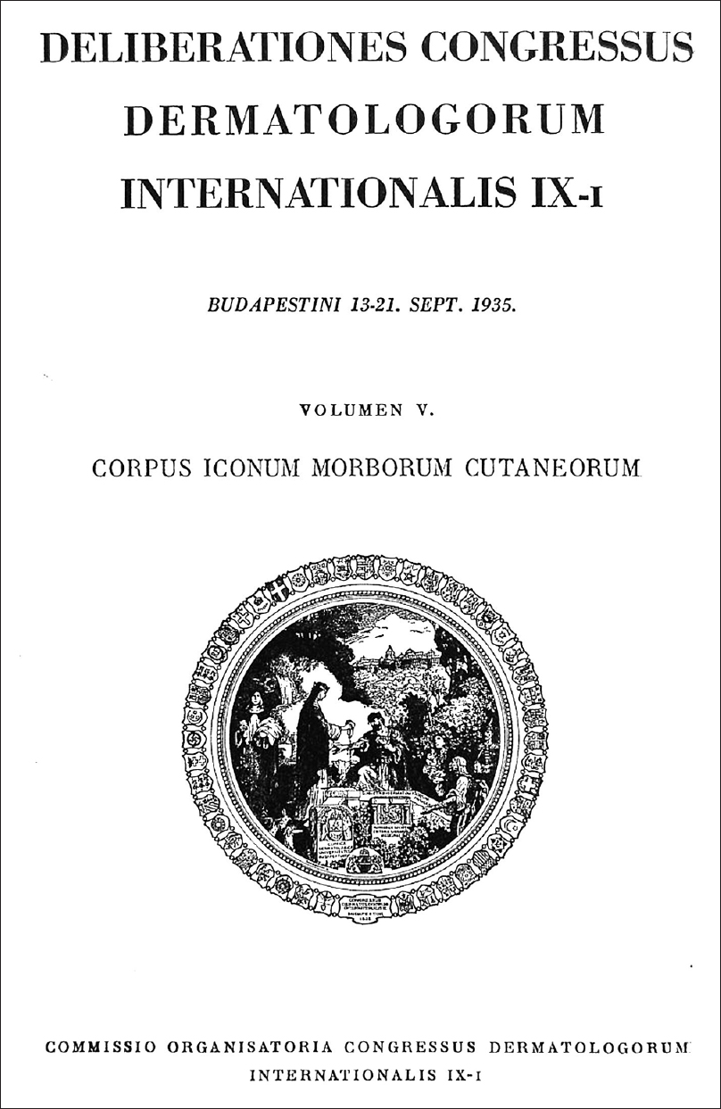 |
| Figure 1: The first introductory page of Corpus Iconum Morborum Cutaneorum (1938) |
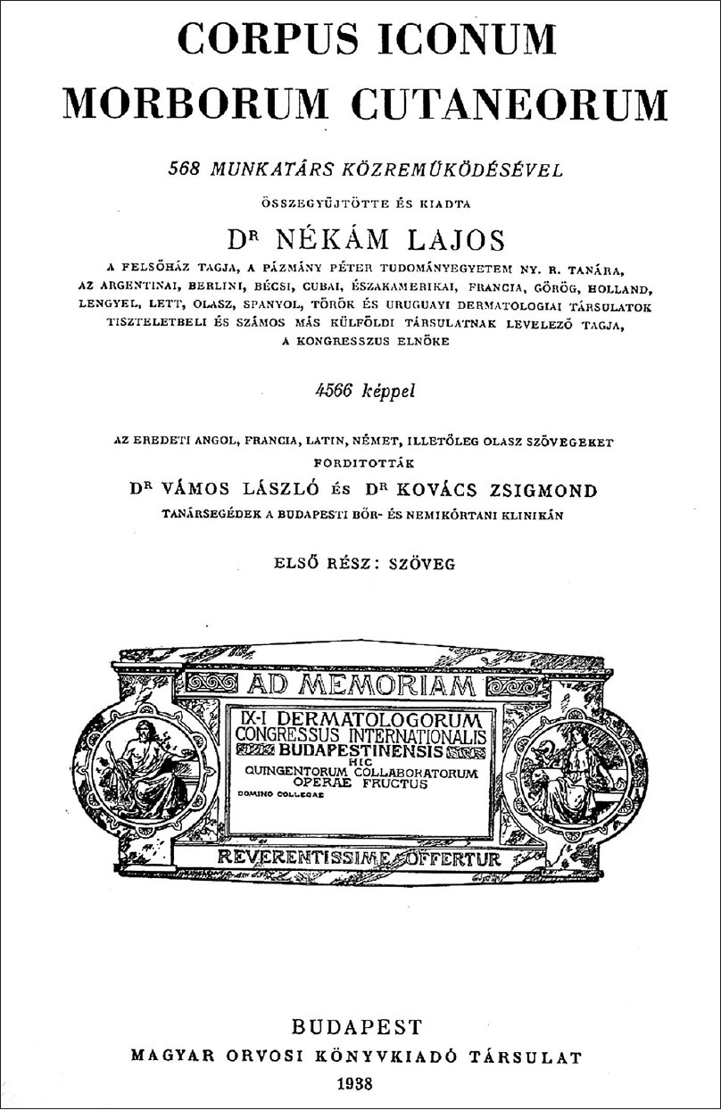 |
| Figure 2: The second introductory page of Corpus Iconum Morborum Cutaneorum (1938) |
Congress preparation and realization was a demanding task in the 1930s, as European political atmosphere was tense. Eastern Europe was under the Communist dictatorship of the Soviet Union represented by Stalin, while in Western Europe the Nazis came into power in 1933 to build a totalitarian Nazi dictatorship under Hitler. Smaller Central European states like Hungary were pressed by both dictatorships with an objective to gain control over them. As Prof. Nékám mentioned, the pre-war tense atmosphere was also present in 1935 during the 9th International Congress of Dermatology and Syphilology, held at Budapest. The atlas was published in 1938, one year before the Second World War. Thus, also from a historical and medical point of view, it is important to highlight and appreciate the friendly and humanistic atmosphere of the Congress. The Congress aimed to unite dermatologists from around the world into an international scientific organization that would allow better scientific collaboration. This idea was directly reflected in the atlas Corpus Iconum Morborum Cutaneorum. The atlas boasts of a rich collection of treasured photographs collected from dermatologists around the world.[2] It is of utmost historical importance to highlight the fact that experts from Nazi Germany, the communist Soviet Union and many other remote countries with different political ideologies also contributed to the atlas. International cooperation and a sense of humanism in the field of science triumphed over the cruel political reality of the 1930s.
The introductory pages show three illustrations. The first illustration is remarkably designed graphically and has a complicated symbolism [Figure - 3].[2] The main image is surrounded by a string of 45 small coats of arms representing the homeland of 568 contributors to the atlas. In the background, we can see the royal palace (the Buda Castle) which was a symbol of the Ninth International Dermatological Congress. To the left of the main image we can see Saint Margaret (one of the greatest saints in the cultural history of Hungary coming from the Hungarian royal dynasty of the Árptáds) treating and healing cutaneous illnesses—symbol of the Hungarian Dermatological Society. The symbol of the Hungarian Publishing House of Medical Books is a sick person holding a torch in his right hand. The University Dermatology Clinic in Budapest is represented by the frontal image on the stone statuette with two angels, one heraldic lion and one griffin with the coat of arms between them representing the capital city of Hungary—Budapest. All the four institutions mentioned above helped in the organization of the congress and printing the atlas.
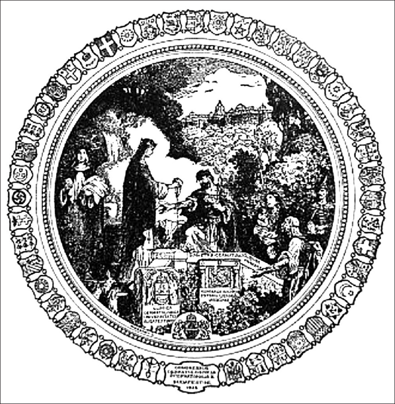 |
| Figure 3: The emblem of the 9th International Dermatological Congress in Budapest 1935 (displayed on the first introductory page of the Corpus Iconum Morborum, Cutaneorum) |
The preparation of Corpus Iconum Morborum Cutaneorum was the result of immense hard work. Invitation for contribution was sent to well-known dermatological clinics and around 3000 dermatologists. Besides, the clinics of internal medicine, surgery, pediatrics, stomatology, ophthalmology, urology, gynecology and obstetrics, neurology and psychiatry, institutes of radiotherapy and radiodiagnosis, pathology, parasitology, tropical medicine, center for seasickness, laboratories of mycology, serology and institutes of veterinary medicine (zoology, veterinary pathology, parasitology, and internal medicine) were also addressed. Additionally, several nonmedical workplaces were also contacted (e.g. London Wellcome Museum) to publish interesting cases from their collections. Most of the institutes mentioned above did not respond. Finally, the illustrations were provided by 568 dermatologists from 45 countries, including well-known and respected figures like Behçet, Besnier, Civatte, Goeckermann, Gottron, Hallopeau, Neisser, Pautrier, Riehl, Sabouraud, Unna, Sulzberger, Vidal, Sutton and many others. Prof. Nékám himself contributed by publishing his 1407 photographs, which accounted for one-third (31%) of the total number (4566 illustrations) in the atlas. Contributions were also received from remote countries like India, Sudan, Venezuela, Japan, Australia and many others.
The original idea of a multilingual atlas could not be realized due to lack of financial support. So, only two language versions were published: one purely Hungarian and one mixed with several foreign languages.
The atlas consists of three separately bound books. The first one contains only texts—short clinical explanations to particular photographs included in the second and third book (4,566 astonishing illustrations), which constitute the dermatovenerological atlas. The majority of illustrations are dedicated to common and rare cutaneous diseases [Figure - 4], including units of tropical dermatology, various genodermatoses and syndromes.[2] The authors focused mainly on relationship between cutaneous and internal diseases—so-called correlative dermatology: a rich collection of illustrations of cutaneous manifestations of various hematological malignancies, avitaminoses, metabolic, rheumatic and endocrine disorders. Several clinical pictures were provided by the London Wellcome Museum. The section of illustrations from historical collections of French authors was prepared by a French photographer and graphic designer Félix Méheux. Also, colored hand drawings by Sir Graham-Little and others are present. The atlas illustrations can be categorized into 20 thematic groups: (1) cutaneous inflammatory diseases and tumors, (2) correlative dermatology, (3) veterinary dermatology, (4) ocular findings and fundus oculi findings, (5) findings pertaining to the oral, rectal and urinary bladder mucosae, (6) stomatological findings, (7) osteological findings, (8) trichologic findings, (9) onychologic findings, (10) histological images, (11) cytology images, (12) autopsy findings, (13) bacteriological images, (14) dermatological mycology, (15) parasitology images, (16) hematological microscopic findings, (17) capillaroscopic findings, (18) radiographic findings, (19) hygienic-epidemiological problems and (20) dermatogrammata.
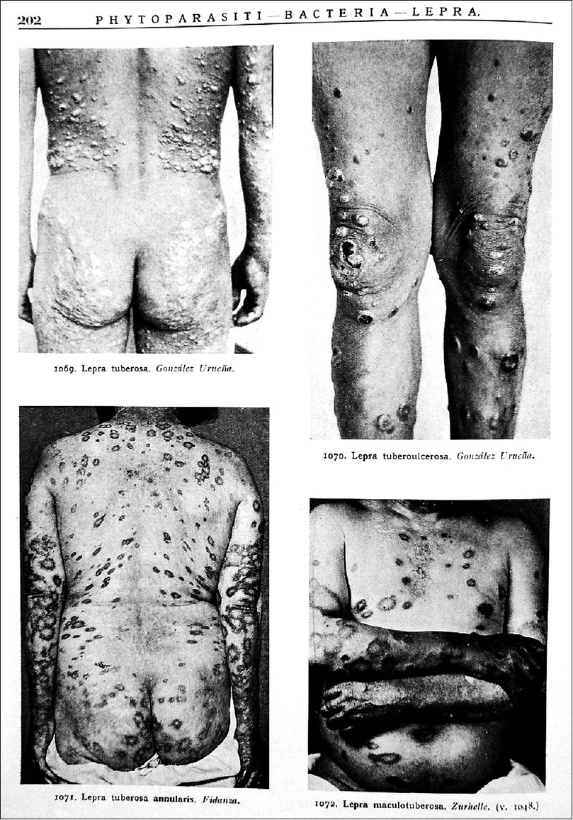 |
| Figure 4: The example of a page from Corpus Iconum Morborum Cutaneorum along with the illustrations of dermatoses (cutaneous manifestations of leprosy) |
To sum up, Corpus Iconum Morborum Cutaneorum is one of the most elaborate and accomplished historical dermatological atlases containing a vast number of scientifically relevant illustrations. It is also a symbol of the exemplary international cooperation achieved by the 9th International Dermatological Congress in the shadow of World War II. This moment is best captured in the words of Prof. Nékám himself [Figure - 5]: “The global warfare captured us for a period of five years – but in this field, we were united. Here, a medical spirit along with a holy sense of humanism have prevailed. We had a goal that inspired all of us: Artis Progressus et Consensus Medicorum! (The progress of the art (of healing) and the consensus (general agreement) of the doctors)”[2]
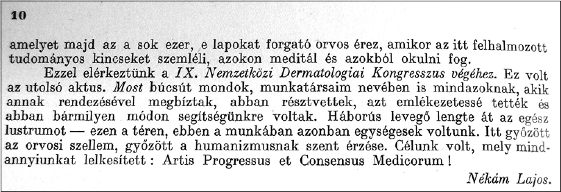 |
| Figure 5: Final words of Prof. Nékám in the Preface of the first volume of Corpus Iconum Morborum Cutaneorum (in Hungarian): “The global warfare captured us for a period of five years – but in this field, we were united. Here, a medical spirit along with a holy sense of humanism have prevailed. We had a goal that inspired all of us: Artis Progressus et Consensus Medicorum! (The progress of the art (of healing) and the consensus (general agreement) of the doctors!)” |
| 1. |
Crissey JT, Parish LC, Holubar K. Historical Atlas of Dermatology and Dermatologists. London: Parthenon Publishing; 2002.
[Google Scholar]
|
| 2. |
Nékám L, editor. Corpus Iconum Morborum Cutaneorum (I, II, III). Budapest: Hungarian Medical Publishing Company; 1938.
[Google Scholar]
|
| 3. |
Foldvari F. Lajos Nékám, 1868-1957. Orv Hetil 1957;98:313-4.
[Google Scholar]
|
Fulltext Views
2,884
PDF downloads
2,058


