Translate this page into:
Sarcomatoid lung carcinoma presenting as alopecia neoplastica
2 Department of Pathology, Government Medical College, Kottayam, Kerala, India
Correspondence Address:
Naveena Jose
Department of Dermatology and Venereology, Government Medical College, Kottayam, Kerala
India
| How to cite this article: Jose N, Isac CM, Kunjumani S, Vilasiniamma L. Sarcomatoid lung carcinoma presenting as alopecia neoplastica. Indian J Dermatol Venereol Leprol 2018;84:188-190 |
Sir,
The primary tumors most commonly metastasizing to the scalp are lung cancer (23.53%), followed by colorectal (11.76%) cancers.[1] Scalp metastases usually present as solitary or multiple nodules or plaques and rarely as alopecia neoplastica.[2] Sarcomatoid carcinomas are a group of poorly differentiated non-small cell lung carcinomas that contain a component of sarcoma or sarcoma-like differentiation. Five subtypes representing a morphologic continuum are currently recognized: pleomorphic carcinoma, spindle cell carcinoma, giant cell carcinoma, carcinosarcoma, and pulmonary blastoma.[3] Here, we present a very rare case of sarcomatoid carcinoma of the lung presenting as alopecia neoplastica. We were unable to find any previous reports of the same.
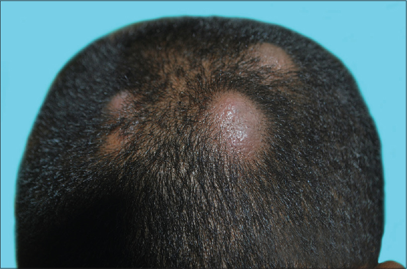
A 32-year-old man was referred with multiple painful nodules over the face and scalp with alopecia [Figure - 1]. On examination there were multiple skin colored firm/firm tender nodules of size 2x2 cm to 5x4 cm with overlying non cicatricial/non cicatricial alopecia. Hepatomegaly was found on abdominal examination but there was no peripheral lymphadenopathy. While in the ward the patient developed excruciating headache, but there was no focal neurological deficit. We proceeded with the clinical possibilities of adnexal tumors and cutaneous metastases from an unknown primary malignancy.
 |
| Figure 1: Multiple nodules over scalp with non-scarring alopecia |
Routine investigations were within normal limits except for the raised ESR (64 mm/1st hour). Chest X-ray showed mediastinal widening while ultrasonography of the abdomen revealed calcified foci in the liver and enlarged periportal lymph nodes. Fine needle aspiration cytology from the cutaneous nodule showed atypical spindle-shaped cells suggestive of invasive carcinoma. For confirmation, we obtained a punch biopsy of the lesion for histopathology and immunohistochemistry. Contrast-enhanced computed tomography revealed the primary tumor as a bronchogenic carcinoma in the posterior basal segment of right lower lobe with metastases in the lung parenchyma, mediastinal lymph nodes, liver, pancreas, and brain.
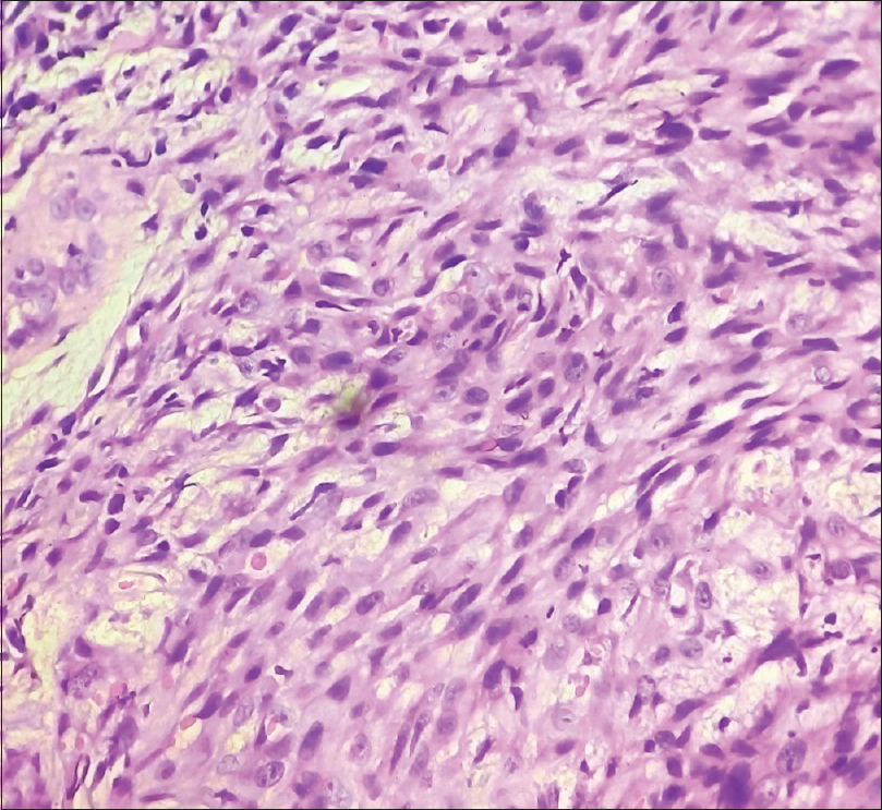
Histopathology of the skin lesion showed metastatic deposits in the dermis from a high-grade malignant neoplasm with cells of epithelial and spindle cell morphology [Figure - 2]. On immunohistochemistry, the cells were positive for cytokeratin and vimentin, and negative for thyroid transcription factor-1 (TTF-1) suggestive of metastasis from sarcomatoid lung carcinoma. Patient was referred to the oncology department where he was treated with cranial irradiation followed by chemotherapy. Unfortunately, the patient succumbed after the second cycle of chemotherapy.
 |
| Figure 2: Histopathology of the scalp biopsy showing pleomorphic cells of epithelial and spindle cell morphology (H and E, ×400) |
Sarcomatoid carcinomas are a group of poorly differentiated non-small cell lung carcinomas with an average age at diagnosis of 60 years. Males are four times more commonly affected, and tobacco smoking is the major risk factor.[3] Though our patient was a heavy smoker with 15 pack years, he was exceptionally young, had no obvious cough or any abnormal lung related signs or symptoms. He originally presented with alopecia neoplastica, which was of nonscarring type. His lesions were remarkable by the presence of non scarring alopecia and tenderness which is unusual as the most common manifestation of scalp metastasis is nontender nodules.[4]
Alopecia neoplastica usually manifests as a single or multiple areas of cicatricial alopecia, however, rarely it can simulate alopecia areata, as in our case.[2],[4],[5] The lesions may have a red-pink color and can evolve from a smooth to an uneven surface with peripheral telangiectasias.[5] Diagnosis of alopecia neoplastica can be confirmed on histologic examination; the dermis is fibrotic with metastatic cells in the reticular dermis or subcutis. The tumor cells may line up in single rows between fibrotic collagen, a finding known as “Indian filing.” There is also loss of the pilosebaceous units.[5] The exact mechanism of alopecia neoplastica is unclear. Fibrosis caused by stromal response or release of cytokines by the tumor cells is proposed to be the probable cause. Factors independent of fibrosis, such as tumor invasion of the hair sheaths, may also play a role in the development of alopecia as there are reports of regrowth of hair in alopecic areas after effective cancer treatment.[4] Archer and Smith had reported a case of alopecia neoplastica secondary to breast carcinoma in whom hair was regrown after treatment with tamoxifen.[6] However, in some patients despite chemotherapy, no hair regrowth was observed, as in a gastric carcinoma patient reported by Kim et al.[4]
The primary malignancy was identified during contrast-enhanced computed tomography scanning and confirmed by the histopathological and immunohistochemistry studies. The histological features and positive cytokeratin and vimentin are indicative of sarcomatoid carcinoma. Terada have previously reported a case of sarcomatoid carcinoma of the lung presenting with cutaneous metastasis on chest.[7]
Undetermined primary tumor accounted for approximately one-third of all metastatic scalp tumors in a study by Chiu et al., indicating that the underlying primary tumor might be poorly differentiated and highly malignant making no detectable internal primary tumor when scalp biopsy was performed.[1]
We report this case to emphasize that careful dermatological examination can provide clues to accurate diagnosis. Moreover, skin provides an easily accessible tissue which can help in diagnosing occult malignancy if seen by a skilled dermatologist and pathologist.
Declaration of patient consent
The authors certify that they have obtained all appropriate patient consent forms. In the form the patient(s) have given their consent for their images and other clinical information to be reported in the journal. The patients understand that their names and initials will not be published and due efforts will be made to conceal their identity, but anonymity cannot be guaranteed.
Financial support and sponsorship
Nil.
Conflicts of interest
There are no conflicts of interest.
| 1. |
Chiu CS, Lin CY, Kuo TT, Kuan YZ, Chen MJ, Ho HC, et al. Malignant cutaneous tumors of the scalp: A study of demographic characteristics and histologic distributions of 398 Taiwanese patients. J Am Acad Dermatol 2007;56:448-52.
[Google Scholar]
|
| 2. |
Kohen L, Kerr H. Alopecia neoplastica. J Am Acad Dermatol 2009; 60:AB42.
[Google Scholar]
|
| 3. |
Corrin B, Wick MR, Chang YL, Rossi G, Koss MN, Geisinger K, et al. Sarcomatoid carcinoma. In: Travis WD, Brambilla E, Muller-Hermelink HK, Harris CC, editors. World Health Organization Classification of Tumours. Pathology and Genetics of Tumours of the Lung, Pleura, Thymus and Heart. Vol. 1. Lyon: IARC Press; 2004. p. 53-8.
[Google Scholar]
|
| 4. |
Kim JH, Kim MJ, Sim WY, Lew BL. Alopecia neoplastica due to gastric adenocarcinoma metastasis to the scalp, presenting as alopecia: A case report and literature review. Ann Dermatol 2014;26:624-7.
[Google Scholar]
|
| 5. |
Rondina A, Kohen LL, Scherschun L, Kerr H. JAAD grand rounds quiz. A woman with focal alopecia. J Am Acad Dermatol 2010;63:545-7.
[Google Scholar]
|
| 6. |
Archer CB, Smith NP. Alopecia neoplastica responsive to tamoxifen. J R Soc Med 1990;83:647-8.
[Google Scholar]
|
| 7. |
Terada T. Sarcomatoid carcinoma of the lung presenting as a cutaneous metastasis. J Cutan Pathol 2010;37:482-5.
[Google Scholar]
|
Fulltext Views
3,245
PDF downloads
1,384


