Translate this page into:
Cellular and biomolecular comparison of a novel, dual-pulsed Q-switched 1064 nm Nd:YAG laser with conventional Q-switched 1064 nm Nd:YAG laser
Correspondence Address:
Sung Eun Chang
Department of Dermatology, University of Ulsan College of Medicine, Asan Medical Center, 86 Asanbyeongwon-Gil, Songpa-gu, Seoul 138-736
Republic of Korea
| How to cite this article: Kim BW, Moon IJ, Chang SE. Cellular and biomolecular comparison of a novel, dual-pulsed Q-switched 1064 nm Nd:YAG laser with conventional Q-switched 1064 nm Nd:YAG laser. Indian J Dermatol Venereol Leprol 2017;83:251-255 |
Sir,
A recent clinical and histopathological study in melasma patients using a low-fluence, 1064 nm Q-switched neodymium-doped yttrium aluminum garnet (QS Nd:YAG) laser has shown effectiveness in reducing the number of melanosomes and expression of melanogenesis associated proteins.[1] Compared to the high-fluence 1064 nm QS Nd:YAG laser treatment, using the low-fluence, multiple pass and repeated version (called 'laser toning') has a lower risk of adverse events in the treatment of melasma. However, the treatment outcomes are inconsistent. Adverse events including post-inflammatory hyperpigmentation and mottled hypopigmentation by conventional laser toning may occur, especially when the melasma lesions are accompanied by erythema.[1],[2],[3] To overcome these pitfalls, a dual-pulsed (or twin-pulsed) mode of 1064 nm QS Nd:YAG laser was devised. This mode delivers the desired fluence in two evenly divided pulses, separated by a very short time interval. In a previous study, the dual-pulsed mode was clinically effective in treating hyperpigmentary disorders such as post inflammatory hyperpigmentation and Riehl's melanosis.[4],[5] However, its effect has not been demonstrated at a histological or molecular level.[1],[4],[5] Therefore, we examined the efficacy of the dual-pulsed 1064 nm QS Nd:YAG laser at a histological and molecular level.
The laser device (Tri-beam ®, Jeisys Medical Inc., Korea) used in this study provides a dual-pulsed mode with a 140 μs pulse interval. For this study in vivo, pre-tanned brown guinea pig skin using UVB handisol (300 mJ/cm [2], 9 times over 3 weeks; National Biological Corporation, OH, USA) and naturally-tanned human skin was used. The right forearm of a 45-year-old Korean male volunteer was chosen for this purpose. For in vitro tests, cultured melanin-rich mouse melanocytes (Melan-A cell line) were used. All of these were irradiated separately by a 1064 nm QS Nd: YAG laser system using the following two parameters, parameter 1: dual-pulsed mode, 3 J/cm [2] (two evenly divided pulses of 1.5 J/cm [2] delivered with a time gap of 140 μs between the pulses), 1 pass and parameter 2: conventional mode, 3 J/cm [2], 1 pass.
After laser irradiation, the guinea pig skin (at day one, and two weeks after laser irradiation) and cultured Melan-A cells (one hour after laser irradiation) were subjected to electron microscopic examination. In addition to this, skin biopsy samples were taken from the treated and non-treated areas of the human skin and evaluated using Fontana-Masson staining and quantitative reverse transcriptase polymerase chain reaction (RT-PCR) for mRNA (seven days after laser irradiation).
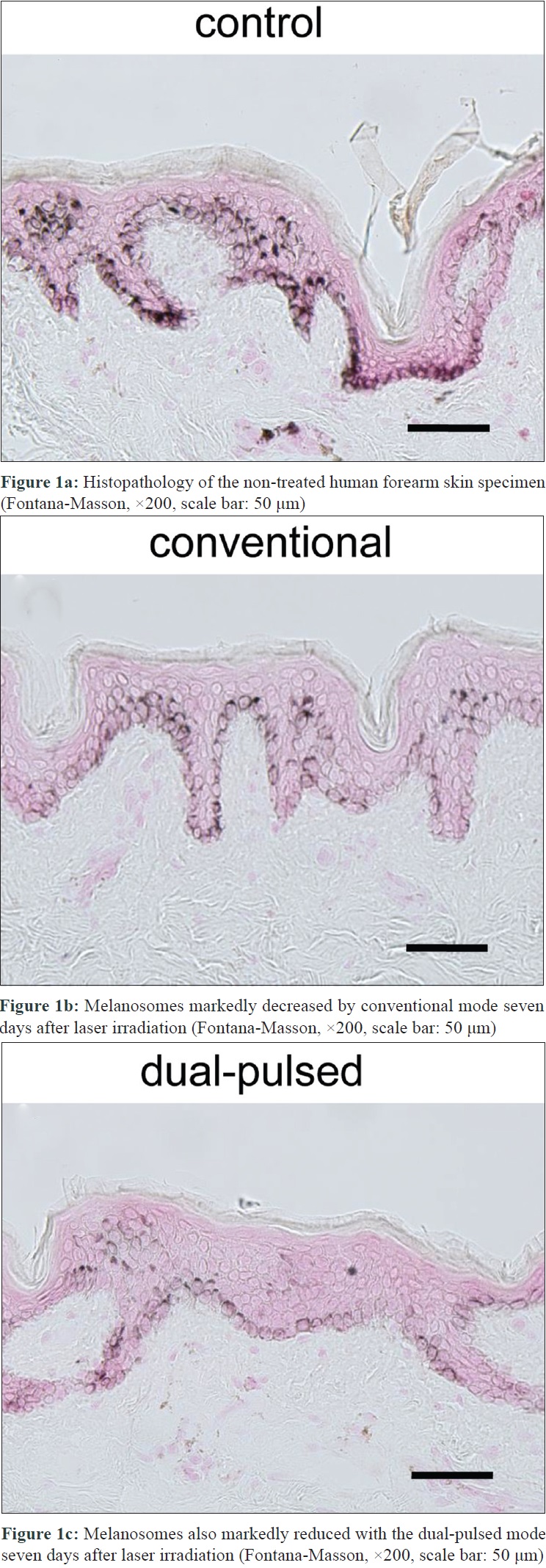
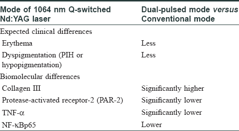
Fontana-Masson staining of the human skin revealed a similar effect of pigment destruction across both parameters used [Figure - 1]. The quantitative RT-PCR showed an increase in type III collagen levels in both modes of laser irradiation. However, the dual-pulsed mode elevated the type III collagen level three times more than the conventional mode [Figure - 2]. The protease-activated receptor (PAR)-2 levels were lower by 50% in the dual-pulsed mode, compared to the conventional mode. Pro-inflammatory transcription factor such as nuclear factor κB p65 subunit (NF-κBp65, RelA) and tumor necrosis factor-α (TNF-α) were also remarkably lesser in the dual-pulsed mode compared to the conventional mode [Figure - 2].
 |
| Figure 1 |
 |
| Figure 2 |
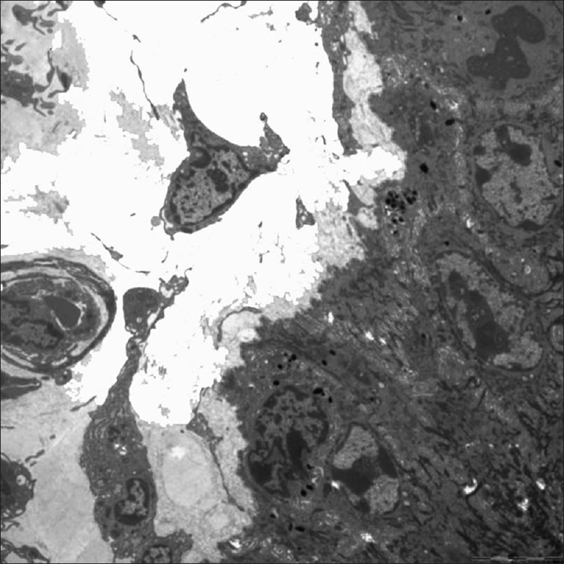
On electron microscopy of guinea pig skin, the dual-pulsed mode was shown to have lesser damage to epidermal keratinocytes and melanocytes compared to the conventional mode. At one day after laser irradiation, spongiosis and damaged mitochondria in keratinocytes were noted in both modes [Figure 3a] and [Figure 3b]. In addition, melanosomes in the upper dermis, with vacuolar changes at the dermo-epidermal junction were observed in specimens irradiated by the conventional mode [Figure 3a]. These are thought to be melanosomes that have fallen into the dermis due to an excessive laser photomechanical effect, clinically appearing as post-inflammatory hyperpigmentation. Interestingly, at two weeks after laser irradiation, the dual-pulsed mode showed less prominent spongiosis and intracellular vacuoles compared to the conventional mode. The basement membranes looked partially damaged only in the conventional mode[Figure 3c] and [Figure 3d]. On electron microscopy of laser-irradiated cultured Melan-A cells at 1 hr after irradiation, the vacuoles were located mainly within the intra-cytoplasmic spaces when irradiated with the conventional mode, whereas it was found within the inter-cellular spaces with the dual-pulsed mode. Disrupted melanosomes and peripheral condensation of chromatin was observed in both modes, with no obvious differences [Figure - 4].
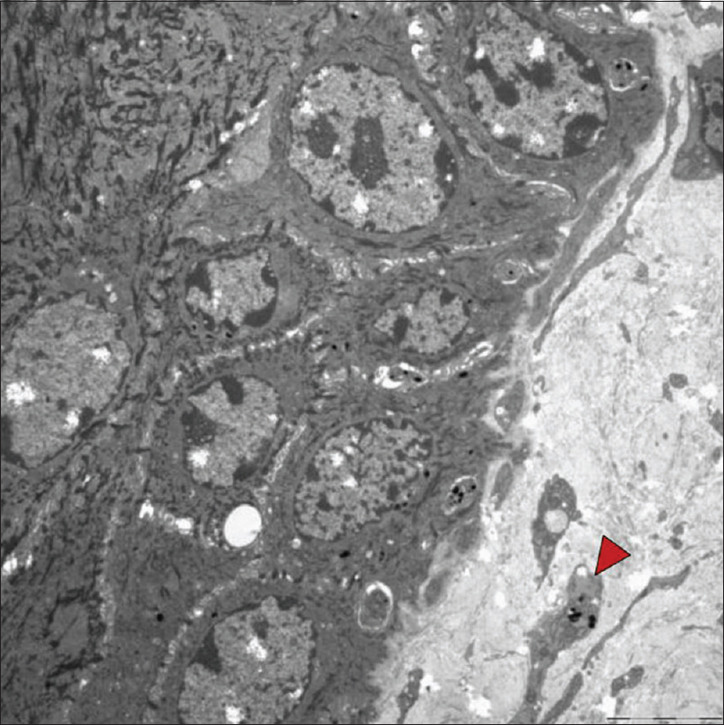 |
| Figure 3a: Electron microscope image (×3000 magnification) of guinea pig epidermis after irradiation with conventional mode of 1064 nm QS Nd:YAG laser at 1 day after laser irradiation. Inter-cellular vacuolar spaces and melanosomes in the upper dermis (arrow), with vacuolar changes at the dermo-epidermal junction are observed |
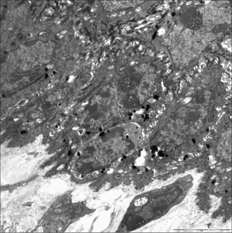 |
| Figure 3b: Electron microscope image (×3000 magnification) of guinea pig epidermis after irradiation with 1064 nm QS Nd:YAG laser at 1 day after laser irradiation in dual-pulsed mode. Inter-cellular vacuolar spaces in keratinocytes were noted |
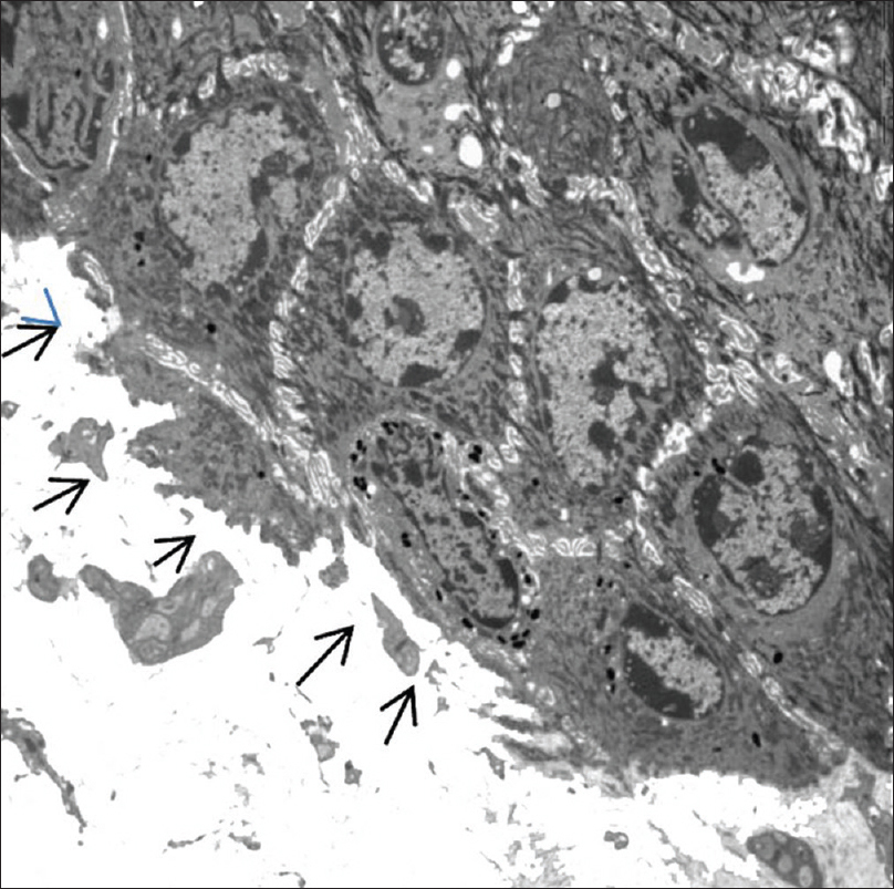 |
| Figure 3c: At 2 weeks after laser irradiation, the conventional mode still showed prominent inter-cellular vacuolar spaces and intra-cytoplasmic vacuoles and the basement membranes are damaged (arrow) (×3000 magnification) |
 |
| Figure 3d: At 2 weeks after laser irradiation, the dual-pulsed mode showed rare inter-cellular vacuolar spaces and intra-cytoplasmic vacuoles (scale bar: 5 μm) (×3000 magnification) |
 |
| Figure 4 |
Our study sheds light on the cellular and biomolecular differences between the dual-pulsed and conventional modes of 1064 nm QS Nd:YAG laser [Table - 1]. The higher levels of type III collagen demonstrated after dual-pulsed mode irradiation is believed to result from the additional photo-acoustic waves generated by dividing the pulse. The relatively lower PAR-2 levels, pro-inflammatory transcription factors and pro-inflammatory cytokines following dual-pulsed mode indicates this mode is less susceptible to skin erythema/inflammation or resulting dyspigmentation. Electron microscopy of the irradiated guinea pig skin shows that the dual-pulsed mode exhibits gentle treatment delivery in terms of reducing damage to epidermal keratinocytes and melanocytes compared to the conventional mode. This fits well into the concept of strict “subcellular selective photothermolysis”, that is the selective elimination of pigment, while simultaneously minimizing the adverse events caused by cellular damage and adjacent inflammatory events.[3] Although the small sample size does not allow generalization of our findings, we propose that the dual-pulsed mode of the 1064 nm QS Nd:YAG laser may be more suitable than the conventional mode for those who are prone to post-inflammatory hyperpigmentation, sensitive skin, or melasma in darker skin types.
Acknowledgment
We would like to express our gratitude to Dr. Un-Cheol Yeo, Dr. Seung-Hyun Bang and Dr. Woo Jin Yun for their considerable contribution to our study design and experiments.
Financial support and sponsorship
Industrial Core Technology Development program through Korea Evaluation Institute of Industrial Technology funded by the Ministry of Trade, Industry and Energy (No. 10048690).
Conflicts of interest
There are no conflicts of interest.
| 1. |
Kim JE, Chang SE, Yeo UC, Haw S, Kim IH. Histopathological study of the treatment of melasma lesions using a low-fluence Q-switched 1064-nm neodymium:yttrium-aluminium-garnet laser. Clin Exp Dermatol 2013;38:167-71.
[Google Scholar]
|
| 2. |
Lee WJ, Kim YJ, Noh TK, Chang SE. Formation of new melasma lesions in the periorbital area following high-fluence, 1064-nm, Q-switched Nd/YAG laser. J Cosmet Laser Ther 2013;15:163-5.
[Google Scholar]
|
| 3. |
Park GH, Lee JH, Choi JR, Chang SE. The degree of erythema in melasma lesion is associated with the severity of disease and the response to the low-fluence Q-switched 1064-nm Nd:YAG laser treatment. J Dermatolog Treat 2013;24:297-9.
[Google Scholar]
|
| 4. |
Kim BW, Lee MH, Chang SE, Yun WJ, Won CH, Lee MW, et al. Clinical efficacy of the dual-pulsed Q-switched neodymium:yttrium-aluminum-garnet laser: Comparison with conservative mode. J Cosmet Laser Ther 2013;15:340-1.
[Google Scholar]
|
| 5. |
Chung BY, Kim JE, Ko JY, Chang SE. A pilot study of a novel dual – Pulsed 1064 nm Q-switched Nd: YAG laser to treat Riehl's melanosis. J Cosmet Laser Ther 2014;16:290-2.
[Google Scholar]
|
Fulltext Views
3,280
PDF downloads
1,686


