Translate this page into:
Dermoscopy: A rapid bedside tool to assess monilethrix
Correspondence Address:
Vinod Kumar Sharma
Department of Dermatology and Venereology, All India Institute of Medical Sciences, New Delhi - 110 029
India
| How to cite this article: Sharma VK, Chiramel MJ, Rao A. Dermoscopy: A rapid bedside tool to assess monilethrix. Indian J Dermatol Venereol Leprol 2016;82:73-74 |
Sir,
Monilethrix is a rare genetic disorder characterized by fragility of hair shafts resulting in alopecia. It is of autosomal dominant inheritance and the genes most commonly implicated are hHB1 and hHB6 genes on chromosome 12. The hair is normal at birth; however, in a few months the patient develops keratotic papules, alopecia and brittle hair. We report a case of monilethrix where dermoscopy helped in the bedside diagnosis and helped in estimating the extent of involvement.
A 12-year-old girl presented to us with a history of decreased hair growth, easy breakability of hair and diffuse alopecia since 3 months of age. She was born at term of a non-consanguineous marriage and had normal hair at birth. Within 2-3 months of age, the parents noticed gradual development of multiple, asymptomatic, keratotic, tiny papules on the scalp along with breakage of hair and alopecia. Later, she developed multiple, itchy, hyperkeratotic papules on the neck, arms, shoulders, and thighs along with progressive alopecia. The parents gave a history of recurrent sneezing, repeated episodes of rhinitis and dryness of skin. There was a history of similar but milder complaints in an older sister.
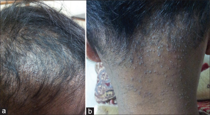
On examination, there was thinning and shortening of hair, most prominent on the temporal and occipital scalp [Figure - 1]a and b]. All the hairs were short and were broken at varying lengths, rough to feel and lusterless. There were follicular keratotic papules on the scalp, nape of neck, shoulders, upper arms and thighs with some scattered lesions elsewhere. The hair on the arms and legs were sparse but those over the eyebrows, eyelashes, axillary and pubic area were within normal limits. Oral cavity, teeth and nails were normal. There was hyperlinearity of palms and soles. A clinical diagnosis of monilethrix with an atopic diathesis was made.
 |
| Figure 1: (a) Diffuse alopecia on the parietal scalp with broken hair. (b) Keratotic papules on the nape of the neck with broken hairs on the occipital scalp |
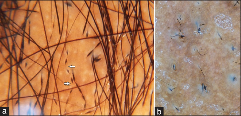
Dermoscopy was performed with a handheld ×10 contact dermoscope (Heine Delta 20). The sites chosen were bilateral temporal scalp, vertex, occipital scalp and the keratotic papules on nape of neck and forearms (devoid of visible hair). The scalp sites showed multiple beaded hair with equidistant nodes and internodes [Figure - 2]a. The hair were of varying lengths; many were broken. Hair with normal morphology were seen interspersed within these beaded hair. The beaded hair showed bending in different directions with a tendency to break at internodes (regularly bended ribbon sign). On the scalp, white dots and a honey comb pigment network was seen. Dermoscopy of the keratotic papules on neck showed beaded hair arising from the papules [Figure - 2]b. The forearm showed no hair clinically but on dermoscopy, beaded short hairs were seen.
 |
| Figure 2: (a) Scalp dermoscopy showing multiple, regularly beaded hairs. Arrows shows “regularly bended ribbon sign.” (b) Dermoscopic image of keratotic papules on the nape of neck showing broken and beaded hair emerging from the surface |
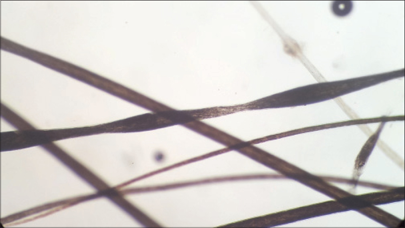
Hair microscopy was performed, and it showed regular beading of hairs [Figure - 3]. Biopsy from the scalp did not reveal any abnormality in the hair follicle. Biopsy from the nape of the neck showed a granulomatous reaction surrounding the hair shaft.
 |
| Figure 3: Hair microscopy view showing beading of hair shaft (×400) |
The patient was counseled regarding the nature of disease. She was treated with 5% minoxidil and antihistamines for pruritus. Emollients were prescribed for her generalized xerosis and the need to avoid trauma to the hair was emphasized.
Monilethrix was first described in 1879 by Walter Smithwho described a rare nodose condition of the hair. Radcliffe Crockersuggested the name monilethrix from monile meaning necklace (Latin), and thrix meaning hair (Greek).[1]
Diagnosis is mostly clinical. Hair microscopy and dermoscopy shows beaded hair. Histology may show abnormal hair follicles with alternating constricted and normal portions, in a few cases. Dermoscopy in monilethrix shows short hair with regular beading.[2] Rakowska et al. described the dermoscopic appearance of beaded monilethrix shafts as a “regularly beaded ribbon.”[3] Because of the tendency to break at internodes, the hair shaft may seem to bend in various directions, and this is described as “regularly bended ribbon sign.” Dermoscopy is much easier and less time consuming than ex vivo microscopic examination and the characteristic dermoscopic features enables one to establish a rapid diagnosis. Direct demonstration of beaded hair in areas of sparse hair such as the forearm and on the keratotic papules helps in diagnosis. Dermoscopy may prevent the misdiagnosis of iatrogenic pseudomonilethrix which is caused by pressed overlapping hair in glass slides prepared for microscopic examination.[4] Another use of dermoscopy would be to quickly assess the extent of involvement in patients and in their relatives. Since the number of beaded hair are quite variable, the diagnosis may be missed if only a few hair are assessed. It will also help to choose a hair for direct microscopy in order to get a maximum yield of abnormal features.
Financial support and sponsorship
Nil.
Conflicts of interest
There are no conflicts of interest.
| 1. |
Dawber RP. An update of hair shaft disorders. Dermatol Clin 1996;14:753-72.
[Google Scholar]
|
| 2. |
Wallace MP, de Berker DA. Hair diagnoses and signs: The use of dermatoscopy. Clin Exp Dermatol 2010;35:41-6.
[Google Scholar]
|
| 3. |
Rakowska A, Slowinska M, Czuwara J, Olszewska M, Rudnicka L. Dermoscopy as a tool for rapid diagnosis of monilethrix. J Drugs Dermatol 2007;6:222-4.
[Google Scholar]
|
| 4. |
Liu CI, Hsu CH. Rapid diagnosis of monilethrix using dermoscopy. Br J Dermatol 2008;159:741-3.
[Google Scholar]
|
Fulltext Views
5,694
PDF downloads
2,434


