Translate this page into:
White fibrous papulosis of the neck
2 Consultant Pathologist, OncQuest Laboratories, New Delhi, India
Correspondence Address:
Rajat Kandhari
11, Munirka Marg, Vasant Vihar, New Delhi - 110 057
India
| How to cite this article: Kandhari R, Kandhari S, Jain S. White fibrous papulosis of the neck. Indian J Dermatol Venereol Leprol 2015;81:224 |
Sir,
White fibrous papulosis of the neck (WFPN) is an under-reported entity. It is characterized by multiple, 2-3 mm sized, discrete, asymptomatic, pale- to skin-colored, non-follicular, firm papular lesions present predominantly on the neck in elderly individuals. The racial prevalence is unclear although it has been reported from various parts of the world, with no sexual predilection. [1]
A 73-year-old woman presented with mildly pruritic, papular lesions on her neck for 1 year. She had no history of prolonged sun exposure and denied rubbing or scratching the affected areas. Her other illnesses included hypertension, paroxysmal atrial fibrillation, chronic kidney disease and hyperuricemia for which she was being treated with doresemide, clopidogrel and aspirin combination, and febuxostat. There was no history suggestive of vascular, gastrointestinal or ocular disorders and no history of similar lesions in other family members. Dermatological examination revealed multiple, discrete, non-confluent, pale- to skin-colored papular lesions predominantly on the lateral side of her neck extending to the nape of the neck. The lesions were firm, non-follicular, and varied in size from 2-4 mm and had progressively increased in number slowly over the past 1 year [Figure - 1]. Examination of the cardiovascular, respiratory and gastrointestinal systems revealed no abnormalities and her peripheral pulses were normal. Examination of the fundus did not demonstrate angioid streaks. Skin biopsy from the papules revealed mild orthokeratotic hyperkeratosis and haphazardly arranged bundles of collagen fibers in the reticular and deep dermis. The elastic fibers were normal but slightly reduced in number [Figure - 2], [Figure - 3], [Figure - 4]. In view of the normal elastic fibers seen on histology, characteristic clinical features and lack of systemic signs a diagnosis of white fibrous papulosis of neck was made. The patient was counseled regarding the condition and no treatment was given. There had been no change in the lesions on follow up 6 months later.
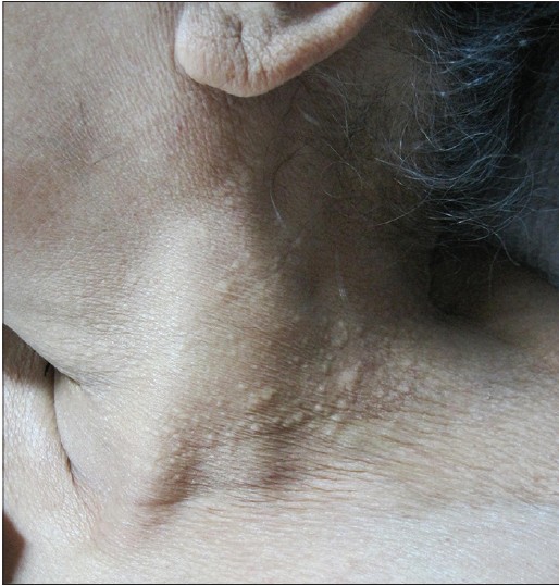 |
| Figure 1: Multiple, discrete, non-follicular, firm papules on the lateral aspect of the neck |
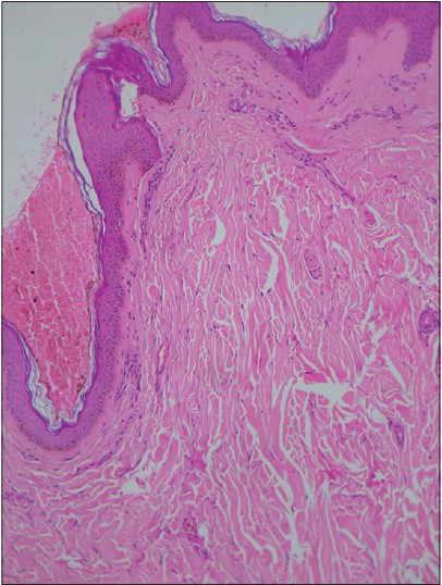 |
| Figure 2: Haphazardly arranged bundles of collagen fibers in the reticular and deep dermis. (H and E, ×100) |
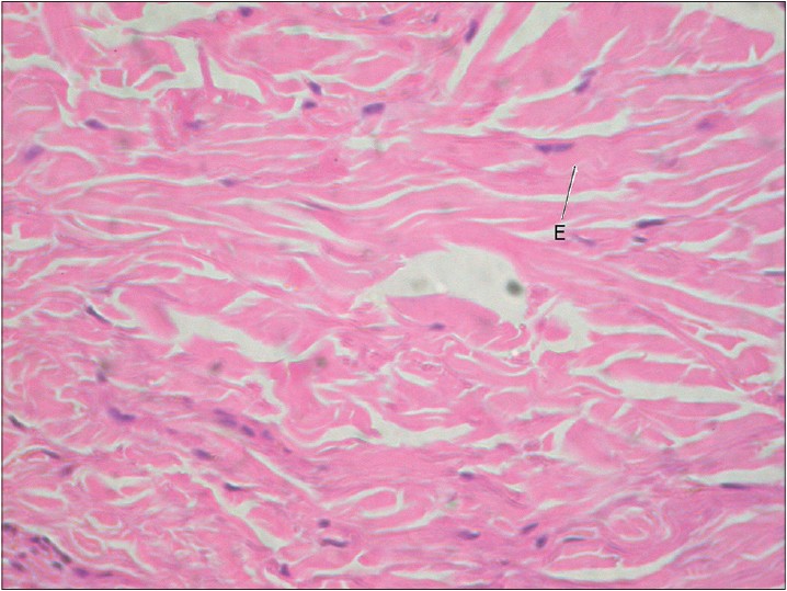 |
| Figure 3: Higher magnification with bundles of collagen with elastic fibers (E) (H and E, ×400) |
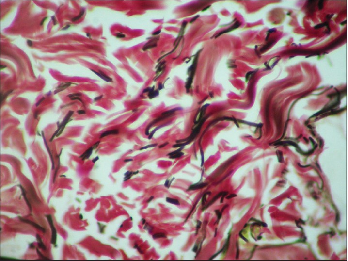 |
| Figure 4: Elastic fibers in black and collagen fibers seen in red (Verhoeff-VanGieson, ×400) |
White fibrous papulosis of neck was described in 1985 by Shimizu and Nishikawa. [2] Clinically, it is characterized by multiple, usually asymptomatic, non-confluent, non-pedunculated, and non-follicular, firm papules occurring on the posterior or lateral sides of the neck. They may vary in number and size and may rarely involve other sites such as the trunk and upper arms. [3] No associated systemic co-morbidities have been reported with this condition and the occurrence of paroxysmal atrial fibrillation and chronic kidney disease in our patient may be purely coincidental. Whether systemic conditions or drug intake have a role in triggering the disease needs further elucidation.
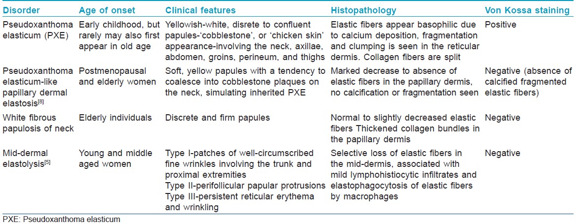
Histologically, white fibrous papulosis of neck is characterized by slight, focal increase and thickening of collagen fibers in the papillary dermis. accompanied by normal to decreased elastic fibers. [4] It has clinical and histological similarities with dermatoses such as pseudoxanthoma elasticum-like papillary dermal elastosis (PXE-PDE) and mid-dermal elastosis (MDE).These are distinguishable on the basis of subtle signs[Table - 1]. In circumstances where there is sharing of clinical and histopathological features, the term "fibroelastolytic papules of the neck" (FEPN) has been used. [5],[6] Ultrastructurally, no abnormalities of the elastic fibers are observed and collagen fibrils seem tightly compacted with a slight increase in their diameter. These changes are considered to be non-specific features of papillary dermal fibrosis, associated with structural collagen alterations and/or abnormalities of the extracellular microenvironment. [7],[8]
Treatment of this condition is unsatisfactory and not necessary considering the benign nature of the condition. Natural regression has not been reported.
| 1. |
Kim HS, Yu DS, Kim JW. White fibrous papulosis of the neck. J Eur Acad Dermatol Venereol 2007;21:419-20.
[Google Scholar]
|
| 2. |
Shimizu H, Nishigawa T, Kimura S. White fibrous papulosis of the neck; review of our 16 cases. Nihon Hifuka Gakkai Zasshi 1985;95:1077-84.
[Google Scholar]
|
| 3. |
Mitoma C, Takahara M, Takeshita T, Kiryu H, Moroi Y, Furue M White fibrous papulosis on the trunk and upper arms. Int J Dermatol 2013;52:337-8.
[Google Scholar]
|
| 4. |
Chan JY, Chu CY, Hsiao CH, Chen JS. Fibroelastolytic patterns of intrinsic skin aging: Pseudoxanthoma elasticum - like papillary dermal elastolysis and white fibrous papulosis of the neck. Dermatol Sin 2003;21:402-7.
[Google Scholar]
|
| 5. |
Rongioletti F, Izakovic J, Romanelli P, Lanuti E, Miteva M. Pseudoxanthoma elasticum-like papillary dermal elastolysis: A large case series with clinicopathological correlation. J Am Acad 2012;67:128-35.
[Google Scholar]
|
| 6. |
Jagdeo J, Ng C, Ronchetti IP, Wilkel C, Bercovitch L, Robinson-Bostom L. Fibroelastolytic papulosis. J Am Acad Dermatol 2004;51:958-64.
[Google Scholar]
|
| 7. |
Zanca A, Contri MB, Carnevali C, Bertazzoni MG. White fibrous papulosis of the neck. Int J Dermatol 1996;35:720-2.
[Google Scholar]
|
| 8. |
Rongioletti F, Rebora A. Pseudoxanthoma elasticum like papillary dermal elastolysis. J Am Acad Dermatol 1992;26:648-50.
[Google Scholar]
|
Fulltext Views
6,283
PDF downloads
2,575


