Translate this page into:
'Swiss cheese' appearance of dilated follicular infundibula in trichotillomania
Correspondence Address:
Rajiv Joshi
14 Jay Mahal, A Road, Churchgate, Mumbai - 400 020, Maharashtra
India
| How to cite this article: Joshi R. 'Swiss cheese' appearance of dilated follicular infundibula in trichotillomania. Indian J Dermatol Venereol Leprol 2014;80:257-258 |
Sir,
A 4-mm punch biopsy from the scalp of a 32-year-old woman was received with the clinical diagnosis of trichotillomania. There was a 2.5 year history of patchy hair loss from the scalp associated with some pruritus. The patches used to recur mainly on the frontal and parietal scalp and individual patches showed broken hair of variable lengths but never was there any complete hair loss as is seen in typical alopecia areata. There were several remissions with complete re-growth of hair followed by recurrences of hair loss.
Sections revealed all follicular infundibula to be markedly dilated and filled with basket weave and laminated keratin [Figure - 1] and [Figure - 2]. Trichomalacia with broken and crumpled hair shafts with clumping of melanin were seen in several dilated infundibula. [Figure - 3] Numerous telogen/catagen hair follicles were seen in the mid and lower dermis [Figure - 4] Based on the clinical picture and histopathological findings of trichomalacia affecting several follicles, a diagnosis of trichotillomania was rendered.
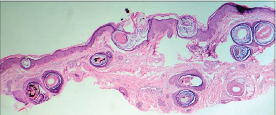 |
| Figure 1: Scanning power showing numerous dilated follicular infundibula, trichomalacia and pigment casts (H and E, ×20) |
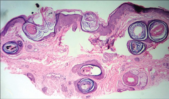 |
| Figure 2: Low power view showing 'swiss cheese' appearance due to dilated follicular infundibula (H and E, ×40) |
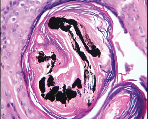 |
| Figure 3: Trichomalacia (H and E, ×400) |
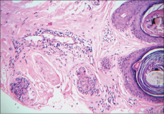 |
| Figure 4: Numerous telogen follicles in the lower dermis adjacent to dilated infundibula (H and E, ×100) |
The appearance of numerous horizontally sectioned dilated follicular infundibula has been likened to ′Swiss cheese′ [Figure - 5] and has been considered to be a useful criterion in the histopathological diagnosis of alopecia areata. [1] In one case series, this finding has been reported to occur in 57% of cases of alopecia areata and along with another common finding of shift of follicles to catagen/telogen (68%) has been regarded as an important and useful criterion for the histopathological diagnosis of alopecia areata [1] because the classic histopathological hallmark of alopecia areata, namely peribulbar ′swarm of bees′ infiltrate of lymphocytes may be seen in only about 38% of biopsies, [1] even less, if a late recovering lesion of alopecia areata has been biopsied. ′Swiss cheese′ appearance, however, has not been described in trichotillomania.
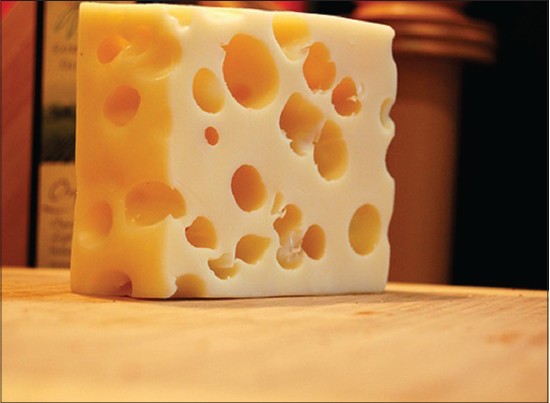 |
| Figure 5: Swiss cheese |
Dermoscopic finding of ′yellow dots′ which represent dilated follicular ostia filled with sebum are very common in alopecia areata and are seen in almost 95% of cases. [2] Yellow dots are distinctly uncommon in female or male pattern androgenetic alopecia and in scarring alopecias. [2]
The mechanisms for dilated follicular infundibula in alopecia areata are not known but it has been suggested that formation of nanogen follicles lacking hair shafts leads to empty follicular infundibula that correspond to the ′yellow dots′ seen on dermoscopy. [3]
Trichomalacia, which refers to the distortion and breaking up of the hair shaft with clumping of melanin, is the most characteristic histological finding in trichotillomania. [4] An unusually high number of catagen/telogen hair are also seen in the early stage of trichotillomania [4] and may be mistaken for alopecia areata, especially if trichomalacia is not present in the sections.
We describe the finding of ′Swiss cheese′ appearance in a case of trichotillomania and suggest that this finding may not be specific for alopecia areata but may be due to repeated rubbing of the skin of the scalp that accompanies the twisting and twirling of the hair which is the main cause of hair breakage in trichotillomania. In trichotillomania, apart from twisting and twirling the hair, the patient often rubs the skin over the affected area and this may lead to epidermal hyperplasia resembling lichen simplex chronicus. Increased numbers of catagen/telogen hair may lead to many nanogen follicles which are devoid of hair shafts and chronic rubbing may lead to dilatation of these empty follicles. Similarly, in long standing alopecia areata, repeated rubbing of the bald skin during application of topical medications may be the cause for the dilation of follicular infundibula as the greatest number of cases with dilated follicular infundibula were seen in patients with ophiasis and resolving lesions of alopecia areata. [1]
To conclude, we describe the histopathologic finding of ′Swiss cheese′ appearance due to dilated follicular infundibula in a case of trichotillomania and suggest that this finding may not be specific for alopecia areata and speculate that repeated rubbing of the skin may be the possible mechanism for inducing follicular dilatation.
| 1. |
Müller CS, El Shabrawi-Caelen L. 'Follicular Swiss cheese' pattern-another histopathologic clue to alopecia areata. J Cutan Pathol 2011;38:185-9.
[Google Scholar]
|
| 2. |
Tosti A, Whiting D, Iorizzo M, Pazzaglia M, Misciali C, Vincenzi C, et al. The role of scalp dermoscopy in the diagnosis of alopecia areata incognita. J Am Acad Dermatol 2008;59:64-7.
[Google Scholar]
|
| 3. |
Whiting DA. Histopathologic features of alopecia areata: A new look. Arch Dermatol 2003;139:1555-9.
[Google Scholar]
|
| 4. |
Muller SA. Trichotillomania: A histopathologic study in sixty-six patients. J Am Acad Dermatol 1990;23:56-62.
[Google Scholar]
|
Fulltext Views
6,106
PDF downloads
1,841


