Translate this page into:
Unilateral asymmetrical double Becker's naevus
2 Department of Pathology, Government Medical College, Kota, Rajasthan, India
Correspondence Address:
Suresh Kumar Jain
Department of Dermatology, Venereology, and Leprology, Government Medical College, Kota - 324 001, Rajasthan
India
| How to cite this article: Mehta P, Kumar R, Jain SK, Rai NN. Unilateral asymmetrical double Becker's naevus . Indian J Dermatol Venereol Leprol 2014;80:470-471 |
Sir,
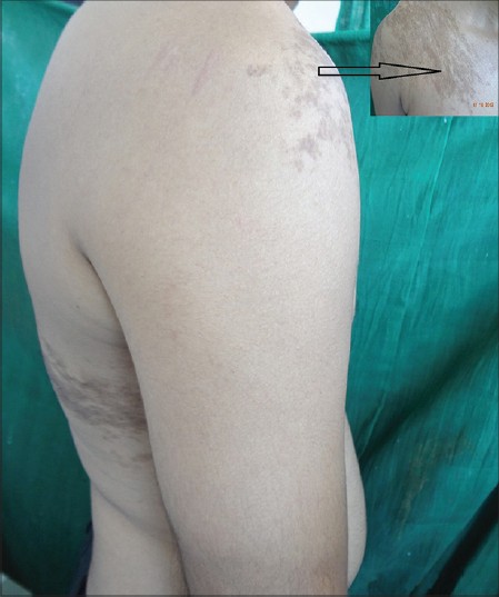
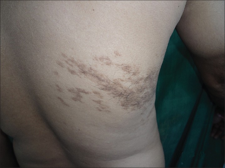
A 35-year-old healthy man presented with two asymptomatic hyperpigmented lesions confined to the right half of his torso. One was a well demarcated brownish patch on the right shoulder with irregular borders, approximately 15 Χ 10 cm in size, with multiple small islands of hyperpigmentation in the surrounding skin [Figure - 1] inset] The other lesion, extending from the right lower scapular area to the lateral chest wall, approximately 7 Χ 15 cm in size, showed a similar morphology with the additional feature of hypertrichosis [Figure - 2]. Both lesions had developed around the onset of puberty and became progressively darker; the shoulder lesion was noticed first and the other one appeared a year later. A clinical diagnosis of Becker′s nevi was made.
General physical examination was within normal limits. We did not find any associated musculoskeletal changes or acneiform lesions. Genital and limb examination were normal. X-ray spine and ultrasound of the abdomen and scrotum did not reveal any abnormality. KOH examination of skin scrapings from the lesion was negative for pityriasis versicolor. Skin biopsy was performed from both lesions. Histopathology revealed epidermal acanthosis along with regular elongation of rete ridges. Increased pigmentation of the basal layer was seen without increase in the number of melanocytes. Some melanophages were seen in the dermis. Based on the clinical and histopathological findings, a diagnosis of Becker′s nevi was made.
 |
| Figure 1: Multiple hyperpigmented coalescing macules over right shoulder (inset) and right lower lateral chest |
 |
| Figure 2: Hypertrichotic variant of Becker's nevus at right lower lateral chest |
In 1949, Samuel William Becker first described a "concurrent melanosis and hypertrichosis in the distribution of nevus unius lateris" which has since been termed Becker′s nevus. [1] Becker′s nevus commonly manifests in peri-pubertal males as a unilateral, solitary, acquired localized hyperpigmented patch composed of coalescing brownish macules. Hyperpigmentation usually increases for the first 2-3 years while hypertrichosis always appears after the pigmentation. However, the non-hypertrichotic variant is more common. "Progressive cribriform and zosteriform hyperpigmentation" may represent the non-hypertrichotic variant of Becker′s nevus. There are a few documented cases with multiple and bilaterally symmetrical Becker′s nevi; multiple and unilateral lesions are reported to occur much more rarely. [2],[3],[4] Two Becker′s nevi on the left side of the face in a segmental distribution with extension onto the oral mucosa have also been reported. [5] The occurrence of two asymmetrical Becker′s nevi in the same person supports the cutaneous mosaicism (somatic) theory of origin of Becker′s nevus, characterized by the presence of two or more populations of genetically different cells derived from the same zygote. Becker′s nevus follows the type 2 clinical pattern (checkerboard pattern) of cutaneous mosaicism described by Happle. [5]
Anomalies associated with Becker′s nevus include smooth muscle hamartoma, pectus excavatum and many other developmental defects. [4] Happle and Koopman [6] coined the term Becker′s naevus syndrome for Becker′s naevus associated with breast hypoplasia or other skin, bone or muscle alterations present on the same side as the nevus. Discoid lupus erythematosus on Becker′s nevus has also been reported. Androgenic pathogenesis of Becker′s nevus has been suggested based on its less frequent occurrence in females; moreover, hypertrichosis and hyperpigmentation are relatively less appreciable in affected women. In support of this hypothesis, increased expression of androgen receptors and androgen receptor mRNA has been found in dermal fibroblasts of Becker′s nevus. [7]
| 1. |
Becker SW. Concurrent melanosis and hypertrichosis in distribution of nevus unius lateris. Arch Derm Syphilol 1949;60:155-60.
[Google Scholar]
|
| 2. |
Bansal R, Sen R. Bilateral Becker's nevi. Indian J Dermatol Venereol Leprol 2008;74:73.
[Google Scholar]
|
| 3. |
Pahwa P, Sethuraman G. Segmental Becker's nevi with mucosal involvement. Pediatr Dermatol 2012;29:670-1.
[Google Scholar]
|
| 4. |
Chung HM, Chang YT, Chen CL, Wang WJ, Wong CK. Becker's melanosis associated with ipsilateral lower limb hyperplasia and pectus excavatum: A case reportand review of the literature. Dermatol Sinica 2002;20:27-32.
[Google Scholar]
|
| 5. |
Happle R. Mosaicism in human skin. Understanding the patterns and mechanisms. Arch Dermatol 1993;129:1460-70.
[Google Scholar]
|
| 6. |
Happle R, Koopman RJ. Becker nevus syndrome. Am J Med Genet 1997;68:357-61.
[Google Scholar]
|
| 7. |
Grande Sarpa H, Harris R, Hansen CD, Callis Duffin KP, Florell SR, Hadley ML. Androgen receptor expression patterns in Becker's nevi: An immunohistochemical study. J Am Acad Dermatol 2008;59:834-8.
[Google Scholar]
|
Fulltext Views
3,539
PDF downloads
1,391


