Translate this page into:
A case of hidroacanthoma simplex with new dermoscopic features
2 Department of Dermatopathology, Tehran University of Medical Sciences, Tehran, Iran
3 Autoimmune Bullous Diseases Research Center, Tehran University of Medical Sciences, Tehran, Iran; Faculty of Medicine, The University of Queensland, Australia,
Correspondence Address:
Hamidreza Mahmoudi
Autoimmune Bullous Diseases Research Center, Razi Hospital, Vahdate-Eslami Square, Tehran 11996
Iran
| How to cite this article: Toosi R, Kamyab K, Rosendahl C, Tavakolpour S, Daneshpazhooh M, Mahmoudi H. A case of hidroacanthoma simplex with new dermoscopic features. Indian J Dermatol Venereol Leprol 2019;85:319-321 |
Sir,
Hidroacanthoma simplex is a rare benign adnexal tumor, first described by Smith and Coburn in 1956. It is recognized as an intraepidermal variant of poroid cell neoplasms that originate from the acrosyringial portion of the eccrine duct. It occurs more often on the extremities, trunk and the head and neck region, in that order. To date, hidroacanthoma simplex has been reported as a pale red to dark brown, plane to verrucous papule or plaque. It may be misdiagnosed as seborrheic keratosis, Bowen's disease, basal cell carcinoma or other adnexal tumors clinically, and as a clonal variant of seborrheic keratosis and other tumors that show the Borst-Jadassohn phenomenon of intraepidermal nesting histologically. In view of the differing management approach to these lesions, it is important to differentiate hidroacanthoma simplex from other tumors.[1]
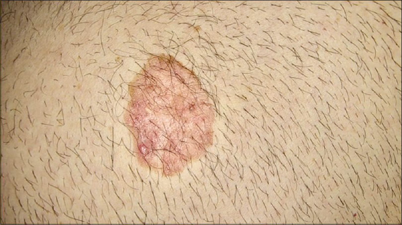
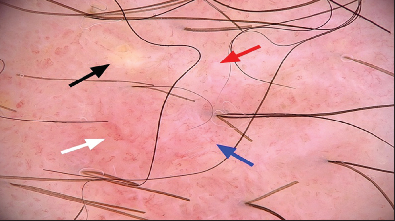
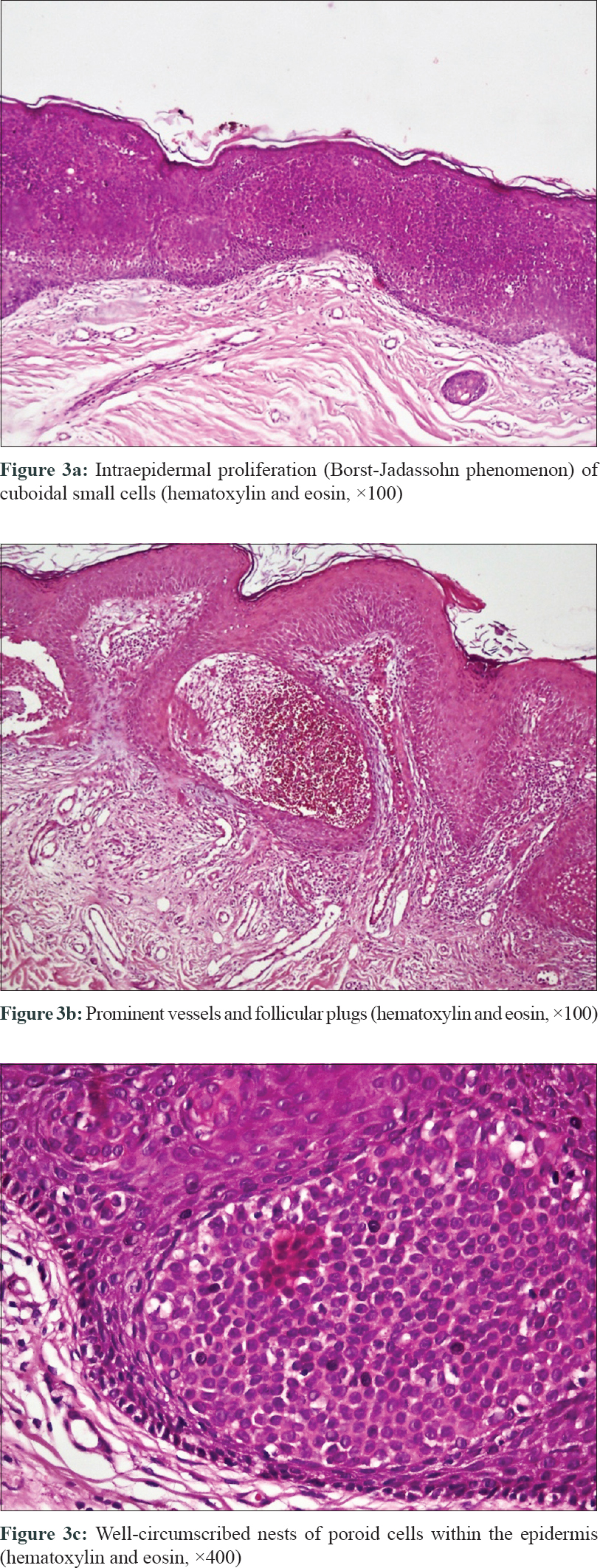
A 61-year-old man was referred to Razi Hospital, Tehran, Iran with a solitary, well-defined, slow-growing, red-to-brown velvety plaque, measuring 3 × 7 cm on the abdomen since 3 years [Figure - 1]. He complained of occasional pruritus but did not have any other signs or symptoms. Dermoscopy revealed diffuse background erythema, an irregular whitish area, yellowish globular structures and densely arranged short linear serpentine and linear-coiled (glomerular) vessels (FotoFinder Medicam 1000, polarized, 20×) [Figure - 2]. The clinical-dermoscopic evaluation was not considered adequate for a definite diagnosis; therefore, a 2 × 4 cm shave biopsy was taken with differential diagnoses of seborrheic keratosis, Bowen's disease, superficial basal cell carcinoma and adnexal tumors. Interestingly, the histologic examination showed some well-circumscribed nests of cuboidal to oval small cells (poroid cells) within the epidermis, with acanthosis, overlying hyperkeratosis and follicular plugs. Prominent vessels were also found. No mitosis, nuclear atypia or pagetoid features were detected [Figure - 3]. Histologic findings confirmed the diagnosis of hidroacanthoma simplex.
 |
| Figure 1: Solitary, well-defined, red-to-brown velvety plaque on abdomen |
 |
| Figure 2: Whitish and yellowish globular structure (red and black arrow). Polymorphous vascular pattern including linear serpentine (blue arrow) and glomerular vessels (white arrow). (FotoFinder Medicam 1000, polarized, 20×) |
 |
| Figure 3: |
We found only very limited number of published reports dwelling on the dermoscopic features of this tumor. Sato et al. published a case of hidroacanthoma simplex with whitish globular structures surrounded by homogenous, pigmented lines in 2014.[2] In a study of four cases of hidroacanthoma simplex, Shiiya et al. described fine black dots/globules, fine scales arranged annularly, nonspecific linear vessels and dotted vessels as dermoscopic features of these tumors.[3] In a recent study, Furlan reported dermoscopic findings of whitish globular structures surrounded by pigmented lines, dotted brown structures and some linear irregular hairpin vessels.[1] In the present case, dermoscopy revealed whitish globular structures which is consistent with previous reports and a polymorphous vascular pattern including linear serpentine and coiled vessels as the new dermoscopic features.
Coiled vessels are one of the characteristic dermoscopic features of Bowen's disease, found in 90% of cases.[4] However, we were unable to find any reports of hidroacanthoma simplex with coiled vessels in the literature. On the contrary, in contrast to some previous reports of hidroacanthoma simplex cases, polymorphous vascular patterns have been reported as prominent dermoscopic features in eccrine poroma. Avilés-Izquierdo described a polymorphous vascular pattern in the dermoscopic examination of non-pigmented eccrine poroma.[5] This polymorphous vascular pattern which is shared by both poroma and our case is consistent with a relationship between these two benign tumors. It is also possible that the higher magnification of the FotoFinder (20×) made the coiled vessels more evident than that achieved by standard 10 × magnification in previous studies.
We speculate that the whitish globular structures seen on dermoscopy may be related either to the nests of poroid cells within the epidermis or to fibrosis, and the yellowish globular structures may be linked to keratin plugs. In addition, prominent vessels in histopathologic examination can be correlated with the polymorphous vascular pattern in the dermoscopic picture.
In conclusion, it is crucial to consider hidroacanthoma simplex as a differential diagnosis of lesions with a dermoscopic polymorphous vascular pattern.
Declaration of patient consent
The authors certify that they have obtained all appropriate patient consent forms. In the form, the patient has given his consent for his images and other clinical information to be reported in the journal. The patient understands that name and initials will not be published and due efforts will be made to conceal identity, but anonymity cannot be guaranteed.
Financial support and sponsorship
Nil.
Conflicts of interest
There are no conflicts of interest.
| 1. |
Furlan KC, Kakizaki P, Chartuni JC, Sittart JA, Valente NY. Hidroacanthoma simplex: Dermoscopy and cryosurgery treatment. An Bras Dermatol 2017;92:253-5.
[Google Scholar]
|
| 2. |
Sato Y, Fujimura T, Tamabuchi E, Haga T, Aiba S. Dermoscopy findings of hidroacanthoma simplex. Case Rep Dermatol 2014;6:154-8.
[Google Scholar]
|
| 3. |
Shiiya C, Hata H, Inamura Y, Imafuku K, Kitamura S, Fujita H, et al. Dermoscopic features of hidroacanthoma simplex: Usefulness in distinguishing it from Bowen's disease and seborrheic keratosis. J Dermatol 2015;42:1002-5.
[Google Scholar]
|
| 4. |
Fargnoli MC, Kostaki D, Piccioni A, Micantonio T, Peris K. Dermoscopy in the diagnosis and management of non-melanoma skin cancers. Eur J Dermatol 2012;22:456-63.
[Google Scholar]
|
| 5. |
Avilés-Izquierdo JA, Velázquez-Tarjuelo D, Lecona-Echevarría M, Lázaro-Ochaita P. Dermoscopic features of eccrine poroma. Actas Dermosifiliogr 2009;100:133-6.
[Google Scholar]
|
Fulltext Views
4,786
PDF downloads
2,098


