Translate this page into:
A case of red ears
2 Paediatric Dermatology, National Skin Centre, Singapore
Correspondence Address:
Yang Sun
673A Fern Grove Yishun Avenue 4 #08-632, 761673
Singapore
| How to cite this article: Sun Y, Yang SS, Tan LS. A case of red ears. Indian J Dermatol Venereol Leprol 2020;86:325-328 |
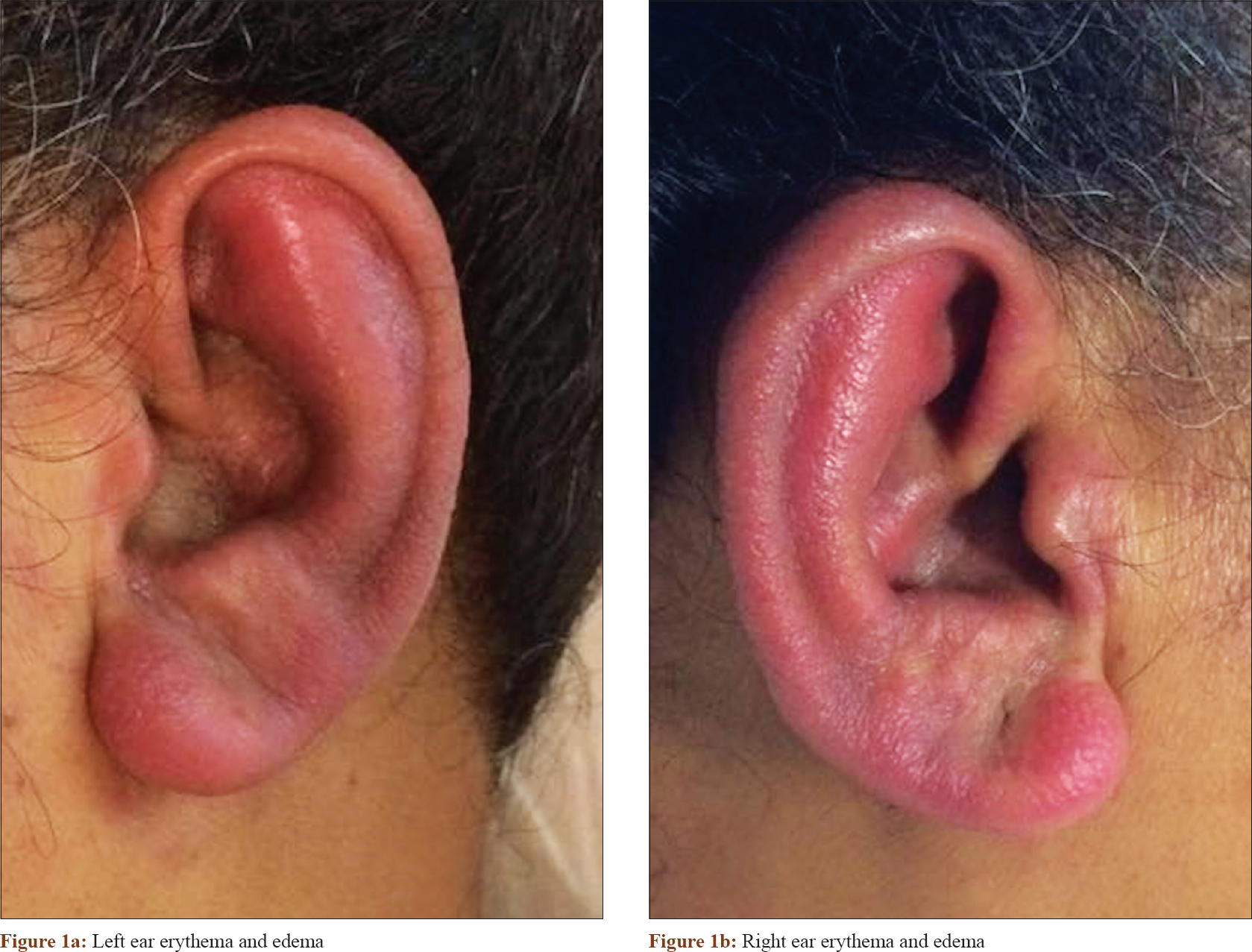
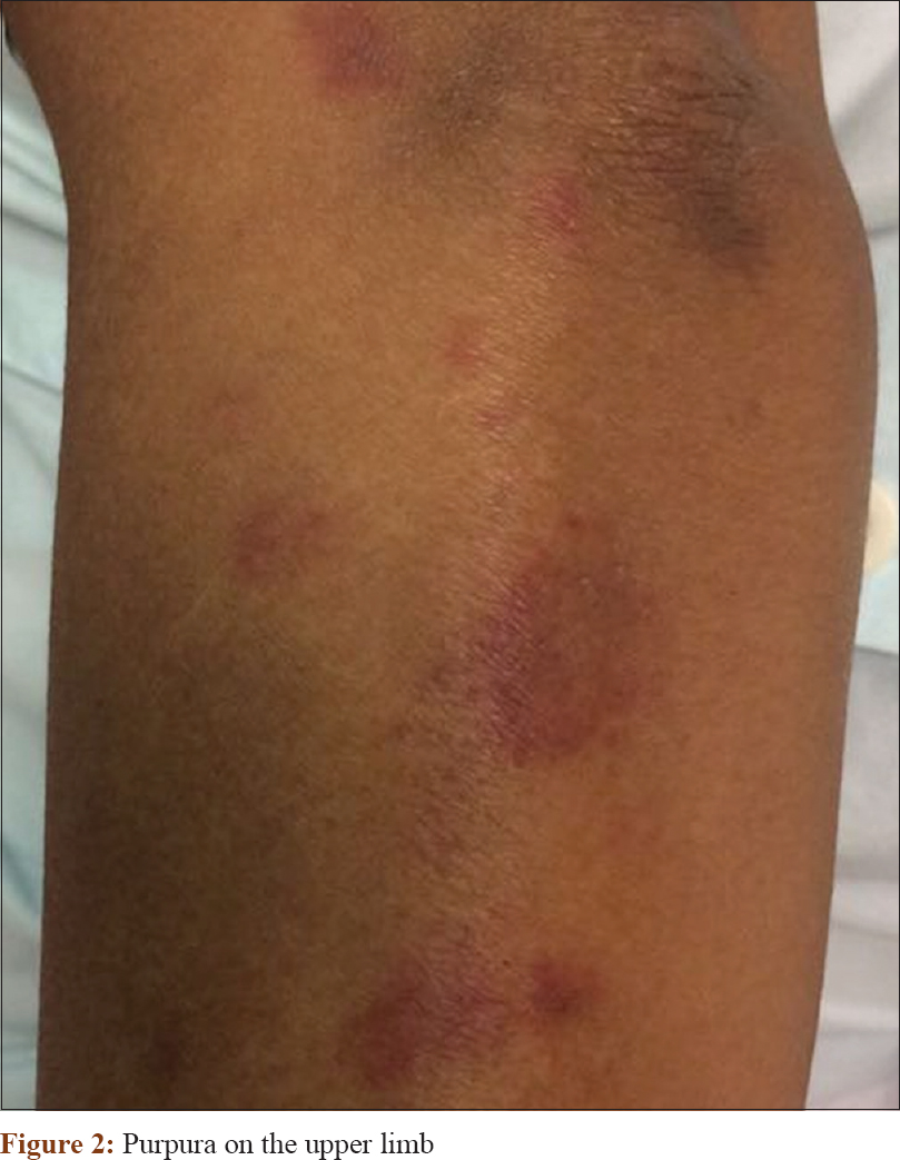
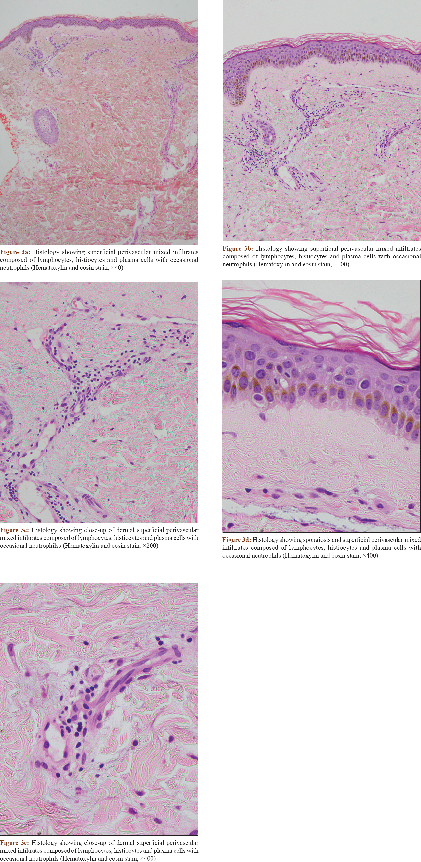
A 49-year-old lady, newly diagnosed with acute myeloid leukemia, was admitted for the first cycle of induction chemotherapy using daunorubicin (days 1 to 3) and cytarabine (days 1 to 7). She had no significant past medical history. On day 7, she developed marked erythema and edema over both her ears [Figure - 1]. There were no auditory complaints, and otoscopic examination reviewed clear tympanic membranes with no effusions. The condition was associated with pruritic purpura over her limbs and trunk, with 5% of body surface area involvement [Figure - 2]. There was no positive Nikolsky's sign or mucositis. Histology of the affected skin over the trunk demonstrated spongiosis and superficial perivascular mixed infiltrates composed of lymphocytes, histiocytes, plasma cells and neutrophils [Figure - 3].
 |
| Figure 1: |
 |
| Figure 2: Purpura on the upper limb |
 |
| Figure 3: |
What Condition Is This?
Declaration of patient consentThe authors certify that they have obtained all appropriate patient consent forms. In the form, the patient has given her consent for her images and other clinical information to be reported in the journal. The patient understands that name and initials will not be published and due efforts will be made to conceal identity, but anonymity cannot be guaranteed.
Financial support and sponsorship
Nil.
Conflicts of interest
There are no conflicts of interest.
| 1. |
Miller KK, Gorcey L, McLellan BN. Chemotherapy-induced hand-foot syndrome and nail changes: A review of clinical presentation, etiology, pathogenesis, and management. J Am Acad Dermatol 2014;71:787-94.
[Google Scholar]
|
| 2. |
Hueso L, Sanmartín O, Nagore E, Botella-Estrada R, Requena C, Llombart B, et al. Chemotherapy-induced acral erythema: A clinical and histopathologic study of 44 cases. Actas Dermosifiliogr 2008;99:281-90.
[Google Scholar]
|
| 3. |
Levine LE, Medenica MM, Lorincz AL, Soltani K, Raab B, Ma A, et al. Distinctive acral erythema occurring during therapy for severe myelogenous leukemia. Arch Dermatol 1985;121:102-4.
[Google Scholar]
|
| 4. |
Baack BR, Burgdorf WH. Chemotherapy-induced acral erythema. J Am Acad Dermatol 1991;24:457.
[Google Scholar]
|
| 5. |
Jucglà A, Sais G, Navarro M, Peyri J. Palmoplantar keratoderma secondary to chronic acral erythema due to tegafur. Arch Dermatol 1995;131:364-5.
[Google Scholar]
|
Fulltext Views
4,932
PDF downloads
5,408


