Generic selectors
Exact matches only
Search in title
Search in content
Post Type Selectors
Search in posts
Search in pages
Filter by Categories
15th National Conference of the IAOMFP, Chennai, 2006
Abstract
Abstracts from current literature
Acne in India: Guidelines for management - IAA Consensus Document
Addendum
Announcement
Art & Psychiatry
Article
Articles
Association Activities
Association Notes
Award Article
Book Review
Brief Report
Case Analysis
Case Letter
Case Letters
Case Notes
Case Report
Case Reports
Clinical and Laboratory Investigations
Clinical Article
Clinical Studies
Clinical Study
Commentary
Conference Oration
Conference Summary
Continuing Medical Education
Correspondence
Corrigendum
Cosmetic Dermatology
Cosmetology
Current Best Evidence
Current Issue
Current View
Derma Quest
Dermato Surgery
Dermatopathology
Dermatosurgery Specials
Dispensing Pearl
Do you know?
Drug Dialogues
e-IJDVL
Editor Speaks
Editorial
Editorial Remarks
Editorial Report
Editorial Report - 2007
Editorial report for 2004-2005
Errata
Erratum
Focus
Fourth All India Conference Programme
From Our Book Shelf
From the Desk of Chief Editor
General
Get Set for Net
Get set for the net
Guest Article
Guest Editorial
History
How I Manage?
IADVL Announcement
IADVL Announcements
IJDVL Awards
IJDVL AWARDS 2015
IJDVL Awards 2018
IJDVL Awards 2019
IJDVL Awards 2020
IJDVL International Awards 2018
Images in Clinical Practice
Images in Dermatology
In Memorium
Inaugural Address
Index
Knowledge From World Contemporaries
Leprosy Section
Letter in Response to Previous Publication
Letter to Editor
Letter to the Editor
Letter to the Editor - Case Letter
Letter to the Editor - Letter in Response to Published Article
LETTER TO THE EDITOR - LETTERS IN RESPONSE TO PUBLISHED ARTICLES
Letter to the Editor - Observation Letter
Letter to the Editor - Study Letter
Letter to the Editor - Therapy Letter
Letter to the Editor: Articles in Response to Previously Published Articles
Letters in Response to Previous Publication
Letters to the Editor
Letters to the Editor - Letter in Response to Previously Published Articles
Letters to the Editor: Case Letters
Letters to the Editor: Letters in Response to Previously Published Articles
Media and news
Medicolegal Window
Messages
Miscellaneous Letter
Musings
Net Case
Net case report
Net Image
Net Images
Net Letter
Net Quiz
Net Study
New Preparations
News
News & Views
Obituary
Observation Letter
Observation Letters
Oration
Original Article
ORIGINAL CONTRIBUTION
Original Contributions
Pattern of Skin Diseases
Pearls
Pediatric Dermatology
Pediatric Rounds
Perspective
Presedential Address
Presidential Address
Presidents Remarks
Quiz
Recommendations
Regret
Report
Report of chief editor
Report of Hon : Treasurer IADVL
Report of Hon. General Secretary IADVL
Research Methdology
Research Methodology
Resident page
Resident's Page
Resident’s Page
Residents' Corner
Residents' Corner
Residents' Page
Retraction
Review
Review Article
Review Articles
Reviewers 2022
Revision Corner
Self Assessment Programme
SEMINAR
Seminar: Chronic Arsenicosis in India
Seminar: HIV Infection
Short Communication
Short Communications
Short Report
Snippets
Special Article
Specialty Interface
Studies
Study Letter
Study Letters
Supplement-Photoprotection
Supplement-Psoriasis
Symposium - Contact Dermatitis
Symposium - Lasers
Symposium - Pediatric Dermatoses
Symposium - Psoriasis
Symposium - Vesicobullous Disorders
SYMPOSIUM - VITILIGO
Symposium Aesthetic Surgery
Symposium Dermatopathology
Symposium-Hair Disorders
Symposium-Nails Part I
Symposium-Nails-Part II
Systematic Review and Meta-Analysis
Systematic Reviews and Meta-analyses
Systematic Reviews and Meta-analysis
Tables
Technology
Therapeutic Guideline-IADVL
Therapeutic Guidelines
Therapeutic Guidelines - IADVL
Therapeutics
Therapy
Therapy Letter
Therapy Letters
View Point
Viewpoint
What’s new in Dermatology
View/Download PDF

Translate this page into:

Case Letter
89 (
4
); 582-584
doi:
10.25259/IJDVL_851_2021
pmid:
37067119

A case of ulcerative cutaneous sarcoidosis

Department of Dermatology, Habib Thameur Hospital, Tunis, Tunisia Research Unit “Genodermatoses and cancers LR12SP03,” Tunis, Tunisia.
Department of Dermatology and Anatomopathology Department, Habib Thameur Hospital, Tunis, Tunisia Research Unit “Genodermatoses and cancers LR12SP03,” Tunis, Tunisia.

Corresponding author: Dr. Amal Chamli, Department of Dermatology, Habib Thameur Hospital, Tunis, Tunisia. amal.fmt@gmail.com

Licence
This is an open-access article distributed under the terms of the Creative Commons Attribution-Non Commercial-Share Alike 4.0 License, which allows others to remix, transform, and build upon the work non-commercially, as long as the author is credited and the new creations are licensed under the identical terms.

How to cite this article: Zaouak A, Chamli A, Ben Samir A, Hlel I, Ben Brahim E, Fenniche S, et al. A case of ulcerative cutaneous sarcoidosis. Indian J Dermatol Venereol Leprol 2023;89:582–4.

Dear Editor,

A 54-year-old woman presented to our department with a 2-month history of progressive painful non-healing ulcero-necrotic plaques and nodules on her legs. Cutaneous examination revealed numerous red-brown nodules and plaques of varying sizes measuring 0.5–2 cm in diameter, with punched-out ulcers covered by necrotic brownish crust and infiltrated rolled borders, localised on the bilateral pretibial areas [Figure 1a]. Dermoscopy of the lesions revealed multiple linear and branching vessels overlying translucent orange-yellowish background [Figure 1b]. The patient was afebrile and the remaining physical examination, including lymph nodes, was normal. There was no clinical evidence of arterial or venous insufficiency. The patient denied any pruritus, respiratory symptoms, ocular symptoms, unexplained weight loss or arthralgias. Histopathological examination of the skin specimens revealed non-caseating epithelioid granulomas in the superficial dermis [Figures 2a and b]. There were no polarizable foreign materials within the granulomas. Periodic acid Schiff, acid-fast and gram stains for fungi and bacteria failed to show micro-organisms. Laboratory evaluations revealed neutropenia, lymphopenia, hypochromic microcytic anaemia, low serum ferritin level and elevated erythrocyte sedimentation rate. Serum angiotensin-converting enzyme level was elevated at 91.78 U/L (normal range: 20–70 U/L). Hepatitis B and C, human immunodeficiency virus, syphilis serologies, and QuantiFERON-TB Gold were negative. The antineutrophil cytoplasmic antibody and antiphospholipid antibodies were unremarkable. Cultures for fungi, bacteria and mycobacteria were negative. The computed tomography scan of chest, abdomen and pelvis revealed bilateral hilar and mediastinal lymphadenopathies and peri lymphatic micronodules. Pulmonary function tests, electrocardiogram and ophthalmological examination were unremarkable. Based on these findings the diagnosis of ulcerative cutaneous sarcoidosis was made. The patient was started on hydroxychloroquine 200 mg twice daily along with daily cleaning with povidone iodine, debridement and topical betamethasone. The clinical course was marked by non-healing of the skin ulcers but without the development of new cutaneous lesions. Topical treatment was continued with an anti-ulcer cream, Mebo (moist exposed burn therapy), an oil-based herbal paste and beta-sitosterol. The ulcers healed within three months leaving flat scars [Figure 3]. The patient was referred to the internal medicine department for the management of systemic component of sarcoidosis.
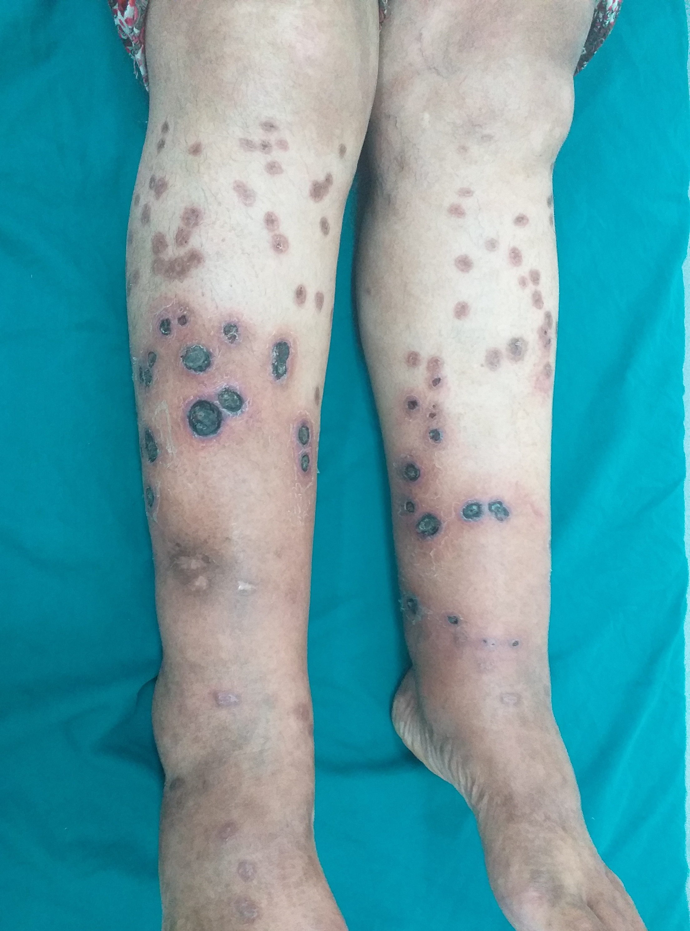
Multiple ulcers covered by necrotic brownish crust with infiltrated erythematous borders on the lower extremities
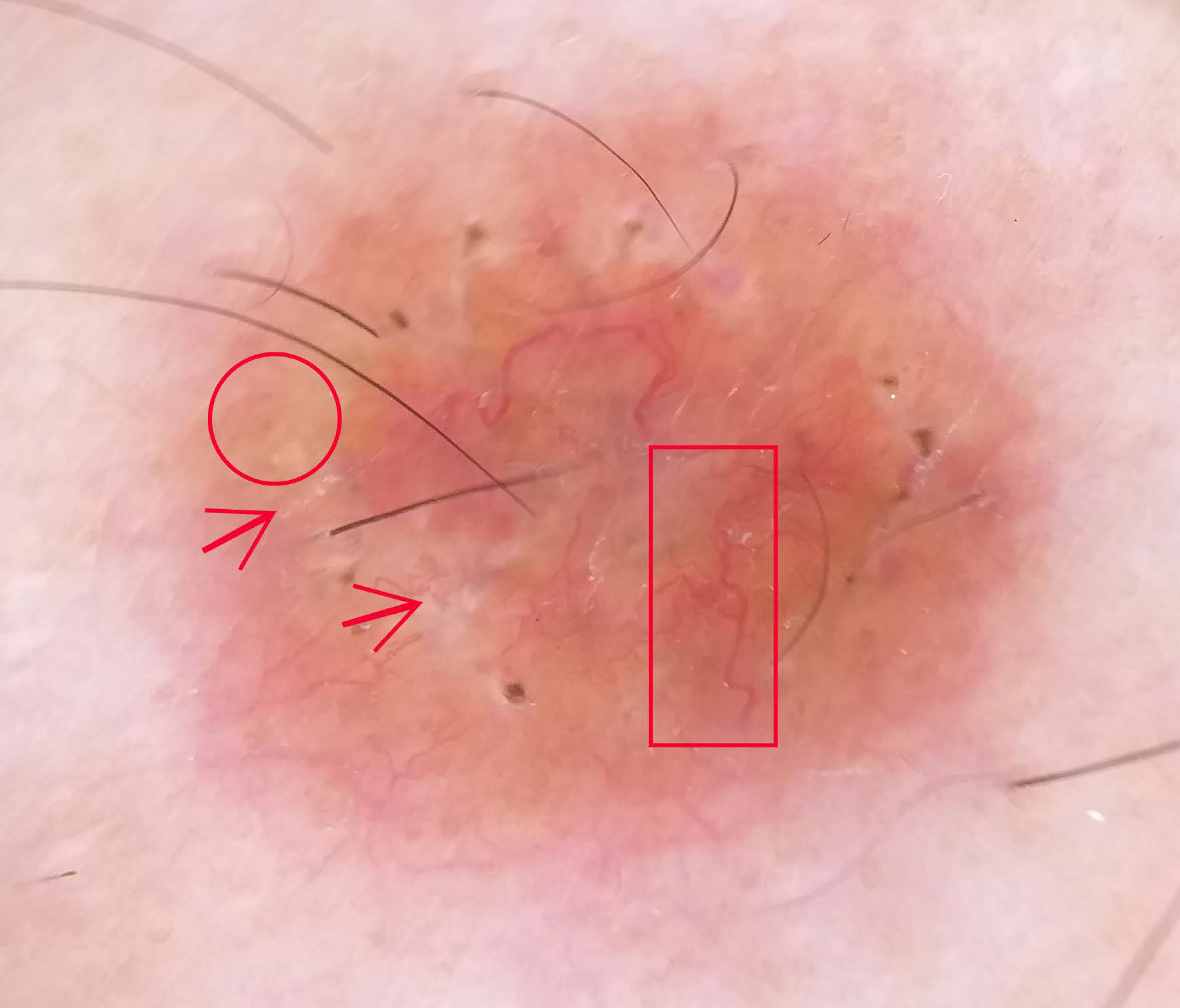
Figure 1a:
Multiple ulcers covered by necrotic brownish crust with infiltrated erythematous borders on the lower extremities
Dermoscopic examination revealed multiple linear and branching vessels (red rectangle) overlying translucent orange-yellowish background (red circle) with scar-like depigmentation areas, white scales (red arrow) and central serous crust (Dermlite DL4)
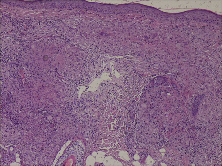
Figure 1b:
Dermoscopic examination revealed multiple linear and branching vessels (red rectangle) overlying translucent orange-yellowish background (red circle) with scar-like depigmentation areas, white scales (red arrow) and central serous crust (Dermlite DL4)
Non-caseating epithelioid granulomas in the dermis (HE ×200)
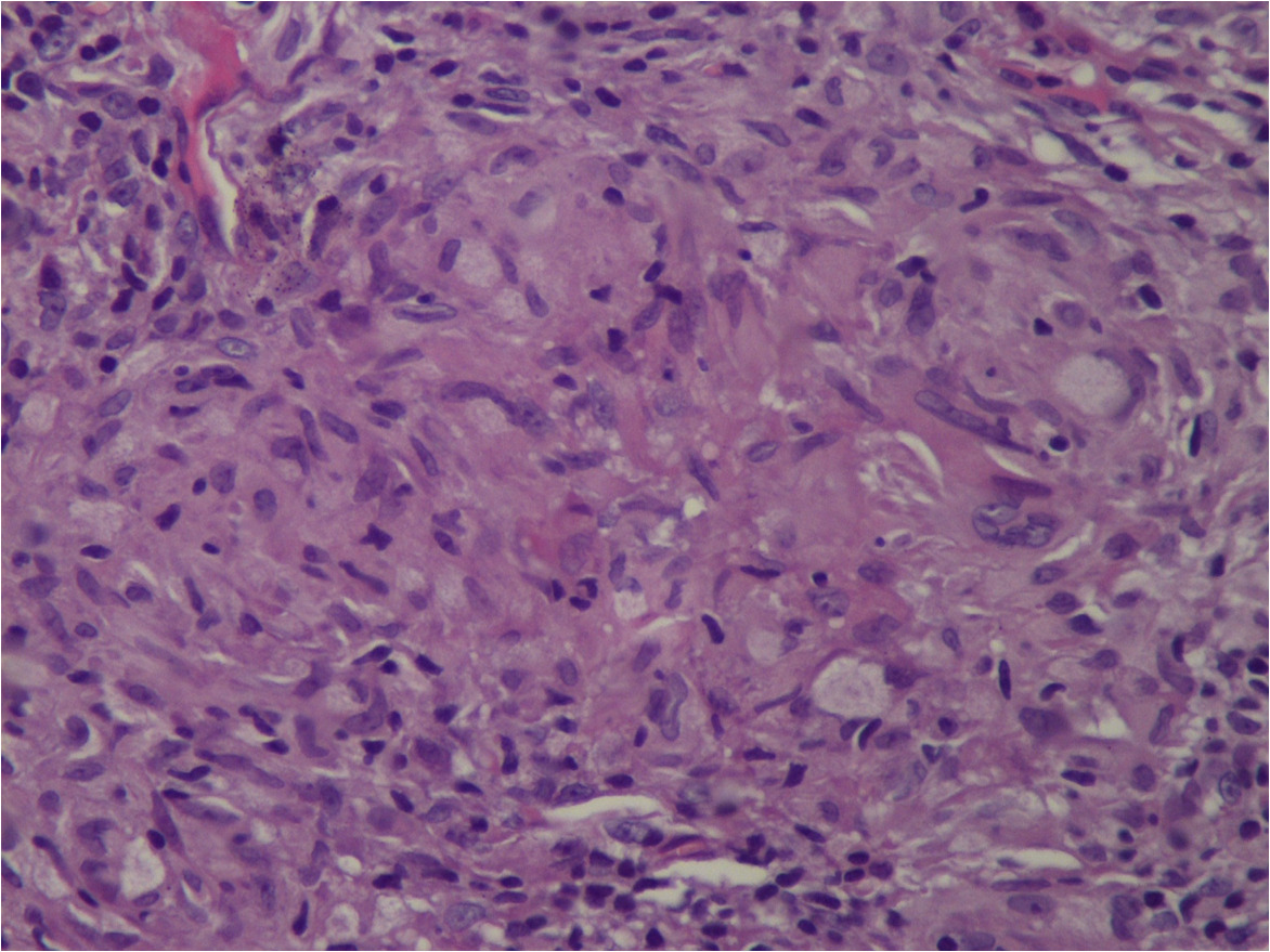
Figure 2a:
Non-caseating epithelioid granulomas in the dermis (HE ×200)
Sarcoid granuloma composed of epithelioid histiocytes and multi-nucleated giant cells (He ×400)
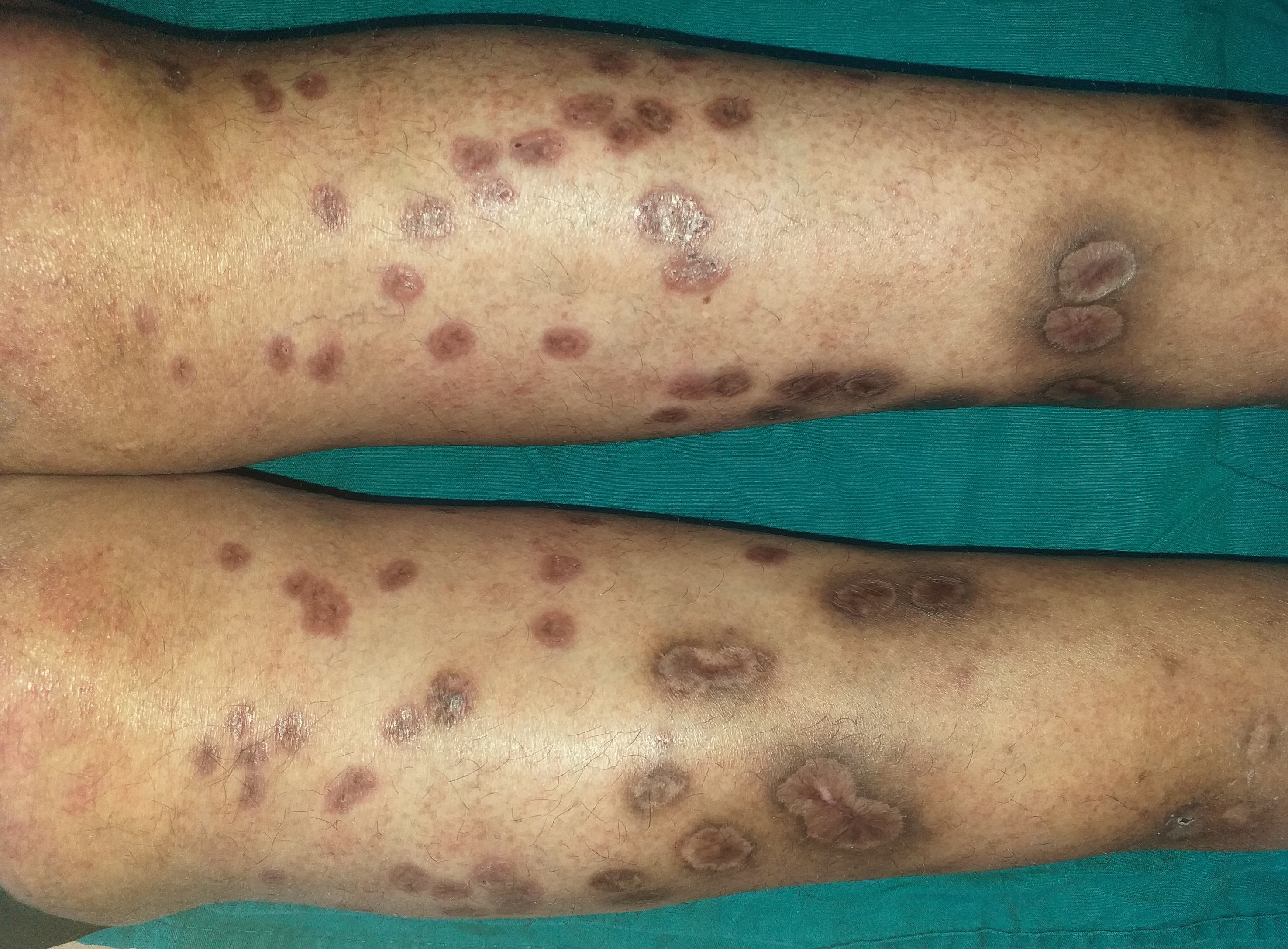
Figure 2b:
Sarcoid granuloma composed of epithelioid histiocytes and multi-nucleated giant cells (He ×400)
Healing ulcers of the legs after 3 months of topical treatment
Figure 3:
Healing ulcers of the legs after 3 months of topical treatment

Discussion

Herein, we describe a rare case of cutaneous sarcoidosis, presenting as punched-out ulcers over the legs of a 54-year-old woman. Cutaneous sarcoidosis is known as the “great imitator” due to variability in manifestations. Indeed, ulcerative cutaneous sarcoidosis is a distinctly unusual manifestation of cutaneous sarcoidosis, affecting 5% of worldwide populations. Blacks and women are more likely to have ulcerative cutaneous sarcoidosis.1 The mechanism of ulceration remains unclear but trauma on atrophic or papulonodular lesions has been reported to be the principal cause of ulceration in sarcoidosis. Granulomatous vasculitis, hyaline degeneration, as well as necrotising granulomas may contribute to other possible mechanisms2. Furthermore, this entity is commonly associated with systemic involvement, as shown in our patient who presented with haematological and mild lung involvement. Ulcers tend to arise within existing papulo-nodules on the lower limbs or de novo. The most frequent location is the pretibial area. In addition, ulcerative cutaneous sarcoidosis may present as ulcerations with purpuric borders, an atrophic base or also necrotic ulcers. Few cases have been described as punched-out ulcers, as in our patient2. In these cases, infectious causes such as syphilis, cryptococcosis, leprosy, cutaneous leishmaniasis, and tuberculosis should be considered in the differential diagnosis. Furthermore, ulcerative cutaneous sarcoidosis of the lower limbs could mimic eschar, superficial pyoderma gangrenosum, necrobiosis lipoidica, perforating diseases, granulomatosis lymphomatoid of Liebow, vascular insufficiency and necrotising vasculitis.3

The presence of non-caseating epithelioid granulomas in histopathology is highly suggestive of ulcerative cutaneous sarcoidosis but is not specific. Therefore, appropriate stains, cultures and polymerase chain reaction (PCR) are crucial to differentiate ulcerative cutaneous sarcoidosis from skin infections such as syphilis, leishmaniasis and tuberculosis.2

In addition, our case may mimic perforating sarcoidosis as well as perforating granuloma annulare or Kyrle disease since the similar presentation. However, in our patient, transepidermal elimination of sarcoid granuloma or collagen was not observed in histopathology.4

Cutaneous manifestations of lymphomatoid granulomatosis of Liebow present with violaceous and hyperpigmented ulcerated nodules of the trunk and extremities and are usually associated with lung involvement. Histology shows characteristically an inflammatory angiocentric infiltrate, composed of polymorphous mononucleated cells often associated with necrosis, unlike our case. Vascular insufficiency could be excluded by arterial and venous Doppler ultrasound and necrotising vasculitis is eliminated by the negativity of antineutrophil cytoplasmic antibodies (ANCAs) and the absence of vasculitis on histology. In addition, as in granulomatous diseases, dermoscopy can be of important diagnostic tool, showing yellow-orange background, and white areas associated with linear vessels. Hence, there is no diagnostic test for sarcoidosis and the diagnosis is made by combining clinic, dermoscopic, histopathological, biological and radiological findings, as in the current case.

In ulcerative cutaneous sarcoidosis, patients are commonly refractory to many types of treatment. Systemic corticosteroids have been reported as standard therapy in ulcerative cutaneous sarcoidosis. Topical treatments alone have been reported to fail.2 Interestingly, our patient was treated successfully with hydroxychloroquine and topical ulcer treatment. We started conventional topical treatment but since there was no improvement, we decided on a trial with Mebo. In fact, Mebo (moist exposed burn ointment) or moist exposed burn treatment/therapy is traditional Chinese medicine. Its main ingredient is beta-sitosterol, a plant-derived steroid with anti-inflammatory and anti-pyretic properties.5 In summary, practitioners should suspect ulcerative cutaneous sarcoidosis in front of ulcero-necrotic plaques of the lower extremities. Furthermore, this case is being reported to draw the attention of practitioners to the importance of local measures in the treatment of ulcerative cutaneous sarcoidosis.

Declaration of patient consent

Patient’s consent not required as patients identity is not disclosed or compromised.

Financial support and sponsorship

Nil.

Conflict of interest

There are no conflicts of interest.

References

  1. . Atypical ulcers: Atypical ulcers. Dermatol Ther. 2013;26:222-35.
    [CrossRef] [PubMed] [Google Scholar]
  2. , , , , , . Clinicopathologic features of ulcerative-atrophic sarcoidosis. Int J Dermatol. 2004;43:108-12.
    [CrossRef] [PubMed] [Google Scholar]
  3. , . Cutaneous sarcoidosis: Differential diagnosis. Clin Dermatol. 2007;25:276-87.
    [CrossRef] [PubMed] [Google Scholar]
  4. , , , . Generalized perforating granuloma annulare. J Am Acad Dermatol. 1992;27:319-22.
    [CrossRef] [PubMed] [Google Scholar]
  5. , , , . Analgesic and anti-inflammatory activity of β-sitosterol isolated from Nyctanthes arbortristis leaves. Inflammopharmacology. 2012;20:219-24.
    [CrossRef] [PubMed] [Google Scholar]

Fulltext Views
3,984

PDF downloads
1,913
View/Download PDF
Download Citations
BibTeX
RIS
Show Sections