Translate this page into:
A case report of buffalopox: A zoonosis of concern
Correspondence Address:
Siva Rami Reddy Karumuri
Flat No. 202, Solitaire Banjara Apartments, Road No. 10, Banjara Hills, Hyderabad - 500 034, Telangana
India
| How to cite this article: Gujarati R, Reddy Karumuri SR, Babu T N, Janardhan B. A case report of buffalopox: A zoonosis of concern. Indian J Dermatol Venereol Leprol 2019;85:348 |
Sir,
A 30-year-old man, resident of Kanakamamidi village, Telangana State, a milkman and owner of a buffalo herd presented with painful, red papules on the dorsae of hands which later developed into fluid-filled lesions over a period of 10 days. Similar lesions developed on forearms and face in the next 3–4 days. There was continuous throbbing pain over the lesions that got aggravated on working. Patient also reported continuous high-grade fever with chills, myalgia, joint pains, redness of eyes, abdominal pain and diarrhea. On examination the patient was febrile (100°F). Multiple, 1–1.5 cm, firm, discrete, tender epitrochlear, axillary, cervical and preauricular lymph nodes were palpable on both sides. Systemic examination was normal. Cutaneous examination revealed ten nodules of size 1 × 1.5 cm, vesicles (few with central umbilication) and pustules on the dorsae of hands [Figure - 1], fingers, flexor aspect of distal forearms, right preauricular area [Figure - 2], right angle of mandible and right ala of nose. Many were intact and few were oozing serosanguinous fluid. Few erosions and crusts were also seen. There was conjunctival congestion in both the eyes [Figure - 3]. Scalp, trunk, palms, soles, nails, oral cavity and genitals were normal. Two buffaloes in his farm had erosions with serosanguinous discharge and crusting over the teats and udders [Figure - 4]. The routine investigations were unremarkable. Histopathological examination of the pustule from the man [Figure - 5] and [Figure - 6] revealed epidermis with intraepidermal vesicle containing neutrophils with elongation of rete ridges and few degenerated keratinocytes. Cytopathic effects were not seen. The dermis revealed lichenoid infiltrate. DNA polymerase chain reaction, done in both human and buffalo scabs, were positive for buffalopox virus. Patient was treated with oral antibiotics and analgesics for one week which led to improvement in pain and pustules and complete healing of lesions in three weeks.
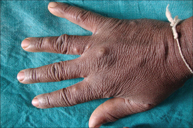 |
| Figure 1: Multiple umbilicated vesicles and nodules on dorsum of right hand |
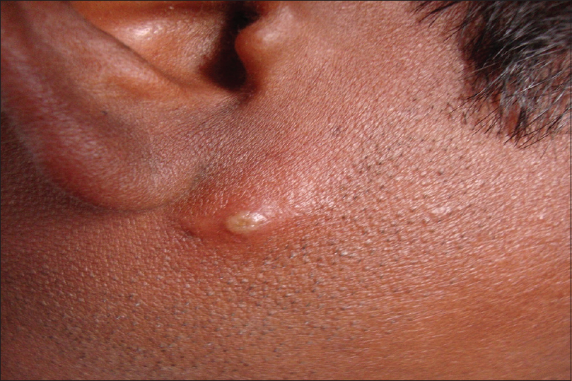 |
| Figure 2: A pustule over right preauricular area |
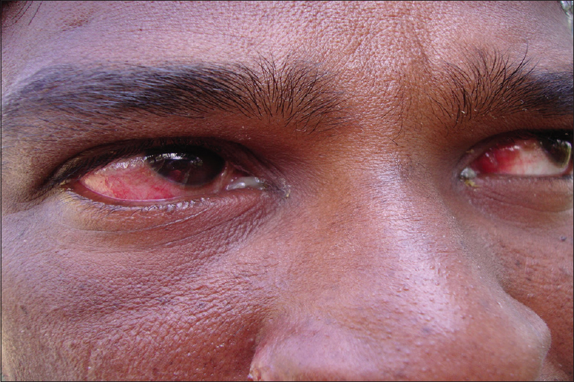 |
| Figure 3: Conjunctival congestion in both eyes |
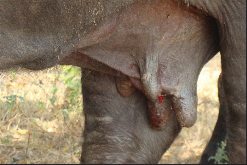 |
| Figure 4: Erosions with serosanguinous discharge over teats of buffalo |
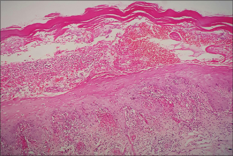 |
| Figure 5: Intraepidermal vesicle formation with inflammatory infiltrates (H and E, ×100) |
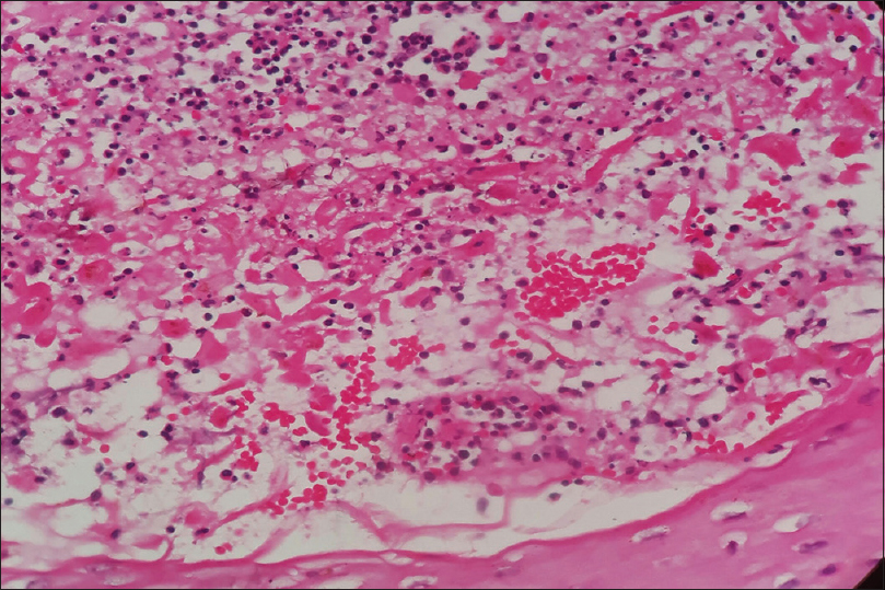 |
| Figure 6: Red blood cells, neutrophils within the vesicle (H and E, ×400) |
Zoonotic diseases affecting humans present uncommonly to the dermatology outpatient department. Buffalopox is a rare disease caused by buffalopox virus, a prototype of member of genus orthopoxvirus, subfamily chordopoxvirinae, family poxviridae and closely related to vaccinia virus.[1],[2] The first recorded incidence of buffalopox occurred in 1934 in Lahore in undivided India.[3] Recently, outbreaks have been reported from villages in Solapur and Kolhapur district, Maharashtra, India.[4] Four outbreaks occurred during 2006–2008 at Nellore (Andhra Pradesh), Aundh (Pune), Aurangabad (Maharashtra) and Sardhar Krishinagar (Gujarat).[5] It affects domestic buffaloes (Bubalus bubalis), cattle and humans. Infection occurs in both sporadic and epidemic forms. Lesions in milch animals are localized on teats and udders.[1],[4] Mastitis due to secondary bacterial infection has been reported in 50% of the affected buffaloes. The appearance of lesions in humans handling pox affected buffaloes, attendants and milkers has been reported from time-to-time.[3] Lesions cannot be clinically differentiated from those of other pox viral infections such as milker's nodule, orf and cowpox. Diagnosis can be confirmed by viral isolation in Vero E6 cell monolayers, plaque reduction neutralization test, electron microscopy, A-type inclusion and Ankyrin repeat protein (C18L) gene-specific polymerase chain reaction and counter immunoelectrophoresis.[2],[5] Polymerase chain reaction done in our case was confirmatory. Apart from reported cases, there may be many cases in the community that go unreported. Awareness and identification of this condition is important and control measures must be undertaken including awareness about hand hygiene and use of gloves while milking. In conclusion, we would like to emphasize the possibility of this zoonosis if there are vesicular, pustular, nodular lesions over extremities in an individual whose occupation involves contact with milch animals.
Declaration of patient consent
The authors certify that they have obtained all appropriate patient consent forms. In the form, the patient has given his consent for his images and other clinical information to be reported in the journal. The patient understand that name and initials will not be published and due efforts will be made to conceal identity, but anonymity cannot be guaranteed.
Acknowledgements
- Dr. A.V. Krishna Mohan, The Joint Director, Veterinary Biologicals and Research Institute, Shanthi Nagar, Hyderabad - 500 028, India
- Dr. Chandra Naik. B. M, Scientist 2 and The Joint Director, Southern Regional Disease Diagnostic Laboratory, Institute of Animal Health and Veterinary Biologicals, Hebbal, Bengaluru - 560 024, India
- Dr. V. Vijay Sreedhar, Prof and HOD, Department of Pathology, Bhaskar Medical College, Yenkapally, Moinabad - 500 075, India
- Dr. Sridevi. P, Consultant Dermatopathologist, Dermpath Institute and Research Centre, Hyderabad, India.
Financial support and sponsorship
Nil.
Conflicts of interest
There are no conflicts of interest.
| 1. |
Damle AS, Gaikwad AA, Patwardhan NS, Duthade MM, Sheikh NS, Deshmukh DG, et al. Outbreak of human buffalopox infection. J Glob Infect Dis 2011;3:187-8.
[Google Scholar]
|
| 2. |
Goyal T, Varshney A, Bakshi SK, Barua S, Bera BC, Singh RK, et al. Buffalo pox outbreak with atypical features: A word of caution and need for early intervention! Int J Dermatol 2013;52:1224-30.
[Google Scholar]
|
| 3. |
Singh RK, Hosamani M, Balamurugan V, Bhanuprakash V, Rasool TJ, Yadav MP, et al. Buffalopox: An emerging and re-emerging zoonosis. Anim Health Res Rev 2007;8:105-14.
[Google Scholar]
|
| 4. |
Gurav YK, Raut CG, Yadav PD, Tandale BV, Sivaram A, Pore MD, et al. Buffalopox outbreak in humans and animals in Western Maharashtra, India. Prev Vet Med 2011;100:242-7.
[Google Scholar]
|
| 5. |
Bhanuprakash V, Venkatesan G, Balamurugan V, Hosamani M, Yogisharadhya R, Gandhale P, et al. Zoonotic infections of buffalopox in India. Zoonoses Public Health 2010;57:e149-55.
[Google Scholar]
|
Fulltext Views
3,432
PDF downloads
1,769


