Translate this page into:
A painful subungual lesion with a surprising diagnosis: Molluscum contagiosum
2 Faculty of Medicine, Harran University, Sanliurfa, Turkey
3 Plastic Surgery Clinic, Sanliurfa Training and Research Hospital, Sanliurfa, Turkey
4 Pathology Clinic, Sanliurfa Training and Research Hospital, Sanliurfa, Turkey
Correspondence Address:
Yeliz Karakoca Basaran
Sanliurfa Training and Research Hospital, Dermatology Clinic, 63300, Sanliurf
Turkey
| How to cite this article: Basaran YK, Turan E, Keklik B, Tarini EZ. A painful subungual lesion with a surprising diagnosis: Molluscum contagiosum. Indian J Dermatol Venereol Leprol 2014;80:278 |
Sir,
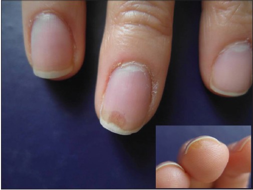
A 33-year-old female dermatologist presented to us with complaints of a painful subungual mass of 2 weeks duration on the middle finger of her right hand. On examination, a brownish, oval, lesion resembling an ′oil spot′ located at the distal part of the nail was observed. In the following 2 weeks, the lesion grew and reached a size of 4 mm across and simultaneously a 2 mm keratotic papule formed on the hyponychium [Figure - 1]. The lesion was significantly tender. Dermoscopic examination revealed brown-black vertical lines resembling splinter hemorrhages on the oil drop-like discoloration. The patient had no similar lesions anywhere else on her body and no history of any significant trauma to her hand. The list of differential diagnoses considered included subungual keratoacanthoma, fibrokeratoma, foreign body tumor, and glomus tumor. The lesion was completely excised by a plastic surgeon; a complete nail plate avulsion was done because of suspicion of a tumor [Figure - 2].
 |
| Figure 1: A brownish oval lesion resembling an 'oil spot' located at the distal part of the nail. A keratotic papule 2 mm in diameter on the hyponychium is observed |
 |
| Figure 2: Complete nail plate avulsion was undertaken |
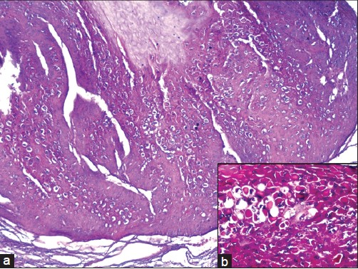
Histological examination revealed the presence of intracytoplasmic hyaline eosinophilic inclusion bodies (Henderson-Paterson bodies), confirming the diagnosis of molluscum contagiosum [Figure - 3].
 |
| Figure 3: Intracytoplasmic hyaline eosinophilic inclusion bodies (Henderson-Paterson bodies; (H and E, ×400) |
Morphologic variants of molluscum contagiosum are common and include giant lesions greater than 5 mm, eczematous lesions, and folliculo-centric lesions with secondary abscess formation. These atypical presentations can closely mimic other dermatologic conditions like lymphangioma, condyloma acuminatum and basal cell carcinoma. [1] Other unusual variants include presentation as a cutaneous horn, [2] on the lip, [3] and giant lesions of the sole. [4] Atypical molluscum contagiosum can be a diagnostic challenge, especially in human immunodeficiency virus type-1 (HIV-1) positive individuals. [5]
To the best of our knowledge, subungual molluscum contagiosum has not been previously described.
Our patient is a dermatologist handling an outpatient clinic and may have acquired the infection from a patient. Molluscum contagiosum infection should be considered a rare possibility in the differential diagnosis of lesions of the nails.
| 1. |
Vanhooteghem O, Henrijean A, de la Brassinne M. Epidemiology, clinical picture and treatment of molluscum contagiosum: Literature review. Ann Dermatol Venereol 2008;135:326-32.
[Google Scholar]
|
| 2. |
Sim JH, Lee ES. Molluscum contagiosum presenting as a cutaneous horn. Ann Dermatol 2011;23:262-3.
[Google Scholar]
|
| 3. |
Matsuura E, Tsunemi Y, Kawashima M. Molluscum contagiosum on the lip. J Dermatol 2013;40:137.
[Google Scholar]
|
| 4. |
Cohen PR, Tschen JA. Plantar molluscum contagiosum: A case report of molluscum contagiosum occurring on the sole of the foot and a review of the world literature. Cutis 2012;90:35-41.
[Google Scholar]
|
| 5. |
Smith KJ, Yeager J, Skelton H. Molluscum contagiosum: Its clinical, histopathologic, and immunohistochemical spectrum. Int J Dermatol 1999;38:664-72.
[Google Scholar]
|
Fulltext Views
3,388
PDF downloads
2,160


