Translate this page into:
A pedunculated nodule on lower lip
Corresponding author: Dr. Somesh Gupta, Department of Dermatology and Venereology, All India Institute of Medical Sciences, New Delhi, India. someshgupta@aiims.edu
-
Received: ,
Accepted: ,
How to cite this article: Anand GRP, Gaurav V, Thirunavukkarasu B, Danish M, Gupta S. A pedunculated nodule on lower lip. Indian J Dermatol Venereol Leprol. 2025;91:123-5. doi: 10.25259/IJDVL_594_2024
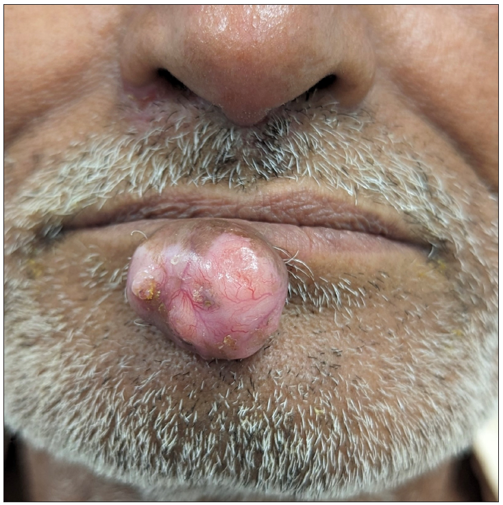
A 55-year-old man presented with an asymptomatic nodule on the right side of his lower lip for 7 years. It started as a small papule which gradually increased to its present size. On examination, we observed a single, pedunculated, skin-coloured, non-tender, firm nodule of size 1.5 cm × 1.5 cm with overlying telangiectasia, on the lower lip [Figure 1].

- A pedunculated, skin-coloured nodule with overlying telangiectasia on the lower lip.
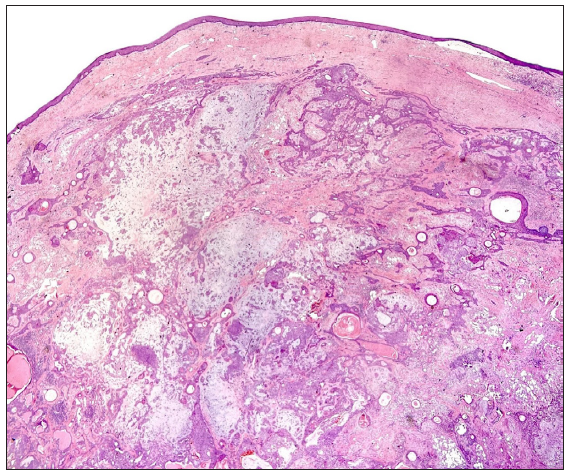
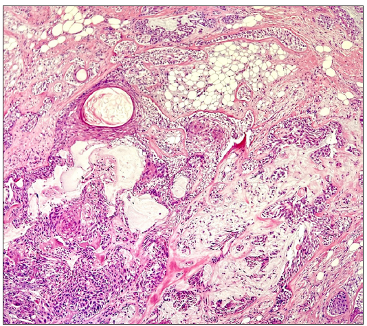
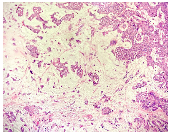
A therapeutic excision of the nodule followed by histopathology revealed tumour islands situated within the upper and mid-dermis, with some islands extending deeper to the deep dermis [Figure 2a]. These tumour islands primarily consisted of uniform cuboidal cells with eosinophilic cytoplasm, and few other cells exhibiting a lighter, clearer cytoplasm. These islands displayed varying shapes and sizes and irregular edges, interconnecting with each other at places. Within these tumour clusters, numerous follicular cysts were observed, many of which contained keratin whorls. The chondromyxoid stroma demonstrated scattered myoepithelial cells with moderate infiltration of lymphocytes, histiocytes, plasma cells, and nests of adipocytes [Figures 2b and 2c]. The tumour cells did not show any cytological atypia, abnormal mitotic figures or necrosis.

- Histopathology showing a circumscribed lesion in the dermis with prominent chondromyxoid stroma. (Haematoxylin and eosin, 20x).

- Histopathology showing a biphasic tumour showing follicular differentiation in the form of keratin cysts representing the epithelial component of the tumour along with cuboidal cell nests, myoepithelial cells and nests of adipocytes representing the mesenchymal components. (Haematoxylin and eosin, 100x).

- Histopathology showing myoepithelial cells admixed in a myxoid background. (Haematoxylin and eosin, 400x).
Question
What is your diagnosis?
Diagnosis
Answer: Chondroid syringoma
Discussion
Mixed tumours of the skin, also called chondroid syringomas (CS), represent an uncommon subset of cutaneous adnexal neoplasms. These tumours exhibit morphologic differentiation towards primary skin appendages, including hair follicles, sebaceous glands and sweat glands (both apocrine and eccrine). Initially described by Billroth in 1859 as a salivary gland tumour, the first documented case of a cutaneous mixed tumour was reported by Nasse in 1892.1,2 The term ‘chondroid syringoma’ was introduced by Hirsch and Helwig in 1961 based on their review of 188 cases.3 The designation ‘chondroid’ reflects the observed cartilaginous characteristics within tumour stroma, particularly in scalp lesions.3 In other locations, the stroma may appear myxoid.4
Headington identified two morphologically distinct types of mixed tumours: apocrine-type and eccrine-type.4 CS reportedly constitute less than 0.36% of all primary skin tumours. They predominantly affect middle-aged males and commonly involve the head and neck, particularly the scalp, nose, cheek and lips.2 Interestingly, upper lip involvement appears to be more frequent than lower lip involvement.5
The clinical diagnosis of CS relies primarily on pathological examination demonstrating both epithelial and mesenchymal components. However, distinguishing CS from other cutaneous adnexal tumours, such as pleomorphic adenoma, can be challenging, especially in areas with minor salivary glands like the lower lip. Recent studies suggest a potential molecular link between CS and pleomorphic adenoma, with shared chromosomal rearrangements involving PLAG1 and HMGA2 genes.5
Immunohistochemistry can facilitate its diagnosis by analysing the distinct protein expression profiles of the epithelial and mesenchymal components within CS. Clinically, CS typically presents as a slow-growing, painless, solid mass, often resembling other cutaneous tumours, necessitating histopathological examination for definitive diagnosis. Surgical excision remains the mainstay of treatment, with careful consideration to prevent recurrence and malignant transformation.3–5
In conclusion, CS, a rare and mostly benign skin lesion, often poses a diagnostic challenge due to its diverse morphological features and varied clinical presentations. Further research is necessary to elucidate its molecular characteristics and improve diagnostic accuracy and management strategies. Inter-departmental collaboration is essential for optimal patient care, emphasising complete excision and long-term follow-up to mitigate potential complications.
Declaration of patient consent
The authors certify that they have obtained all appropriate patient consent.
Financial support and sponsorship
Nil.
Conflicts of interest
There are no conflicts of interest.
Use of artificial intelligence (AI)-assisted technology for manuscript preparation
The authors confirm that there was no use of artificial intelligence (AI)-assisted technology for assisting in the writing or editing of the manuscript and no images were manipulated using AI.
References
- Observations on tumors of the salivary glands. Virchows Arch Pathol Anat.. 1859;17:357-75.
- [Google Scholar]
- Die Geschwülste der Speicheldrüsen und verwandte Tumoren des Kopfes [Tumors of the salivary glands and related head tumors] Arch Klin Chir.. 1892;44:233-302. German
- [Google Scholar]
- Chondroid syringoma: Mixed tumor of skin, salivary gland type. Arch Dermatol. 1961;84:835-47.
- [CrossRef] [PubMed] [Google Scholar]
- Mixed tumors of skin: Eccrine and apocrine types. Arch Dermatol. 1961;84:989-96.
- [CrossRef] [PubMed] [Google Scholar]
- Mixed tumour of the skin of the lower lip: A case report and review of the literature. Mol Clin Oncol. 2022;16:69.
- [CrossRef] [PubMed] [PubMed Central] [Google Scholar]


