Translate this page into:
A rare case of multiple Becker's nevi without systemic involvement
Correspondence Address:
Lin Wang
Department of Dermatovenereology, West China Hospital, Sichuan University, 37# Guoxue Alley, Wuhou District, Chengdu, Sichuan
People's Republic of China
| How to cite this article: Li F, Wang T, Wang L. A rare case of multiple Becker's nevi without systemic involvement. Indian J Dermatol Venereol Leprol 2018;84:194-197 |
Sir,
Becker's nevus is a cutaneous hamartoma, usually characterized by a single hyperpigmented patch with or without hypertrichosis. We are presenting a case of multiple giant Becker's nevi in a 14-year-old male, which is extremely rare.
A 14-year-old boy born of non-consanguineous marriage was referred to us with multiple asymptomatic hyperpigmented patches over his trunk and lower limbs for 2 years [Figure - 1]a and [Figure - 1]b. Initially, he developed a light brown patch on the right anterior thigh, which gradually increased in size and turned darker, and continued to expand to the trunk and legs over the last 2 years. He also noticed increasing hairs on some pigmentary patches during recent months. He had no significant past or present medical history or family history of similar lesions. Physical examination revealed multiple light to dark-brown patches in variable size distributed across the left anterior chest and abdominal wall, inguinal regions, lower back, buttocks, right thigh, left leg, anterior aspects of proximal left thigh and medial parts of both lower limbs, some of which were rough and corrugated. The left and right lesions coalesced at the lower abdomen and genital area. The lesions' margins were well defined and irregular, mostly presenting as geographic or island-like appearance. There were increased hairs on the lesions of extensor aspect of the right thigh [Figure - 2] and left leg. No other accompanied change of skin or musculoskeletal system was found. The examination of breast and genitalia were also normal.
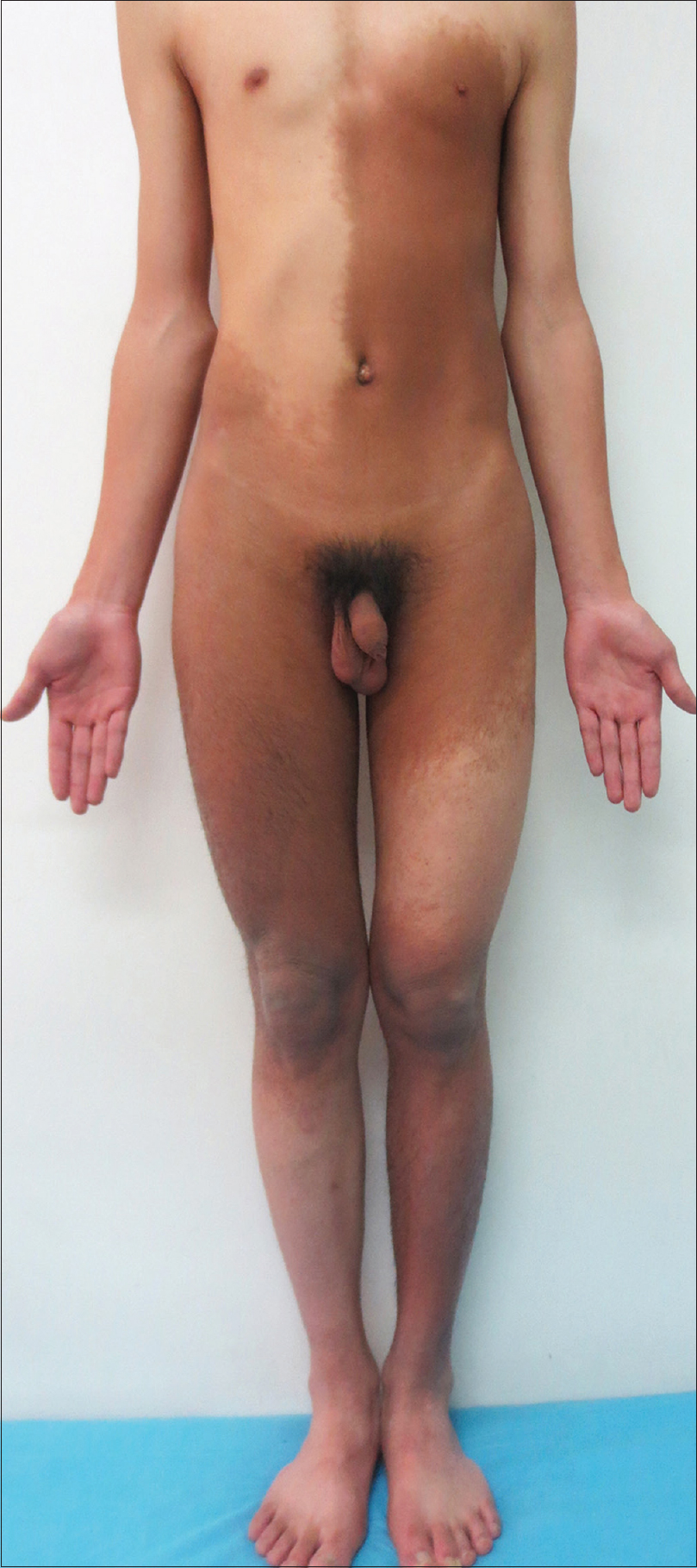 |
| Figure 1a: Hyperpigmented patches distributing across the patient's chest, abdominal wall, and lower limbs |
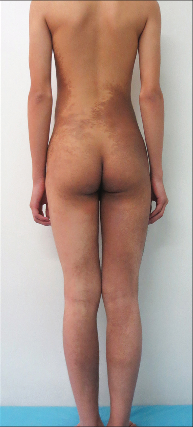 |
| Figure 1b: Multiple light to dark-brown patches in different size involving the lower back, buttocks, and posteromedial sides of lower limbs |
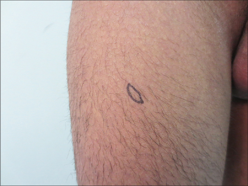 |
| Figure 2: There were increased hairs on the lesions of extensor side of the right thigh |
Laboratory investigations including complete blood cell count, liver and renal function, urine and stool analysis were all normal. X-ray of the chest, spine, and lower extremities also showed no abnormality, as were the findings of abdominal and pelvic cavity ultrasound. Two biopsies were performed from the left upper buttock and extensor side of the right thigh, respectively, both of which showed slight hyperkeratosis, acanthosis, elongation and fusion of the epidermal ridges, with hyperpigmentation of the basal layer [Figure - 3]. A mild increase in the amount of smooth muscle in the reticular dermis [Figure - 4] was also noticed. Thus, the diagnosis of Becker's nevus was made. The patient did not receive any treatment.
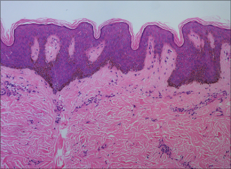 |
| Figure 3: Hyperkeratosis, acanthosis, elongation and fusion of the neighboring rete ridges with hyperpigmentation of the basal layer (H and E, ×100) |
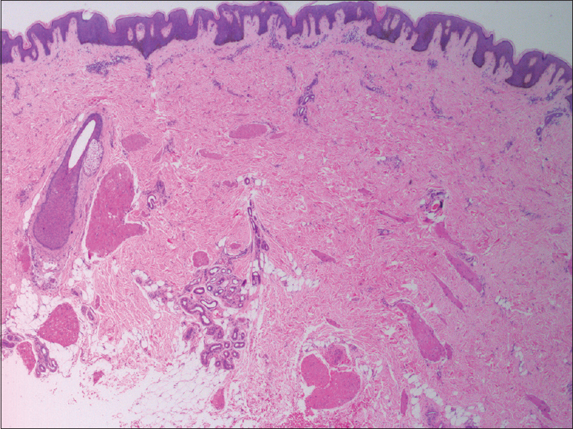 |
| Figure 4: Increased smooth muscle in the dermis was noticed besides the epidermal change (H and E, ×25) |
Becker's nevus usually manifests as a solitary lesion that predominantly involves the upper trunk, scapular region, or upper arm unilaterally.[1] Though multiple Becker's nevi have been described sporadically in literature [1],[2], there have been only few reports wherein the lesions involved trunk and lower limbs concurrently. In our case, approximately 25% of the total body surface was involved, which is extremely rare and we found only one similar case in literature.[2] What's more interesting, in our case was that, most lesions were distributed following a checkerboard configuration, which had been observed by Ramot et al., confirming again that Becker's nevus can follow a mosaic pattern.[1]
The differential diagnoses for our case mainly included café-au-lait spot and acquired smooth muscle hamartoma. The former may be confused with early Becker's nevus, but it is rarely more than 20 cm in diameter. Histopathology can help differentiate them easily. It is more difficult to discriminate the latter from Becker's nevus, for they can share similar clinical and histological changes and they can occur concurrently. Some authors considered that they may represent a spectrum of disease.[3] In our case, the lesions were not as firm as smooth muscle hamartoma usually is, and there was only a mild increase of smooth muscle histologically, so we made a diagnosis of Becker's nevus.
The pathogenesis of Becker's nevus has not been interpreted completely, but many authors believed that heredity, increased androgen receptor density, and androgen sensitivity may play an important role.[4] Onset at puberty, hypertrichosis, and male predominance can be explained by the latter. The possible reason for the relative lack of hypertrichosis in our case might be the young age of this patient. Occasionally, Becker's nevus may accompany some other cutaneous and musculoskeletal anomalies, such as hypoplasia of breast, spinal bifida, and so on, which has been known as Becker nevus syndrome. It may relate to the dysplasia of ectoderm and mesoderm development. Therefore, for each patient with Becker's nevus, especially multiple ones, we should investigate carefully.
Becker's nevus is a benign melanosis without any report of malignant transformation so far. The patients do not need any treatment unless there is a cosmetic demand. Though various cosmetic laser therapies have been tried, the results are not satisfactory so far. Topical use of flutamide, a kind of nonsteroidal antiandrogen, was reported to be effective to some degree.[5] Anyways, further randomized controlled trials and comparative studies are needed to evaluate these different therapies in Becker's nevus patients.
Declaration of patient consent
The authors certify that they have obtained all appropriate patient consent forms. In the form the patient(s) has/have given his/her/their consent for his/her/their images and other clinical information to be reported in the journal. The patients understand that their names and initials will not be published and due efforts will be made to conceal their identity, but anonymity cannot be guaranteed.
Financial support and sponsorship
Nil.
Conflicts of interest
There are no conflicts of interest.
| 1. |
Ramot Y, Maly A, Zlotogorski A. A rare case of multiple Becker's nevi in a checkerboard mosaic pattern. J Eur Acad Dermatol Venereol 2014;28:1573-4.
[Google Scholar]
|
| 2. |
Khaitan BK, Manchanda Y, Mittal R, Singh MK. Multiple Becker's naevi: A rare presentation. Acta Derm Venereol 2001;81:374-5.
[Google Scholar]
|
| 3. |
ul Bari A, Rahman SB. Acquired smooth muscle hamartoma. Indian J Dermatol Venereol Leprol 2006;72:178.
[Google Scholar]
|
| 4. |
Kim YJ, Han JH, Kang HY, Lee ES, Kim YC. Androgen receptor overexpression in Becker nevus: Histopathologic and immunohistochemical analysis. J Cutan Pathol 2008;35:1121-6.
[Google Scholar]
|
| 5. |
Taheri A, Mansoori P, Sandoval LF, Feldman SR. Treatment of Becker nevus with topical flutamide. J Am Acad Dermatol 2013;69:e147-8.
[Google Scholar]
|
Fulltext Views
3,272
PDF downloads
2,161


