Translate this page into:
A solitary hyperkeratotic papule on the palm
2 Department of Pathology, Ramon y Cajal Hospital, 28034 Madrid, Spain
Correspondence Address:
Natalia S�nchez Neila
Department of Dermatology, Ramon y Cajal Hospital, Ctra. de Colmenar Viejo, Km. 9.1, 28034 Madrid
Spain
| How to cite this article: Neila NS, Pascual PF, Zarza EM, del Real CM, de la Vega MU. A solitary hyperkeratotic papule on the palm. Indian J Dermatol Venereol Leprol 2016;82:237-238 |
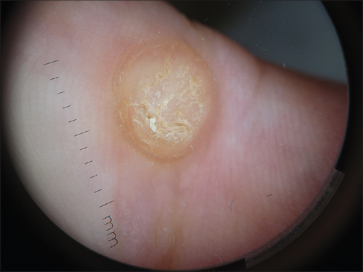
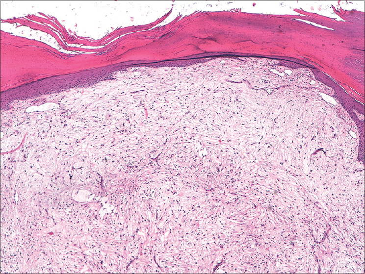
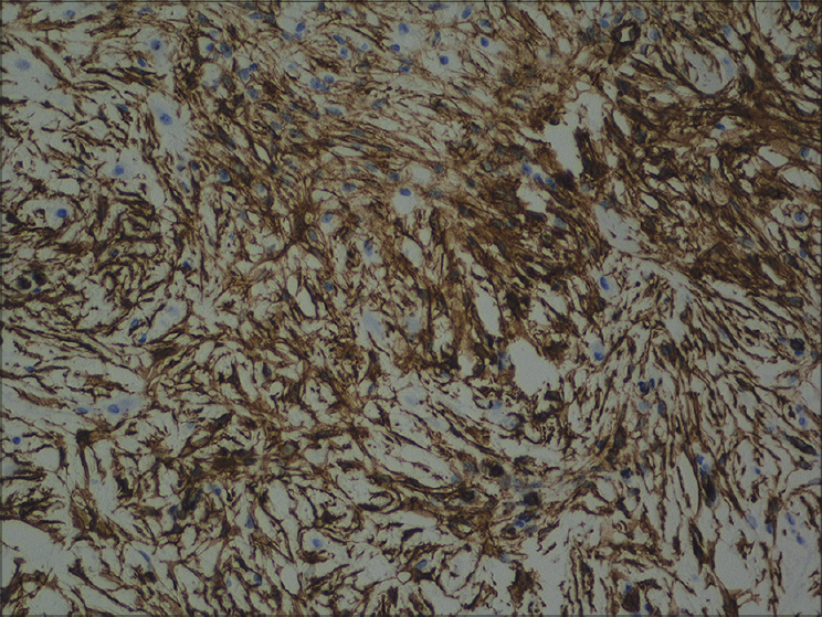
A 42-year-old woman presented to us with an asymptomatic lesion on her left palm. It was present since three years and was gradually increasing in size. Clinical examination revealed a well-defined papule measuring 1 cm × 0.8 cm, surrounded by an epidermal collarette [Figure - 1]. Dermoscopy showed a hyperkeratotic yellowish papule without any vascular pattern [Figure - 2]. The patient was earlier diagnosed as verruca vulgaris and was treated with cryotherapy on three occasions. Due to absence of clinical improvement, an excisional biopsy was done. Histopathological examination showed spindle shaped and stellate cells in a fibromyxoid background stroma, with no obvious architectural growth pattern [Figure - 3]. There were no signs of mitotic activity or nuclear pleomorphism. Immunohistochemical analysis revealed CD34 positive cells, while other antigens such as desmin, S100 and epithelial membrane antigen were negative [Figure - 4].
 |
| Figure 1: Well-defined papule measuring 1 cm × 0.8 cm surrounded by an epidermal collarette |
 |
| Figure 2: Hyperkeratotic yellowish papule without any vascular pattern on dermoscopy |
 |
| Figure 3: Spindle and stellate cells in a fibromyxoid stroma that did not follow any architectural growth pattern (H and E stain, ×400) |
 |
| Figure 4: Immunohistochemistry showing positivity for CD34 (×400) |
What is Your Diagnosis?
| 1. |
Fetsch JF, Laskin WB, Miettinen M. Superficial acral fibromyxoma: A clinicopathologic and immunohistochemical analysis of 37 cases of a distinctive soft tissue tumor with a predilection for the fingers and toes. Hum Pathol 2001;32:704-14.
[Google Scholar]
|
| 2. |
Messeguer F, Nagore E, Agustí-Mejias A, Traves V. Superficial acral fibromyxoma: A CD34+ periungual tumor. Actas Dermosifiliogr 2012;103:67-9.
[Google Scholar]
|
| 3. |
Aguado M, Meseguer C, Tardío JC, Borbujo J. Dermoscopy of acral fibromyxoma. J Am Acad Dermatol 2014;70:e5-6.
[Google Scholar]
|
| 4. |
Carranza C, Molina-Ruiz AM, Pérez de la Fuente T, Kutzner H, Requena L, Santonja C. Subungual acral fibromyxoma involving the bone: A mimicker of malignancy. Am J Dermatopathol 2015;37:555-9.
[Google Scholar]
|
| 5. |
Hollmann TJ, Bovée JV, Fletcher CD. Digital fibromyxoma (superficial acral fibromyxoma): A detailed characterization of 124 cases. Am J Surg Pathol 2012;36:789-98.
[Google Scholar]
|
Fulltext Views
5,797
PDF downloads
3,490


