Generic selectors
Exact matches only
Search in title
Search in content
Post Type Selectors
Search in posts
Search in pages
Filter by Categories
15th National Conference of the IAOMFP, Chennai, 2006
Abstract
Abstracts from current literature
Acne in India: Guidelines for management - IAA Consensus Document
Addendum
Announcement
Art & Psychiatry
Article
Articles
Association Activities
Association Notes
Award Article
Book Review
Brief Report
Case Analysis
Case Letter
Case Letters
Case Notes
Case Report
Case Reports
Clinical and Laboratory Investigations
Clinical Article
Clinical Studies
Clinical Study
Commentary
Conference Oration
Conference Summary
Continuing Medical Education
Correspondence
Corrigendum
Cosmetic Dermatology
Cosmetology
Current Best Evidence
Current Issue
Current View
Derma Quest
Dermato Surgery
Dermatopathology
Dermatosurgery Specials
Dispensing Pearl
Do you know?
Drug Dialogues
e-IJDVL
Editor Speaks
Editorial
Editorial Remarks
Editorial Report
Editorial Report - 2007
Editorial report for 2004-2005
Errata
Erratum
Focus
Fourth All India Conference Programme
From Our Book Shelf
From the Desk of Chief Editor
General
Get Set for Net
Get set for the net
Guest Article
Guest Editorial
History
How I Manage?
IADVL Announcement
IADVL Announcements
IJDVL Awards
IJDVL AWARDS 2015
IJDVL Awards 2018
IJDVL Awards 2019
IJDVL Awards 2020
IJDVL International Awards 2018
Images in Clinical Practice
Images in Dermatology
In Memorium
Inaugural Address
Index
Knowledge From World Contemporaries
Leprosy Section
Letter in Response to Previous Publication
Letter to Editor
Letter to the Editor
Letter to the Editor - Case Letter
Letter to the Editor - Letter in Response to Published Article
LETTER TO THE EDITOR - LETTERS IN RESPONSE TO PUBLISHED ARTICLES
Letter to the Editor - Observation Letter
Letter to the Editor - Study Letter
Letter to the Editor - Therapy Letter
Letter to the Editor: Articles in Response to Previously Published Articles
Letters in Response to Previous Publication
Letters to the Editor
Letters to the Editor - Letter in Response to Previously Published Articles
Letters to the Editor: Case Letters
Letters to the Editor: Letters in Response to Previously Published Articles
Media and news
Medicolegal Window
Messages
Miscellaneous Letter
Musings
Net Case
Net case report
Net Image
Net Images
Net Letter
Net Quiz
Net Study
New Preparations
News
News & Views
Obituary
Observation Letter
Observation Letters
Oration
Original Article
ORIGINAL CONTRIBUTION
Original Contributions
Pattern of Skin Diseases
Pearls
Pediatric Dermatology
Pediatric Rounds
Perspective
Presedential Address
Presidential Address
Presidents Remarks
Quiz
Recommendations
Regret
Report
Report of chief editor
Report of Hon : Treasurer IADVL
Report of Hon. General Secretary IADVL
Research Methdology
Research Methodology
Resident page
Resident's Page
Resident’s Page
Residents' Corner
Residents' Corner
Residents' Page
Retraction
Review
Review Article
Review Articles
Reviewers 2022
Revision Corner
Self Assessment Programme
SEMINAR
Seminar: Chronic Arsenicosis in India
Seminar: HIV Infection
Short Communication
Short Communications
Short Report
Snippets
Special Article
Specialty Interface
Studies
Study Letter
Study Letters
Supplement-Photoprotection
Supplement-Psoriasis
Symposium - Contact Dermatitis
Symposium - Lasers
Symposium - Pediatric Dermatoses
Symposium - Psoriasis
Symposium - Vesicobullous Disorders
SYMPOSIUM - VITILIGO
Symposium Aesthetic Surgery
Symposium Dermatopathology
Symposium-Hair Disorders
Symposium-Nails Part I
Symposium-Nails-Part II
Systematic Review and Meta-Analysis
Systematic Reviews and Meta-analyses
Systematic Reviews and Meta-analysis
Tables
Technology
Therapeutic Guideline-IADVL
Therapeutic Guidelines
Therapeutic Guidelines - IADVL
Therapeutics
Therapy
Therapy Letter
Therapy Letters
View Point
Viewpoint
What’s new in Dermatology
View/Download PDF

Translate this page into:

Observation Letter
ARTICLE IN PRESS
doi:
10.25259/IJDVL_116_2024

Agminated pigmented papules on the face: A rare presentation of neurofibroma

Department of Dermatology, All India Institute of Medical Sciences, Sijua, Patrapada, Bhubaneswar, Odisha, India
Department of Pathology and Lab Medicine, All India Institute of Medical Sciences, Sijua, Patrapada, Bhubaneswar, Odisha, India.

Corresponding author: Dr. Biswanath Behera, Department of Dermatology, All India Institute of Medical Sciences, Sijua, Patrapada, Bhubaneswar, Odisha, India. biswanathbehera61@gmail.com

Licence
This is an open-access article distributed under the terms of the Creative Commons Attribution-Non Commercial-Share Alike 4.0 License, which allows others to remix, transform, and build upon the work non-commercially, as long as the author is credited and the new creations are licensed under the identical terms.

How to cite this article: Gowda KS, Behera B, Thakur V, Sahu DK, Ayyanar P, Sethy M. Agminated pigmented papules on the face: A rare presentation of neurofibroma. Indian J Dermatol Venereol Leprol. doi: 10.25259/IJDVL_116_2024

Dear Editor,

Pigmented papular lesions can have varied differentials, especially in skin of colour, including seborrheic keratosis, melanocytic naevus, dermatofibroma, leiomyoma, pigmented skin tag, pigmented poroma, nevoid basal cell carcinoma, pigmented trichoblastoma and others. Besides, the face is a relatively common location for verruca plana, milia and syringoma; the last two entities can have pigmented morphology in the skin of colour.

A 55-year-old female presented with multiple mildly itchy skin lesions over her right cheek for two years. On examination, multiple monomorphic, brown, dome-shaped, 2–5 mm sessile papules were noted on the right cheek [Figure 1a]. Dermoscopy revealed fingerprint-like structures on a background of a brown homogeneous area [Figure 1b]. There was no evidence of freckles or café au lait macules (CALM) and the button-hole sign was negative. Systemic and mucocutaneous examinations were normal. The possibility of pigmented syringoma, pigmented trichoepithelioma and pigmented milia were considered. Histopathology showed epidermal hyperpigmentation with a non-encapsulated dermal tumour composed of short spindle-shaped cells with wavy s-shaped nuclei [Figures 2a-c] with immunohistochemistry revealing diffuse positivity for S-100 [Figure 2d] and negative for HMB45. The epidermis had increased melanin in the basal and suprabasal layers. A diagnosis of neurofibroma (NF) was considered, and a detailed evaluation for any syndromic association yielded negative results.
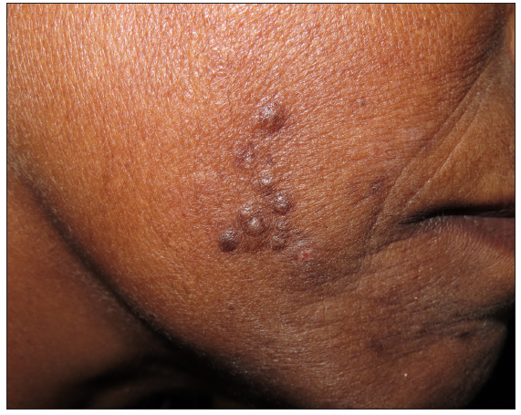
Clinical image shows multiple grouped hyperpigmented papules on the right cheek.
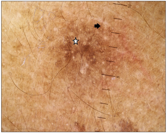
Figure 1a:
Clinical image shows multiple grouped hyperpigmented papules on the right cheek.
Fingerprint pattern on dermoscopy characterised by brown, fine, parallel structures (white star) and a background of homogeneous brown pigmentation (black arrow) (Heine 20T, polarised mode, 10x).
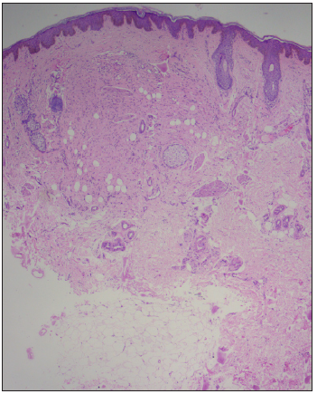
Figure 1b:
Fingerprint pattern on dermoscopy characterised by brown, fine, parallel structures (white star) and a background of homogeneous brown pigmentation (black arrow) (Heine 20T, polarised mode, 10x).
Epidermis is mildly acanthotic and hyperpigmented. The dermis shows a non-encapsulated tumour composed of spindle-shaped cells with wavy nuclei along with bubblegum pink cytoplasm (Haematoxylin & Eosin, 50x).
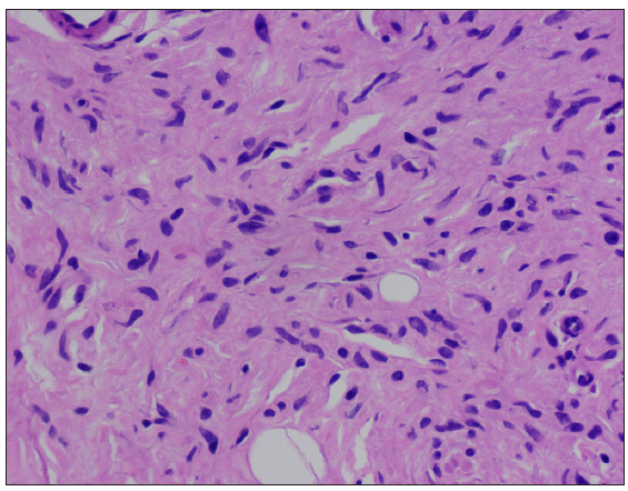
Figure 2a:
Epidermis is mildly acanthotic and hyperpigmented. The dermis shows a non-encapsulated tumour composed of spindle-shaped cells with wavy nuclei along with bubblegum pink cytoplasm (Haematoxylin & Eosin, 50x).
The spindle-shaped cells with wavy nuclei along with bubblegum pink cytoplasm (Haematoxylin & Eosin, 100x).
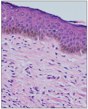
Figure 2b:
The spindle-shaped cells with wavy nuclei along with bubblegum pink cytoplasm (Haematoxylin & Eosin, 100x).
Increased basal pigmentation (Haematoxylin & Eosin, 400x).
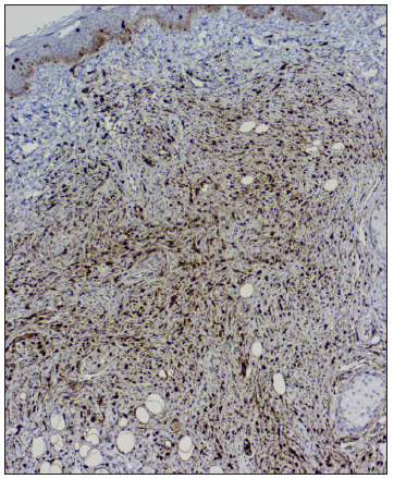
Figure 2c:
Increased basal pigmentation (Haematoxylin & Eosin, 400x).
Tumour cells are immunopositive for S100 (IHC, 100x). (IHC: immunohistochemistry)
Figure 2d:
Tumour cells are immunopositive for S100 (IHC, 100x). (IHC: immunohistochemistry)

NF is a benign tumour and originates from a nerve sheath. Multiple NF associated with CALM and freckles are not difficult to diagnose; however, the solitary form or non-syndromic NF can be a diagnostic challenge. Pigmented NF is a rare entity that constitutes less than 1% of all NF, and it presents as solitary or multiple pigmented papules or nodules predominantly over the scalp, usually in the second or third decade. In addition, pigmented NF acquires a brown colour, either due to the increase in epidermal melanin or pigment deposition in between tumour cells, requiring it to be excluded from other pigmented skin papules.1 Our case was unique due to the arrangement of the pigment papules in an agminate fashion. The dermoscopic features of syndromic NF are a peripheral pigment network (regular or irregular brown network extending beyond the borders of the lesion); a peripheral halo of brown homogeneous pigmentation (continuous or discontinuous dark or light brown homogeneous pigmentation at the borders); fingerprint-like structures (brown, fine, parallel cord-like structures); pink-red structureless areas (pink to red areas); fissures (irregular linear crypts); and scar-like areas (opaque white distinct areas).1

The agminate arrangements of facial papules are described in the literature in blue naevus, spitz naevus and colloid milia. Agminate blue naevi present as blue clustered dome-shaped papules at birth or the second decade. Dermoscopy reveals homogeneous blue or blue-black pigmented structures. Histopathology shows pigmented dendritic bipolar spindle melanocytes admixed with melanophages in the superficial dermis with melan A and HMB-45 positivity.2 Agminate spitz naevi present as grouped, pink to brown, dome-shaped papules and nodules over the face and trunk in the second decade. Biopsy demonstrates naevus cells embedded in a desmoplastic stroma diffusely in the dermis with little junctional activity, and dermoscopy showing a starburst pattern with reticular depigmentation.3

Cells of neural, muscle, histiocytes, fibroblasts and endothelial origin have spindle cell morphology. Smooth muscle cells have deep eosinophilic cytoplasm with a thick cigar-shaped, round-ended nucleus with positivity to smooth muscle actin and vimentin, fibroblasts show less eosinophilic cytoplasm with nuclei with tapered ends,while neural tissue has pale eosinophilic bubblegum cytoplasm with wavy nuclei with S100 positivity. Facial papules with spindle cell morphology are described in neurotised congenital melanocytic naevi (NCMN), fibrous papules, eruptive collagenoma, desmoplastic cellular neurothekeoma (DCN), dermatofibroma and trichodiscoma.

NCMN and pigmented NF pose diagnostic challenges as both have a neural crest cell origin. On histopathology, prominent clustering of melanocytes in cords and nests with S100, Melan A and HMB 45 positivity is described in NCMN.4 DCN on histopathology shows nests and cords of bland epithelioid to spindle cells with myxoid and dense fibrotic collagenous stroma with IHC negativity for S100, positive for S100A6, CD 63 and PGP 9.5.4 Biopsy of fibrous papules shows dermal proliferation of stellate fibroblasts with collagen arranged concentrically around hair follicles and blood vessels with thin-walled dilated vasculature.5 Multiple clustered dermatofibroma on histopathology reveal a hyperpigmented basal layer, Grenz zone, spindle-shaped fibrous cells admixed with histiocytoid cells and IHC shows factor XIIIa and CD68 positivity.6 Trichodiscomas are fibrovascular hamartoma of hair disks that shows proliferative connective tissue with anastomosing strands of follicular epithelium on biopsy.7

We are reporting this case due to the rarity of pigmented, non-syndromic variant of NF, with a unique arrangement in agminate fashion, where dermoscopic and histopathological features aid in arriving at the diagnosis.

Declaration of patient consent

The authors certify that they have obtained all appropriate patient consent.

Financial support and sponsorship

Nil.

Conflicts of interest

There are no conflicts of interest.

Use of artificial intelligence (AI)-assisted technology for manuscript preparation:

The authors confirm that there was no use of AI-assisted technology for assisting in the writing or editing of the manuscript and no images were manipulated using AI.

References

  1. , . Dermoscopy of cutaneous neurofibromas associated with neurofibromatosis type 1. J Am Acad Dermatol. 2015;73:529-31.
    [CrossRef] [PubMed] [Google Scholar]
  2. , , , , . Agminated blue nevus with a GNAQ mutation: A case report and review of the literature. J Cutan Pathol. 2019;46:130-3.
    [CrossRef] [PubMed] [Google Scholar]
  3. , , , , , , et al. International dermoscopy society update on dermoscopy of spitz/Reed naevi and management guidelines by the international dermoscopy society. Br J Dermatol. 2017;177:645-55.
    [CrossRef] [PubMed] [Google Scholar]
  4. , , , , . Neurotized congenital melanocytic nevus resembling a pigmented neurofibroma. Indian J Dermatol. 2015;60:46-50.
    [CrossRef] [PubMed] [PubMed Central] [Google Scholar]
  5. , , , , , , et al. Challenging patterns of atypical dermatofibromas and promising diagnostic tools for differential diagnosis of malignant lesions. Diagnostics (Basel). 2023;13:671.
    [CrossRef] [PubMed] [PubMed Central] [Google Scholar]
  6. , , , , . Histopathological characteristics and CD163 immunostaining pattern in fibrous papule of the face. J Cutan Pathol. 2021;48:274-80.
    [CrossRef] [PubMed] [Google Scholar]
  7. , , . CD-34-reactive trichodiscoma. J Cutan Pathol. 2006;33:709.
    [CrossRef] [PubMed] [Google Scholar]

Fulltext Views
1,264

PDF downloads
373
View/Download PDF
Download Citations
BibTeX
RIS
Show Sections