Generic selectors
Exact matches only
Search in title
Search in content
Post Type Selectors
Search in posts
Search in pages
Filter by Categories
15th National Conference of the IAOMFP, Chennai, 2006
Abstract
Abstracts from current literature
Acne in India: Guidelines for management - IAA Consensus Document
Addendum
Announcement
Art & Psychiatry
Article
Articles
Association Activities
Association Notes
Award Article
Book Review
Brief Report
Case Analysis
Case Letter
Case Letters
Case Notes
Case Report
Case Reports
Clinical and Laboratory Investigations
Clinical Article
Clinical Studies
Clinical Study
Commentary
Conference Oration
Conference Summary
Continuing Medical Education
Correspondence
Corrigendum
Cosmetic Dermatology
Cosmetology
Current Best Evidence
Current Issue
Current View
Derma Quest
Dermato Surgery
Dermatopathology
Dermatosurgery Specials
Dispensing Pearl
Do you know?
Drug Dialogues
e-IJDVL
Editor Speaks
Editorial
Editorial Remarks
Editorial Report
Editorial Report - 2007
Editorial report for 2004-2005
Errata
Erratum
Focus
Fourth All India Conference Programme
From Our Book Shelf
From the Desk of Chief Editor
General
Get Set for Net
Get set for the net
Guest Article
Guest Editorial
History
How I Manage?
IADVL Announcement
IADVL Announcements
IJDVL Awards
IJDVL AWARDS 2015
IJDVL Awards 2018
IJDVL Awards 2019
IJDVL Awards 2020
IJDVL International Awards 2018
Images in Clinical Practice
Images in Dermatology
In Memorium
Inaugural Address
Index
Knowledge From World Contemporaries
Leprosy Section
Letter in Response to Previous Publication
Letter to Editor
Letter to the Editor
Letter to the Editor - Case Letter
Letter to the Editor - Letter in Response to Published Article
LETTER TO THE EDITOR - LETTERS IN RESPONSE TO PUBLISHED ARTICLES
Letter to the Editor - Observation Letter
Letter to the Editor - Study Letter
Letter to the Editor - Therapy Letter
Letter to the Editor: Articles in Response to Previously Published Articles
Letters in Response to Previous Publication
Letters to the Editor
Letters to the Editor - Letter in Response to Previously Published Articles
Letters to the Editor: Case Letters
Letters to the Editor: Letters in Response to Previously Published Articles
Media and news
Medicolegal Window
Messages
Miscellaneous Letter
Musings
Net Case
Net case report
Net Image
Net Images
Net Letter
Net Quiz
Net Study
New Preparations
News
News & Views
Obituary
Observation Letter
Observation Letters
Oration
Original Article
ORIGINAL CONTRIBUTION
Original Contributions
Pattern of Skin Diseases
Pearls
Pediatric Dermatology
Pediatric Rounds
Perspective
Presedential Address
Presidential Address
Presidents Remarks
Quiz
Recommendations
Regret
Report
Report of chief editor
Report of Hon : Treasurer IADVL
Report of Hon. General Secretary IADVL
Research Methdology
Research Methodology
Resident page
Resident's Page
Resident’s Page
Residents' Corner
Residents' Corner
Residents' Page
Retraction
Review
Review Article
Review Articles
Reviewers 2022
Revision Corner
Self Assessment Programme
SEMINAR
Seminar: Chronic Arsenicosis in India
Seminar: HIV Infection
Short Communication
Short Communications
Short Report
Snippets
Special Article
Specialty Interface
Studies
Study Letter
Study Letters
Supplement-Photoprotection
Supplement-Psoriasis
Symposium - Contact Dermatitis
Symposium - Lasers
Symposium - Pediatric Dermatoses
Symposium - Psoriasis
Symposium - Vesicobullous Disorders
SYMPOSIUM - VITILIGO
Symposium Aesthetic Surgery
Symposium Dermatopathology
Symposium-Hair Disorders
Symposium-Nails Part I
Symposium-Nails-Part II
Systematic Review and Meta-Analysis
Systematic Reviews and Meta-analyses
Systematic Reviews and Meta-analysis
Tables
Technology
Therapeutic Guideline-IADVL
Therapeutic Guidelines
Therapeutic Guidelines - IADVL
Therapeutics
Therapy
Therapy Letter
Therapy Letters
View Point
Viewpoint
What’s new in Dermatology
View/Download PDF

Translate this page into:

Quiz
89 (
4
); 606-608
doi:
10.25259/IJDVL_1165_2021
pmid:
35962489

An asymptomatic erythematous plaque on the cheek

Department of Dermatology, Wuhan No 1 Hospital, Wuhan, Hubei Province, People’s Republic of China

Corresponding author: Dr. Jing Xiao, Department of Dermatology, Wuhan No 1 Hospital, Wuhan, Hubei Province, People’s Republic of China. mickey9610@163.com

Licence
This is an open-access article distributed under the terms of the Creative Commons Attribution-Non Commercial-Share Alike 4.0 License, which allows others to remix, transform, and build upon the work non-commercially, as long as the author is credited and the new creations are licensed under the identical terms.

How to cite this article: Xiao J, Zhang L, Chen L. An asymptomatic erythematous plaque on the cheek. Indian J Dermatol Venereol Leprol 2023;89:606–8.

A woman in her 80s presented with an erythematous plaque on her left cheek of more than two years’ duration. The lesion was asymptomatic and slow-growing. Physical examination revealed an ill-demarcated, irregular erythematous plaque with slight scaling and crusting [Figure 1a]. Reflectance confocal microscopy image shows suspicious cells with dark cytoplasm and a mild bright nucleus; these cells were twice the size of normal keratinocytes or larger. A large number of cells merged into nests and formed dark glandular structures [Figure 1b]. Histopathologic examination revealed epidermal infiltration by large atypical cells that had eosinophilic cytoplasm and prominent nuclei [Figure 2a]. Immunohistochemically, the cells were strongly positive for cytokeratin stains cytokeratin 7 (CK7) and epithelial membrane antigen (EMA) [Figures 2b and 2c], but negative for CK20, S100 protein, human melanoma black 45 (HMB-45) and p63 (not shown).
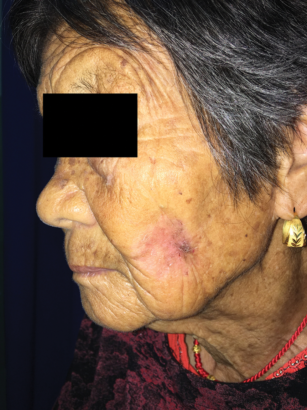
An erythematous plaque on the cheek with ill-demarcated margins
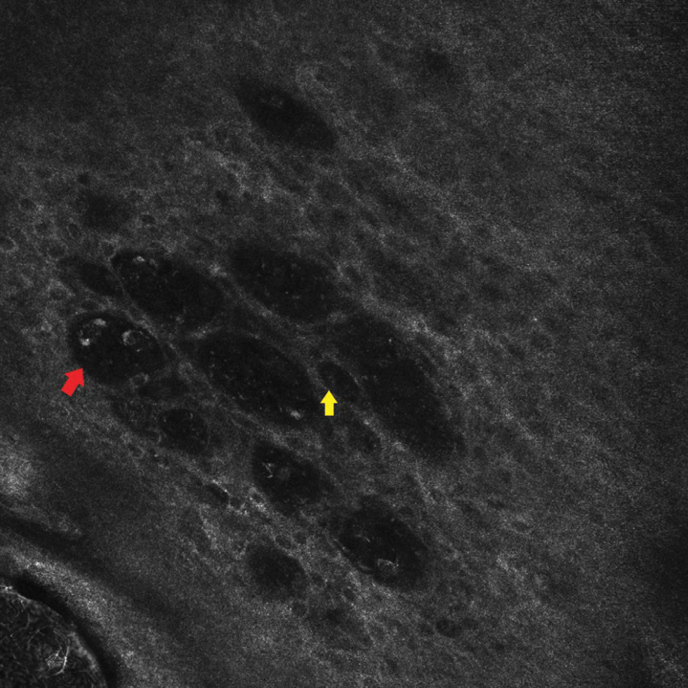
Figure 1a:
An erythematous plaque on the cheek with ill-demarcated margins
Dark cytoplasm and mild bright nucleus (yellow arrows) and dark glandular nest structures in epidermal layer (red arrows) (750 ×750 μm)
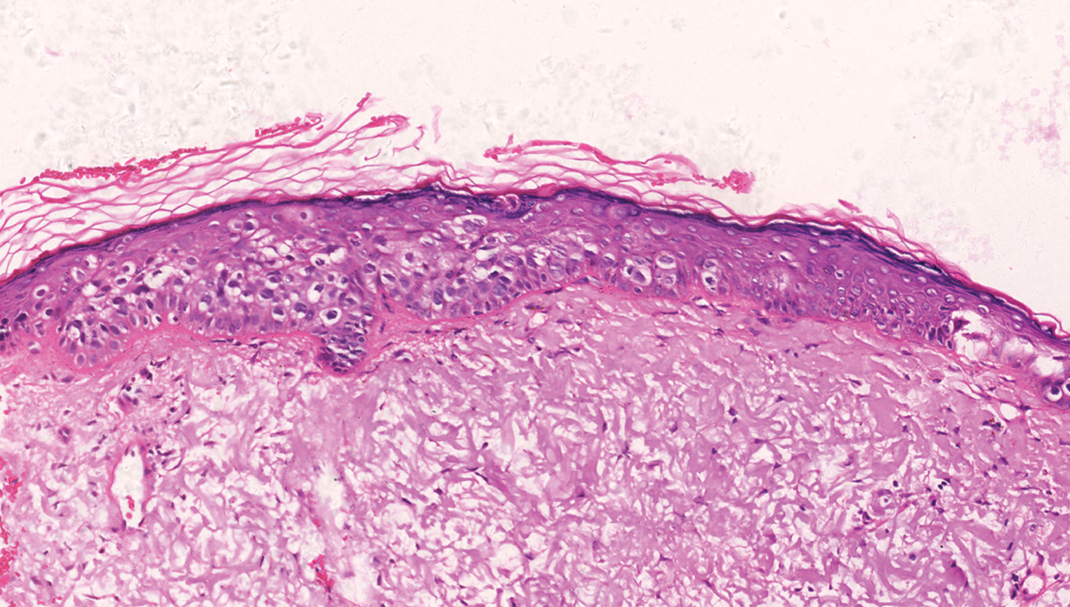
Figure 1b:
Dark cytoplasm and mild bright nucleus (yellow arrows) and dark glandular nest structures in epidermal layer (red arrows) (750 ×750 μm)
Paget cells throughout epidermis (H&E, ×200)
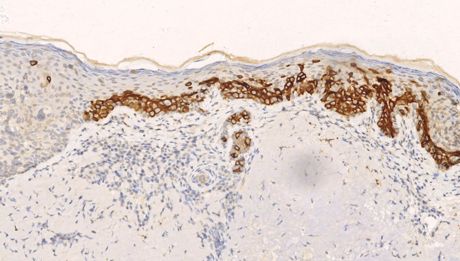
Figure 2a:
Paget cells throughout epidermis (H&E, ×200)
The clear cells were positive for CK7 (Original magnification, ×200)
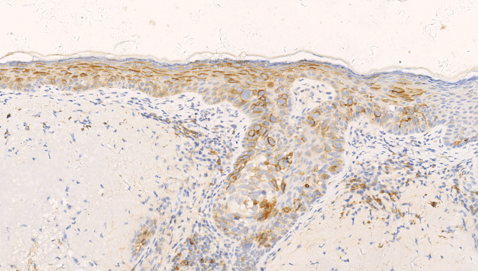
Figure 2b:
The clear cells were positive for CK7 (Original magnification, ×200)
Positive staining for epithelial membrane antigen (Original magnification, ×200)
Figure 2c:
Positive staining for epithelial membrane antigen (Original magnification, ×200)

What is Your Diagnosis?

Diagnosis

Ectopic extramammary Paget disease.

DISCUSSION

Extramammary Paget disease is a rare tumour that generally occurs in areas with a high density of apocrine glands.1 It presents with an erythematous spread in elderly patients, mainly in the anogenital region and in the axilla.1 In our study, we found that the disease occurred in apocrine-poor areas such as the cheek. Saida et al. proposed such cases to be called ectopic extramammary Paget disease, which are exceedingly rare, with more than 40 case reports on the buttock, back, chest, hypochondrium, abdomen, scalp and face, mostly in Asian patients (approximately 60%).2 The tumour location of ectopic extramammary Paget disease appears to be more common on the trunk.2 The risk of ectopic extramammary Paget disease-associated malignancy is not well known. There is only one case identified in the literature of association with underlying malignancy of adenocarcinoma of colon.3 Our patient did not reveal a concurrent malignancy.

Clinically and histologically, ectopic and “ordinary” extramammary Paget disease have similar manifestations except for the location of the disease,2 and in our study ectopic extramammary Paget disease also had a similar presentation on reflectance confocal microscopy. Researchers have proved the Paget cells present in reflectance confocal microscopy images, along with the characteristic nest structures at the dermo-epidermal junction, offer significant clues when it comes to identifying extramammary Paget disease.4 Reflectance confocal microscopy images of our patient correlating well with histopathology, contribute to the early diagnosis of extramammary Paget disease.

Immunohistochemistry has been used both to diagnose Paget disease and to differentiate it from other intraepidermal neoplasms with clear cells, such as pagetoid Bowen disease (CK7 negative/positive, EMA negative, p63 positive) and pagetoid melanoma (S100 positive, HMB-45 positive, CK7 negative).1 Especially for pagetoid Bowen’s disease, there are some in which it is difficult to make a definite histopathological differentiation from extramammary Paget disease; because there is considerable immunophenotypic overlap between pagetoid Bowen disease and extramammary Paget disease. CK7, especially, is an extremely sensitive marker of extramammary Paget disease, but occasional cases of pagetoid Bowen disease have been unexpectedly positive, limiting the value of this marker in this diagnostic scenario. In addition, there is a scarce report of pagetoid Bowen disease that expresses immunohistochemical markers of secretory cells like EMA. The presence of p63 in the neoplastic cells of pagetoid Bowen’s disease has been demonstrated, in contrast to its total absence in the Paget cells of extramammary Paget disease.1 The specific expression of p63 in pagetoid Bowen disease indicates that p63 constitutes a useful marker for its histopathological diagnosis.

Immunohistochemistry also helps to identify the likely cell of origin, as there are antigenic differences between primary intraepidermal Paget disease (CK7 positive, CK20 negative, gross cystic disease fluid protein (GCDFP-15) positive) and Paget disease that has spread from an associated internal carcinoma (CK7 positive, CK20 positive, GCDFP-15 negative).2

Surgical excision with wide margins and Mohs micrographic surgery remains the mainstay of treatment for extramammary Paget disease.1 In literature, there was only one case identified of lymph node metastasis in ectopic extramammary Paget disease.5 Our patient was treated with Mohs micrographic surgery and has been followed up for 5 years without recurrence, lymphadenopathy or metastasis. It seems that ectopic extramammary Paget disease appears to have a more benign biologic activity compared to “ordinary” extramammary Paget disease, and there is an associated malignancy in 20-30% of cases in “ordinary” extramammary Paget disease.2

Declaration of patient consent

The authors certify that they have obtained all appropriate patient consent.

Financial support and sponsorship

Nil.

Conflict of interest

There are no conflicts of interest.

References

  1. . Mammary and extramammary Paget’s disease. J Eur Acad Dermatol Venereol. 2007;21:581-90.
    [CrossRef] [PubMed] [Google Scholar]
  2. , , . Definition, association with malignancy, biologic behavior, and treatment of ectopic extramammary Paget’s disease: A review of the literature. J Clin Aesthet Dermatol. 2019;12:40-4.
    [PubMed] [Google Scholar]
  3. , . Pigmented extramammary Paget disease of the abdomen: A potential mimicker of melanoma. Dermatol Online J. 2011;17:13.
    [PubMed] [Google Scholar]
  4. , , , , . In vivo reflectance confocal microscopy of extramammary Paget disease: Diagnostic evaluation and surgical management. J Am Acad Dermatol. 2012;66:e47-53.
    [CrossRef] [PubMed] [Google Scholar]
  5. , , . Extramammary Paget disease. Dermatol Clin. 2019;37:261-7.
    [CrossRef] [PubMed] [Google Scholar]

Fulltext Views
4,738

PDF downloads
3,808
View/Download PDF
Download Citations
BibTeX
RIS
Show Sections