Translate this page into:
An asymptomatic swelling on the neck
2 Department of Surgery, M. G. M. Medical College and Hospital, Kamothe, Navi Mumbai, India
3 Consultant Radiologist at UBM Institute and Bhatt Sonography Center, Dadar - East, Mumbai, India
Correspondence Address:
Swagata Tambe
19/558, Udyan Society, Nehru Nagar, Kurla, Mumbai - 400 024, Maharashtra
India
| How to cite this article: Tambe S, Someshwar S, Dedhia A, Jadhav R, Bhatt K, Jerajani H. An asymptomatic swelling on the neck. Indian J Dermatol Venereol Leprol 2015;81:221-223 |
A 24-year-old male presented to our clinic for evaluation of a single, asymptomatic swelling present on the posterior aspect of his neck for the last six months. The swelling was initially small, approximately around 0.5 cm, but gradually increased in size over a period of 6 months. There was a history of a similar lesion at the same site 1 year ago, which was excised under local anesthesia.
Cutaneous examination revealed a non-compressible cystic swelling of 3 cm × 4 cm size on the left side of the nape of neck [Figure - 1]a. The overlying skin had an erythematous hue. [Figure - 1]b.The transillumination test was positive [Figure - 1]c. Within the cystic swelling, a solid, non-tender, firm mass of approximately 2 × 3 cm was palpated.
 |
| Figure 1: (a) Translucent, non-compressible cystic swelling of size 3 × 4 cm on the left side of the nape of neck. (b) The overlying skin had an erythematous hue with normal skin markings. The surface of the tumor showed multiple facets and angles (c) Transillumination test was positive |
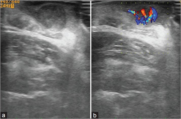
A high-resolution ultrasonography of the lesion showed a well-defined hypoechoic round mass lesion in the - subcutaneous tissue with a hyperechoic center and specks of calcification [Figure - 2]a. A color Doppler study showed increased vascularity in the lesion and a branching tree pattern of blood vessels [Figure - 2]b.
 |
| Figure 2: (a) High-resolution ultrasonography of the lesion showed a well-defined hypoechoic round mass lesion in the subcutaneous tissue with hyperechoic center and specks of calcification. (b) Color Doppler study showed increased vascularity in the lesion with branching tree pattern of blood vessels |
On histopathological examination, the tumor mass was seen separate from the dermis and did not have a cellular lining. The solid tumor mass showed basophilic nucleated cells in the periphery and anucleated cells with pink keratinized cytoplasm in the center, showing abrupt keratinization with foci of calcification [Figure - 3]a-d.
 |
| Figure 3: (a) The tumor mass was separated from dermis without any cellular lining. There was evidence of inflammatory infiltrate surrounding the tumor. (H and E, ×40) (b and c) The solid tumor mass showed basophilic nucleated cells in the periphery and anucleated cells with pink keratinized cytoplasm in the center, showing abrupt keratinization with foci of calcification (H and E, ×100). (d) Higher magnification showed basophilic nucleated cells in the periphery and anucleated cells with pink keratinized cytoplasm in the center (H and E, ×400) |
What is Your Diagnosis?
| 1. |
Malherbe A, Chenanatis J. Note sur I'epithelioma calcifiedes glandes sebacees. Prog Med 1880;8:826-37.
[Google Scholar]
|
| 2. |
Dubreuilh W, Cazenave E. De I' epithelioma calcifie: Etude histolgique. Ann Dermatol Syphilol 1922;3:257-68.
[Google Scholar]
|
| 3. |
Forbis R Jr, Helwig EB. Pilomatrixoma (calcifying epithelioma). Arch Dermatol 1961;83:606-18.
[Google Scholar]
|
| 4. |
Arnold HL. Pilomatricoma. Arch Dermatol 1977;113:1303.
[Google Scholar]
|
| 5. |
Garg LN, Arora S, Gupta S, Gupta S, Singh P. Pilomatricoma: Forget me not. Indian Dermatol Online J 2011;2:75-7.
[Google Scholar]
|
| 6. |
Simi CM, Rajalakshmi T, Correa M. Pilomatricoma: A tumor with hidden depths. Indian J Dermatol Venereol Leprol 2010;76:543-6.
[Google Scholar]
|
| 7. |
Kaddu S, Soyer HP, Hödl S, Kerl H. Morphological stages of pilomatricoma. Am J Dermatopathol 1996;18:333-8.
[Google Scholar]
|
| 8. |
Wortsman X, Wortsman J, Arellano J, Oroz J, Giugliano C, Benavides MI, et al. Pilomatrixomas presenting as vascular tumors on color Doppler ultrasound. J Pediatr Surg 2010;45:2094-8.
[Google Scholar]
|
| 9. |
Choo HJ, Lee SJ, Lee YH, Lee JH, Oh M, Kim MH, et al. Pilomatricomas: The diagnostic value of ultrasound. Skeletal Radiol 2010;39:243-50.
[Google Scholar]
|
| 10. |
Hwang JY, Lee SW, Lee SM. The common ultrasonographic features of pilomatricoma. J Ultrasound Med 2005;24:1397-402.
[Google Scholar]
|
Fulltext Views
3,187
PDF downloads
3,166


