Translate this page into:
An exaggerated manifestation of penile cancer: a report from Japan
Correspondence Address:
Masaki Okano
Department of Dermatology, Aiezembashi Hospital, Higashi 3-1-11, Nihombashi, Naniwa-ku, Osaka 556
Japan
| How to cite this article: Okano M. An exaggerated manifestation of penile cancer: a report from Japan. Indian J Dermatol Venereol Leprol 1997;63:194-195 |
Abstract
This report describes a Japanese case of penile carcinoma characterized by an extravagantly harsh clinical presentation. Report of penile cancer from Japan is extremely rare. |
 |
 |
 |
Penile cancer is a rare neoplasm and there has been few reports of this disorder in the dermatological literature from Japan. The author presents, herein, a Japanese case of penile cancer with an exaggerated clinical manifestation.
Case Report
A 55-year-old man visited the Dermatology Clinic for evaluation of progressively destructive disorder of the penis for a years′s duration. He confessed that clinical signs initiated at the left side of the shaft by a traumatic chance. The penile lesion was mixture of a deep erosion around the distal portion and lumpy thickening around the proximal, and the shape of the penis was markedly deformed [Figure - 1]. The irregularly ulcerative lesion invaded the underlying tissues, discharged a purulent exudate and was hemorrhagous. The proliferative nodules were stiffly indurated and pain was violent.
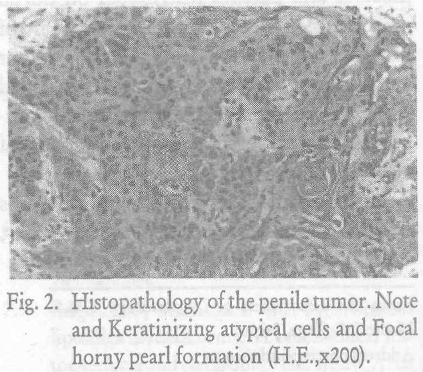
A foul odor was noticed. There was no urinary retention. The bilateral lymph nodes were indurately palpated. Histopathological study revealed irregular masses of atypical cells with variable keratinization invading the dermis. The tumour cells were composed predominantly of well-differentiated polygonal cells with large nuclei, prominent nucleoli and abundant basophilic cytoplasms containing numerous tonofibrils. Focal parakeratotic horny pearls were present [Figure - 2]. Mitotic figures were also observed. The neoplasm excited an inflammatory infiltrate of mononuclear cells in the dermis. The capillaries were dilated and increased in number. A routine laboratory examination disclosed no prominent abnormal data except for an increased serum SCC antigen level of 7.5 ng/ ml (normal < 1.5) and an elevated titer of serum C-reacuve protein of 2.07 mg/dl (normal < 0.20). Several bacteria were also cultured from the penile lesion. A diagnosis of squamous cell carcinoma (SCC) of the penis was made. Thorough systemic inspections including a CT scan detected no apparent distant metastases to the visceral or skeletal organs. The author introduced the patient to an university hospital, and expert plastic surgeons there, together with urologists, excised the penile cancer and reconstructed the penis. The patient has remained clinically free of disease from then to date, 15 months later.
Discussion
Penile carcinoma constitutes less than 1 per cent of all malignancies among the United States′ male population; and incidence of 1 to 2 cases per 100,000 per annum.[1] Most patients are over 50 years of age, and many present with advanced disease, presumably due to a reluctance to seek medical therapy. Penile carcinoma occurs frequently in uncircumcised individuals, and has been associated with sexually transmitted human papilloma virus (HPV). A study from Brazil found HPV 18 DNA sequences in seven of 18 cases.[2] Carcinoma of the penis is characterized by a relentless progressive course, causing death for the majority of untreated patients within 2 years.[3][4] The lesion is most frequent on the glans (48%), the prepuce (21%) and the coronal sulcus(6%) in an uncircumcissd individual It is less commonly found on the shaft (<2%).[5] The tumours form nodular, ulcerated, often secondarily infected masses. Invasion into the corpora and urethra may pro-dace urinary obstruction or fistula formation. Inguinal lymph nodes are frequently enlarged, sometimes due to metastatic involvement but most commonly because of infection.[6] Over 95 percent of all penile cancers are squamous cell carcinomas demonstrating keratinization and horny pearl formation.[7] The Jackson staging system,[8] is most frequently applied to penile carcinomas; stage I: Tumor confined to the penis; operable inguinal nodal metastases. Stage II: Invasion into the shaft or the copore, but without metastases. Stage III: Tumour confined to the penis ; operabe inguinel nodal metastase Stage IV: Invasion of the shaft with inoperative regional and / or distant metastases.
Amputation by partial or total penectomy has been the gold standard of therapy.
This case represents penile carcinoma (SCC) in developing of the tumor on the penile shaft and clinical manifestations are characterized by their exaggerated macroscopical appearance, with an advanced clinical stage III in the Jackson staging system.
Penile carcinoma accounts for as many as 10% to 20% of male cancers in the countries of Asia, Africa and south America.[9][10] Its reports from Japan have, however, been extremely infrequent in the English literature. The present paper should be an exceedingly rare report from Japan describing cancer.
| 1. |
Shellhammer PF, Jordan GH, Schlossberg SM. Tumors of the penis, in: Cambell's Urology, Editors Walsh PC, Retik AB, Stamey TA et al, WB Saunders, Philadelphia, 1991;1264-1298.
[Google Scholar]
|
| 2. |
Villa LL, Lopes A. Human papillomavirus DNA sequences in penile carcinomas in Brazil, Int J cancer, 1986;37:853-855.
[Google Scholar]
|
| 3. |
Beggs JH, Spratt JS. Epidermoid carcinoma of the penis, J Urol, 1961;91:166-172.
[Google Scholar]
|
| 4. |
Skinner DG, Leadbetter WF, Kelly SB. The surgical management of squamous cell carcinoma of the penis, J Urol, 1972;107:273-277.
[Google Scholar]
|
| 5. |
Burgers JK, Badalament RA, Drago JR. Penile cancer. Clinical presentation, diagnosis, and staging, Urol Clin North Am, 1992;19:247-256.
[Google Scholar]
|
| 6. |
Weiss MA, Mills SE. Neoplastic lesions of the penis, scrotum, and urethra, in : Atlas of Genitourinary Tract Disorders, JB Lippincott, Philadelphia, 1988;19.1-19.15.
[Google Scholar]
|
| 7. |
Chesney TM, Murphy WM. Disease of the penis and scrotum, in: Urological pathology Edited by Murphy WM, WB Saunders, Philadelphia, 1989;380-408.
[Google Scholar]
|
| 8. |
Jackson SM : The treatment of caricinoma of the penis, Br J Surg, 1966;53:33-35.
[Google Scholar]
|
| 9. |
Narayana AS, Olney LE, Loening SA, et al. Carcinoma of the penis. Analysis of 219 cases, Cancer, 1982;49:2185-2191.
[Google Scholar]
|
| 10. |
Persky L Epidemiology of cancer of the penis. Recent results, Cancer Res, 1977;60:97-109.
[Google Scholar]
|


