
Translate this page into:
Asymptomatic digital swelling with nail changes- Multiple subungual osteochondromas

Corresponding author: Dr. Prakhar Srivastava, Department of Dermatology & STD, Vardhman Mahavir Medical College and Safdarjung Hospital, New Delhi, India. sriprakhar1996@gmail.com
-
Received: ,
Accepted: ,
How to cite this article: Srivastava P, Singh P, Sharma P, Srivastava P, Khunger N. Asymptomatic digital swelling with nail changes- Multiple subungual osteochondromas. Indian J Dermatol Venereol Leprol. doi: 10.25259/IJDVL_1802_2024
Dear Editor,
Osteochondromas are benign osteocartilaginous tumours rarely encountered in subungual locations. These lesions commonly arise in children and young adults, with a female-to-male ratio of 2:1. While typically solitary, cases involving multiple digits are exceedingly rare. This report highlights an 18-year-old male presenting with multiple subungual osteochondromas, emphasising unique clinical, onychoscopic and radiological findings.
An 18-year-old male presented with a painless swelling on the left second finger since one year. There was no history of trauma and his medical and family histories were unremarkable. Examination revealed a firm, non-tender swelling on the distal phalanx of 2x2 cm size, with lateral onycholysis of the nail plate [Figure 1]. On onychoscopy, abundant tortuous dilated vessels resembling red-coral with tentacle-like arms dancing in the sea were seen over the swelling [Figure 2a]. Onychoscopy of the right second and fourth fingernails also showed triangular lunula, onychorrhexis and onycholysis. [Figures 2b and 2c].
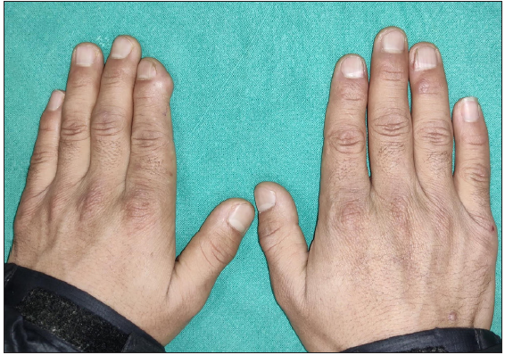
- Clinical photograph showing subungual swelling in the distal phalanx of left 2nd finger.
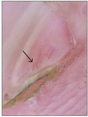
- Onychoscopy showing a red-coral sign in which abundant tortuous dilated vessels, resembling red-coral with tentacle-like arms are dancing in the sea, seen in left 2nd finger nail (black arrow) (DermLite DL3N, contact, polarised, 10x).
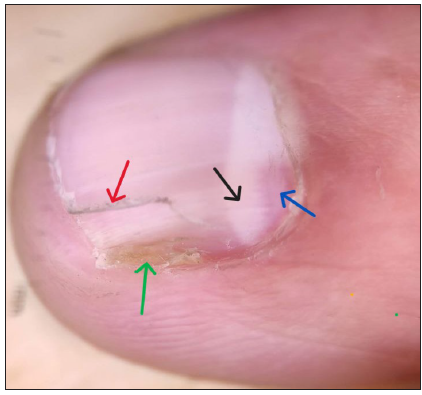
- Onychoscopy showing triangular deformed lunula (black arrow) with red areas (blue arrow), onycholysis (green arrow) and onychorrhexis (red arrow) in the right 2nd finger nail (DermLite DL3N, contact, polarised, 10x).
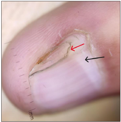
- Onychoscopy showing triangular deformed lunula (black arrow) and onychorrhexis (red arrow) in right 4th finger nail (DermLite DL3N, contact, polarised, 10x).
Ultrasonography of the hard swelling showed a hyperechoic mass protruding from the distal phalanx of the left second finger. No obvious subungual soft tissue swellings were seen [Figure 3a]. X-ray survey of the hands revealed many sessile bony lesions, the cortices and medullae of which were contiguous with the host bones, suggestive of osteochondromas. These lesions were seen on the left second distal phalanx, right second and fourth distal phalanx, left first metacarpal, interosseous margins of bilateral radius and interosseous and lateral borders of the right ulna [Figure 3b]. These findings confirmed the diagnosis of multiple subungual osteochondromas. The patient declined surgical intervention and was managed conservatively with adequate counselling on the need for monitoring for progression.
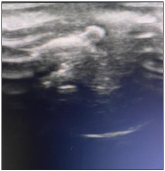
- Ultrasound showing hyperechoic subungual swelling suggestive of osteochondroma on the distal phalanx of left 2nd finger.
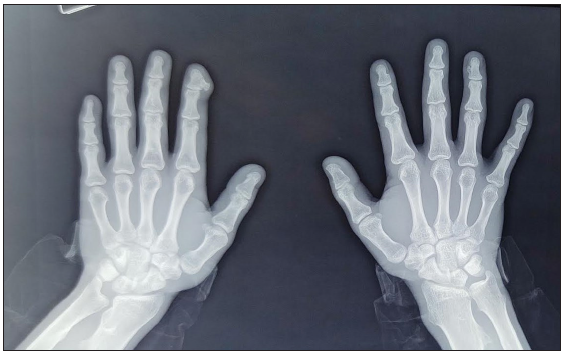
- X-ray showing osteochondromas on the left 2nd distal phalanx, right 2nd and 4th distal phalanx and left 1st metacarpal, with characteristic coat-hanger sign.
Osteochondromas are the most common benign skeletal neoplasms, accounting for 20-50% of the benign bone tumours.1 Dupuytren first described them as benign osteo-cartilaginous tumours attached to the bone of the distal phalanx.2 Mostly asymptomatic, they may cause pain, deformity and nail dystrophy. The mass effect of subungual osteochondromas may cause onycholysis and their pressure on the nail matrix may lead to deformed lunula and onychorrhexis. Chronic infections, hereditary anomalies and trauma are considered as risk factors for their development.3 The differential diagnoses include glomus-tumours, ingrown toenails, tenosynovial giant cell tumours, subungual warts, squamous-cell carcinomas and subungual malignant-melanomas.4 The occurrence of multiple osteochondromas could be sporadic or they could be a part of genetic autosomal dominant disorders caused by mutations in the Exostosin Glycosyltransferase (EXT- 1 and 2) genes, disrupting heparan sulphate synthesis. This impairs growth plate signalling pathways such as Indian hedgehog homolog (Ihh), Parathyroid hormone-related protein (PTHrP), Bone morphogenetic protein (BMP), and Wingless-related integration site (Wnt) leading to abnormal osteochondral growth.
The onychoscopic features of subungual osteochondromas are onycholysis, hyperkeratosis, ulceration and vascular ectasia- which looks like red-corals.5 Radiological investigations are diagnostic for osteochondromas as the lesions show both marrow and cortical continuity with the underlying parent bone. X-rays often demonstrate a ‘coat-hanger’ sign if the osteochondromas point away from the joint due to muscle pull.6 Histopathology shows mature trabecular bone covered by a fibrocartilaginous cap.7
Surgical excision is the treatment of choice. The mass lesion is removed until the spongy bone tissue is visible.7 This case highlights the importance of recognising rare benign subungual tumours and utilising multidisciplinary collaboration for accurate diagnosis and management. Awareness of characteristic clinical and imaging findings can prevent unnecessary interventions and improve patient outcomes.
Declaration of patient consent
The authors certify that they have obtained all appropriate patient consent.
Financial support and sponsorship
Nil.
Conflicts of interest
There are no conflicts of interest.
Use of artificial intelligence (AI)-assisted technology for manuscript preparation
The authors confirm that there was no use of artificial intelligence (AI)-assisted technology for assisting in the writing or editing of the manuscript and no images were manipulated using AI.
References
- Cartilaginous bone tumors. Radiol Clin North Am. 1993;31:237-59.
- [CrossRef] [PubMed] [Google Scholar]
- Subungual osteochondroma. Differential diagnosis and treatment. Arch Dermatol. 1979;115:472-3.
- [CrossRef] [Google Scholar]
- Subungual exostosis of the toes: A systematic review. Clin Orthop Relat Res. 2014;472:1251-9.
- [CrossRef] [PubMed] [PubMed Central] [Google Scholar]
- Subungual exostosis with an unusual dermoscopic feature. JAAD Case Rep. 2020;6:725-6.
- [CrossRef] [PubMed] [PubMed Central] [Google Scholar]
- Management of knee deformity in hereditary multiple exostoses: A case report. Br J Chiropr. 2000;4:68-73.
- [CrossRef] [Google Scholar]
- Subungual exostosis and subungual osteochondromas: A description of 25 cases. Int J Dermatol. 2018;57:872-81.
- [CrossRef] [PubMed] [Google Scholar]


