Translate this page into:
Asymptomatic nodule on the tongue
2 Department of Dermatology, Seth G S Medical College and KEM Hospital, Parel, Mumbai, Maharashtra, India
Correspondence Address:
Atul Dongre
Department of Dermatology, Government Medical College, Aurangabad, Maharashtra
India
| How to cite this article: Dongre A, Khopkar U. Asymptomatic nodule on the tongue. Indian J Dermatol Venereol Leprol 2011;77:112 |
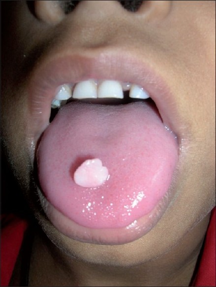
An 8-year-old female child presented with an asymptomatic nodular lesion of mucosal color on the dorsum of the tongue since the last 1 year. The lesion gradually increased in size since it was first noticed. There was neither history of bleeding from the lesion nor any history of trauma. Examination revealed a single, pink, sessile, firm and smooth-surfaced nodule of size 0.5 cm x 0.5 cm on the dorsum of the tongue [Figure - 1]. There was no significant lymphadenopathy in the cervical region.
 |
| Figure 1: Pink-colored nodule on the dorsum of the tongue |
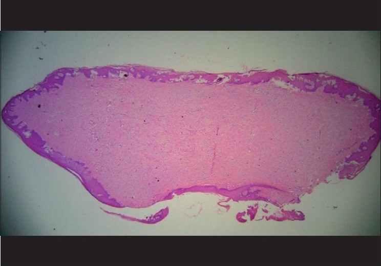
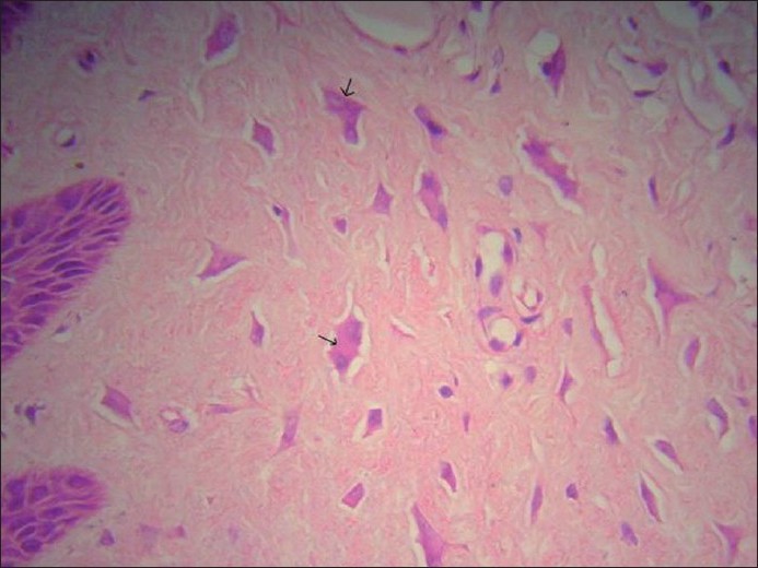
An excision biopsy of the nodule was performed. The histopathological examination showed hyperplastic epidermis and densely packed collagen fibres in the dermis [Figure - 2]. Other features seen were parakeratosis and dermal tissue containing many stellate-shaped cells in the vascular and fibrous connective tissue [Figure - 3]. Also, there were multiple multinucleated cells (marked with arrow in [Figure - 4]) with oval nuclei and abundant eosinophilic cytoplasm just beneath the hyperplastic epidermis [Figure - 4].
 |
| Figure 2: Hyperplastic epidermis and densely packed collagen in the dermis (H and E, ×25) |
 |
| Figure 3: Epidermal hyperplasia along with parakeratosis and stellate-shaped cells in the vascular and fibrous connective tissue in the dermis (H and E, ×200) |
 |
| Figure 4: Multinucleated cells in the dermis (H and E, ×400) |
What is your diagnosis?
| 1. |
Weathers DR, Callihan MD. Giant cell fibroma. Oral Surg Oral Med Oral Pathol 1974;37:374-84.
[Google Scholar]
|
| 2. |
Houston GD. The giant cell fibroma. A review of 464 cases. Oral Surg Oral Med Oral Pathol 1982;53:582-7.
[Google Scholar]
|
| 3. |
Bakos LH. The giant cell fibroma: a review of 116 cases. Ann Dent 1992;51:32-5.
[Google Scholar]
|
| 4. |
Wang Z, Levy B. Clinico-pathological study on giant cell fibroma of oral mucosa. Zhonghua Kou Qiang Yi Xue Za Zhi 1995;30:332-3.
[Google Scholar]
|
| 5. |
Swan RH. Giant cell fibroma: a case presentation and review. J Periodontol 1988;59:338-40.
[Google Scholar]
|
| 6. |
Odell EW, Lock C, Lombardi TL. Phenotypic characterisation of stellate and giant cells in giant cell fibroma by immunocytochemistry. J Oral Pathol Med 1994;23:284-7.
[Google Scholar]
|
| 7. |
Magnusson BC, Rasmusson LG. The giant cell fibroma: a review of 103 cases with immunohistochemical findings. Acta Odontol Scand 1995;53:293-6.
[Google Scholar]
|
| 8. |
Regezi JA, Courtney RM, Kerr DA. Fibrous lesions of the skin and mucous membranes which contain stellate and multinucleated cells. Oral Surg Oral Med Oral Pathol 1975;39:605-14.
[Google Scholar]
|
| 9. |
Katsikeris N, Kakarantza-Angelopoulou E, Angelopoulos AP. Peripheral giant cell granuloma. Clinicopathologic study of 224 new cases and review of 956 reported cases. Int J Oral Maxillofac Surg 1988;17:94-9.
[Google Scholar]
|
| 10. |
Lee NC, Norton JA. Multiple endocrine neoplasia type 2B--genetic basis and clinical expression. Surg Oncol 2000;9:111-8.
[Google Scholar]
|
Fulltext Views
3,713
PDF downloads
2,323


