Generic selectors
Exact matches only
Search in title
Search in content
Post Type Selectors
Search in posts
Search in pages
Filter by Categories
15th National Conference of the IAOMFP, Chennai, 2006
Abstract
Abstracts from current literature
Acne in India: Guidelines for management - IAA Consensus Document
Addendum
Announcement
Art & Psychiatry
Article
Articles
Association Activities
Association Notes
Award Article
Book Review
Brief Report
Case Analysis
Case Letter
Case Letters
Case Notes
Case Report
Case Reports
Clinical and Laboratory Investigations
Clinical Article
Clinical Studies
Clinical Study
Commentary
Conference Oration
Conference Summary
Continuing Medical Education
Correspondence
Corrigendum
Cosmetic Dermatology
Cosmetology
Current Best Evidence
Current Issue
Current View
Derma Quest
Dermato Surgery
Dermatopathology
Dermatosurgery Specials
Dispensing Pearl
Do you know?
Drug Dialogues
e-IJDVL
Editor Speaks
Editorial
Editorial Remarks
Editorial Report
Editorial Report - 2007
Editorial report for 2004-2005
Errata
Erratum
Focus
Fourth All India Conference Programme
From Our Book Shelf
From the Desk of Chief Editor
General
Get Set for Net
Get set for the net
Guest Article
Guest Editorial
History
How I Manage?
IADVL Announcement
IADVL Announcements
IJDVL Awards
IJDVL AWARDS 2015
IJDVL Awards 2018
IJDVL Awards 2019
IJDVL Awards 2020
IJDVL International Awards 2018
Images in Clinical Practice
Images in Dermatology
In Memorium
Inaugural Address
Index
Knowledge From World Contemporaries
Leprosy Section
Letter in Response to Previous Publication
Letter to Editor
Letter to the Editor
Letter to the Editor - Case Letter
Letter to the Editor - Letter in Response to Published Article
LETTER TO THE EDITOR - LETTERS IN RESPONSE TO PUBLISHED ARTICLES
Letter to the Editor - Observation Letter
Letter to the Editor - Study Letter
Letter to the Editor - Therapy Letter
Letter to the Editor: Articles in Response to Previously Published Articles
Letters in Response to Previous Publication
Letters to the Editor
Letters to the Editor - Letter in Response to Previously Published Articles
Letters to the Editor: Case Letters
Letters to the Editor: Letters in Response to Previously Published Articles
Media and news
Medicolegal Window
Messages
Miscellaneous Letter
Musings
Net Case
Net case report
Net Image
Net Images
Net Letter
Net Quiz
Net Study
New Preparations
News
News & Views
Obituary
Observation Letter
Observation Letters
Oration
Original Article
ORIGINAL CONTRIBUTION
Original Contributions
Pattern of Skin Diseases
Pearls
Pediatric Dermatology
Pediatric Rounds
Perspective
Presedential Address
Presidential Address
Presidents Remarks
Quiz
Recommendations
Regret
Report
Report of chief editor
Report of Hon : Treasurer IADVL
Report of Hon. General Secretary IADVL
Research Methdology
Research Methodology
Resident page
Resident's Page
Resident’s Page
Residents' Corner
Residents' Corner
Residents' Page
Retraction
Review
Review Article
Review Articles
Reviewers 2022
Revision Corner
Self Assessment Programme
SEMINAR
Seminar: Chronic Arsenicosis in India
Seminar: HIV Infection
Short Communication
Short Communications
Short Report
Snippets
Special Article
Specialty Interface
Studies
Study Letter
Study Letters
Supplement-Photoprotection
Supplement-Psoriasis
Symposium - Contact Dermatitis
Symposium - Lasers
Symposium - Pediatric Dermatoses
Symposium - Psoriasis
Symposium - Vesicobullous Disorders
SYMPOSIUM - VITILIGO
Symposium Aesthetic Surgery
Symposium Dermatopathology
Symposium-Hair Disorders
Symposium-Nails Part I
Symposium-Nails-Part II
Systematic Review and Meta-Analysis
Systematic Reviews and Meta-analyses
Systematic Reviews and Meta-analysis
Tables
Technology
Therapeutic Guideline-IADVL
Therapeutic Guidelines
Therapeutic Guidelines - IADVL
Therapeutics
Therapy
Therapy Letter
Therapy Letters
View Point
Viewpoint
What’s new in Dermatology
View/Download PDF

Translate this page into:

Quiz
89 (
3
); 469-471
doi:
10.25259/IJDVL_369_2021
pmid:
35841352

Asymptomatic papules and nodules of the vulva

Department of Dermatology, Hospital Universitario Ramon y Cajal, IRYCIS, Madrid, Spain
Department of Pathology, Hospital Universitario Ramon y Cajal, IRYCIS, Madrid, Spain

Corresponding author: Dr. Emilio De Dios Berna-Rico, Department of Dermatology, Hospital Universitario Ramon y Cajal, IRYCIS, Madrid, Spain. emilioberna2a@gmail.com

Licence
This is an open-access article distributed under the terms of the Creative Commons Attribution-Non Commercial-Share Alike 4.0 License, which allows others to remix, transform, and build upon the work non-commercially, as long as the author is credited and the new creations are licensed under the identical terms.

How to cite this article: Berna-Rico EDD, Jimenez-Cauhe J, Guevara-Barona KE, Gonzalez-Cantero A, del Real CMG. Asymptomatic papules and nodules of the vulva. Indian J Dermatol Venereol Leprol 2023;89:469-71.

An otherwise healthy 74-year-old woman presented with a 2-year history of asymptomatic lesions that appeared gradually on her vulva. Clinical examination revealed multiple yellowish and blue-grey papules and nodules located on both labia majora, some with an open-comedo-like appearance [Figure 1a]. Dermoscopy confirmed these findings, also showing an erythematous background and few radiating capillaries at the periphery of some papules [Figure 1b]. There was neither a history of trauma nor inflammation in the vulvar skin prior to the development of lesions. She denied any family history of cutaneous disorders. A punch biopsy from a yellowish papule with a peripheral pore was obtained.

Histological examination revealed a mid-dermal cyst [Figure 2a] lined by stratified squamous epithelium with a granular layer [Figure 2b]. The lumen of the cyst was occupied by orthokeratin with a laminated appearance, admixed with various calibre vellus hair shafts and keratin debris [Figure 2c].

What is your diagnosis?
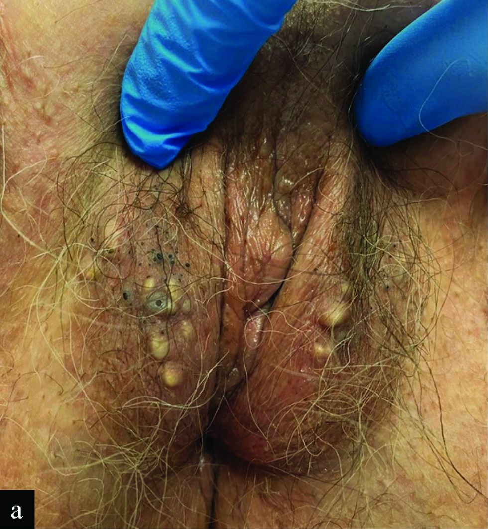
Papules and nodules on both labia majora, some of them resembling open comedones
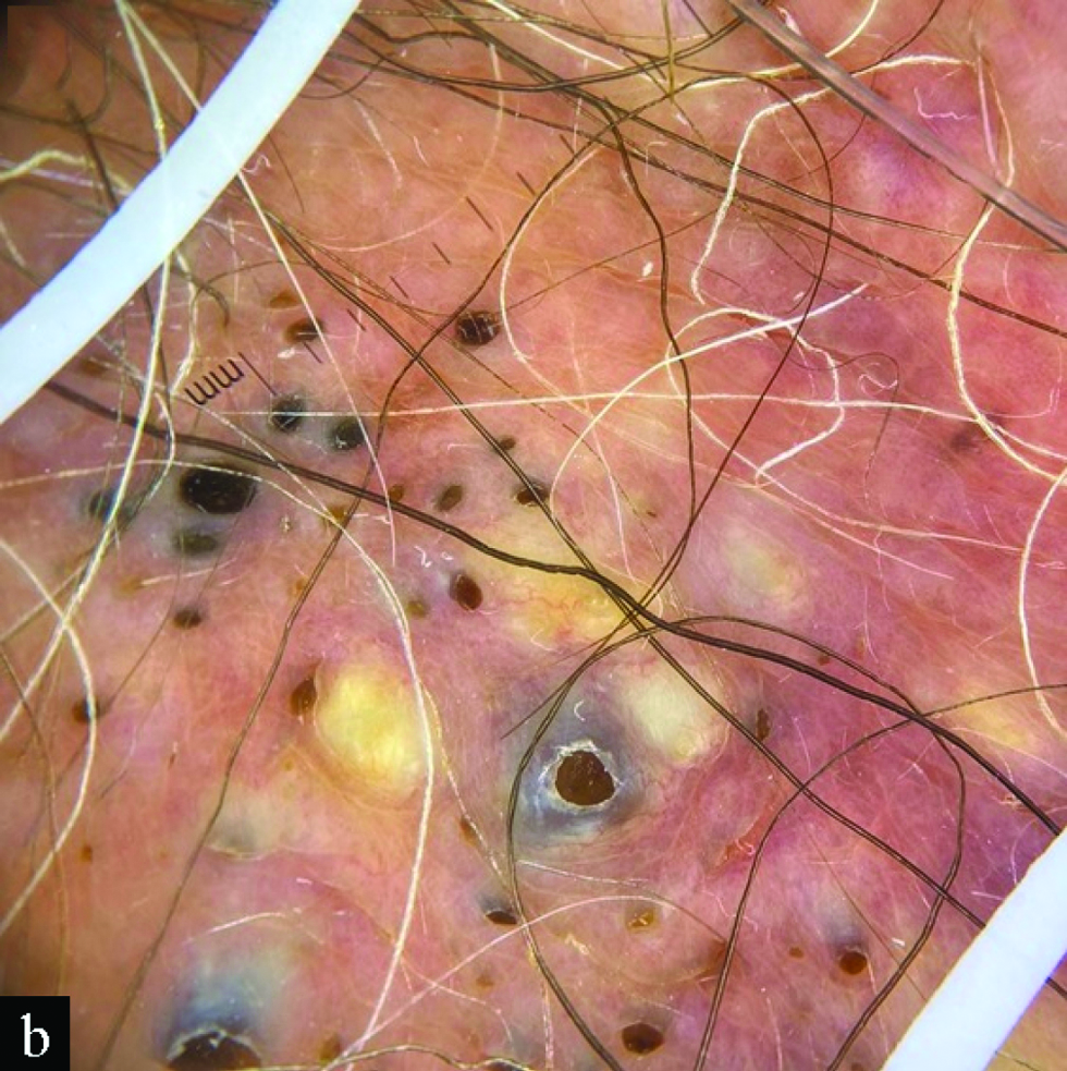
Figure 1a:
Papules and nodules on both labia majora, some of them resembling open comedones
Dermoscopy shows multiple non-melanocytic round yellowish or blue-grey papules with central or peripheral comedo-like openings
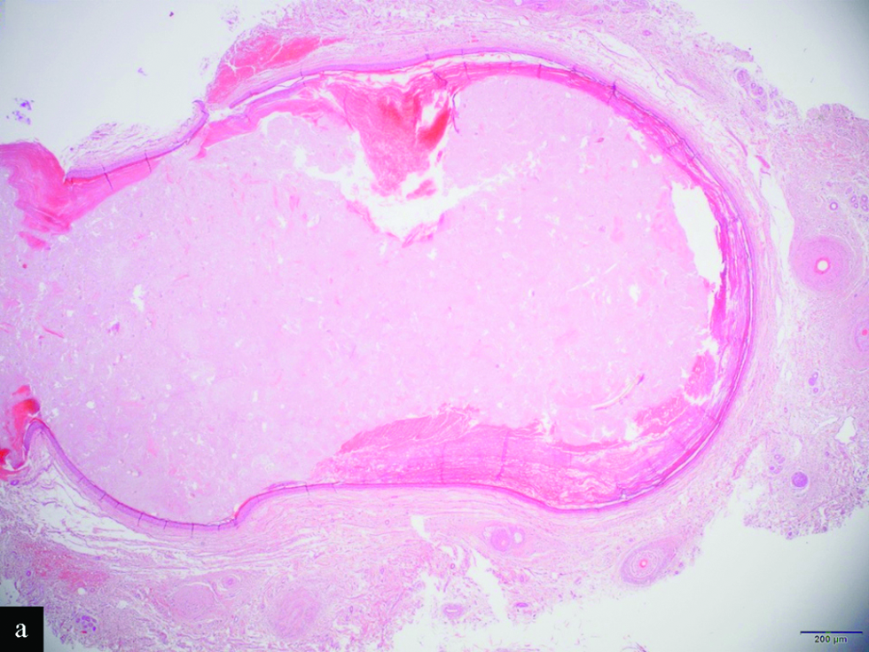
Figure 1b:
Dermoscopy shows multiple non-melanocytic round yellowish or blue-grey papules with central or peripheral comedo-like openings
Scanning magnification shows a mid-dermal cyst (H&E, x2)
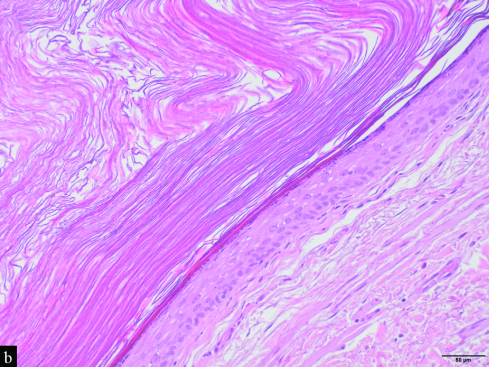
Figure 2a:
Scanning magnification shows a mid-dermal cyst (H&E, x2)
Detail of the cystic wall, which is lined by a stratified squamous epithelium with a granular layer. Note the laminated keratin within the cyst (H&E, x20)
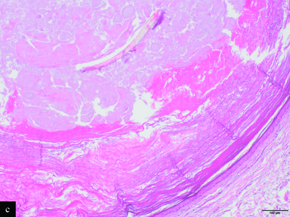
Figure 2b:
Detail of the cystic wall, which is lined by a stratified squamous epithelium with a granular layer. Note the laminated keratin within the cyst (H&E, x20)
The lumen is occupied by orthokeratin with laminated appearance, admixed with a large vellus hair shaft and keratin debris (H&E, x10)
Figure 2c:
The lumen is occupied by orthokeratin with laminated appearance, admixed with a large vellus hair shaft and keratin debris (H&E, x10)

Answer

Eruptive vellus hair cysts

Discussion

Eruptive vellus hair cysts are an uncommon condition caused by abnormal development of the infundibular portion of the hair follicle, leading to its occlusion. Clinically, they are characterized by multiple asymptomatic, 1–7 mm dome-shaped papules or nodules that are skin-coloured, erythematous, blue-grey or yellowish. Papules resembling open comedones could also be encountered. Most cases are sporadic, but 19 families affected by eruptive vellus hair cysts have been reported, with a suggested autosomal dominant inheritance with incomplete penetrance.1 More than 91% of eruptive vellus hair cysts cases appear before the age of 35, typically occurring symmetrically on the chest, abdomen or upper limbs. There are also presentations with limited involvement of the face.1 However, isolated involvement of the labia majora is rare, with very few cases published in the literature.1-3 Including ours, two of those six (33.3%) published cases showed multiple polyporous comedones, suggesting this presentation could be particularly frequent in the vulva.2

Dermoscopy of eruptive vellus hair cysts usually shows non-melanocytic, round, light yellow or blue-grey homogeneous structures, some of them surrounded by an erythematous halo and/or radiating capillaries.4 Our case additionally showed a striking open-comedo-like pattern. Oiso et al., reported bright yellow openings of these cysts to the epidermis,3 and Zhu et al., described comet-like bleary blue-grey lines next to follicular openings.5 We suggest that this presentation could represent a later stage of eruptive vellus hair cysts resulting from the oxidation of keratin and/or melanin pigment present in the exposed material.

Differential diagnosis of eruptive vellus hair cysts includes eruptive syringomas, trichogenic tumours, molluscum contagiosum, milia and comedonal acne, among others. The particular presentation in our patient should also include idiopathic calcinosis of the vulva and given the open-comedo-like appearance, epidermal inclusion cysts and a nevus comedonicus.1,2 The presence of hair and keratin within a dermal cyst lined by stratified squamous epithelium could also be suggestive of a dermoid cyst. However, they are usually congenital benign tumours that present at birth or during the first years of life, as slowly growing subcutaneous nodules located on the head and neck. The main diagnostic challenge of eruptive vellus hair cysts is their differentiation from steatocystoma multiplex. Both have an overlapping clinical appearance, may co-exist in the same patient and there are reports of hybrids cysts. Nevertheless, its histopathologic features are usually distinctive. Eruptive vellus hair cysts are lined by stratified squamous epithelium with a granular layer and contain laminated keratin and vellus hair shafts, that represent its diagnostic hallmark.1-3,5 Steatocystoma multiplex originate from the sebaceous duct rather than the infundibulum, and usually show sebaceous glands within the cyst wall, which is formed by a thin stratified squamous epithelium with undulating configuration and without or minimal granular layer. There is also a constant prominent hyaline cuticle lining the inner surface of the cyst wall. Finally, the keratin expression profile is also different between them. While steatocystoma multiplex express both K17 and K10, eruptive vellus hair cysts only express K17.1

Regarding its treatment, several options have been reported to be effective, especially topical retinoids, surgical approaches and CO2 laser.1,2,5 In our case, the larger lesions were successfully treated with CO2 laser, without recurrence at a 2-months follow-up visit.

In conclusion, eruptive vellus hair cysts should be considered in the differential diagnosis of asymptomatic papules and nodules of the vulva. Aside from yellow or blue-grey structures, open-comedo-like plugs could be encountered in the dermoscopy of eruptive vellus hair cysts and could represent a diagnostic clue in locations where acne and variants are uncommon, as in the vulva.

Declaration of patient consent

Patient’s consent is not required as patient’s identity is not disclosed or compromised.

Financial support and sponsorship

Nil.

Conflicts of interest

There are no conflicts of interest.

References

  1. , , . Eruptive vellus hair cysts: A systematic review. Am J Clin Dermatol. 2012;13:19-28.
    [CrossRef] [PubMed] [Google Scholar]
  2. , , . Eruptive vellus hair cysts of the vulva. Australas J Dermatol. 2017;58:e254-55.
    [CrossRef] [PubMed] [Google Scholar]
  3. , , . Eruptive vellus hair cysts of the labia majora: Detection of openings of the cysts to the epidermis by dermoscopy. Eur J Dermatol. 2013;23:417-18.
    [CrossRef] [PubMed] [Google Scholar]
  4. , , , , . Dermoscopy distinction of eruptive vellus hair cysts with molluscum contagiosum and acne lesions. Pediatr Dermatol. 2012;29:772-73.
    [CrossRef] [PubMed] [Google Scholar]
  5. , , , , . Eruptive vellus hair cysts diagnosed using dermatological imaging technique. Australas J Dermatol. 2021;62:86-88.
    [CrossRef] [PubMed] [Google Scholar]

Fulltext Views
5,013

PDF downloads
2,121
View/Download PDF
Download Citations
BibTeX
RIS
Show Sections