Translate this page into:
Auricular cutaneous leishmaniasis: An unusual presentation of Leishmania major from Southwest Iran
Corresponding author: Dr. Reza Yaghoobi, Department of Dermatology, Imam Khomeini Hospital, Azadegan Street, Ahvaz, Iran. yaghoobi_rz@yahoo.com
-
Received: ,
Accepted: ,
How to cite this article: Yaghoobi R, Pazyar N, Rafiei A, Saki N. Auricular cutaneous leishmaniasis: An unusual presentation of Leishmania major from Southwest Iran. Indian J Dermatol Venereol Leprol. 2024;90:349-50. doi: 10.25259/IJDVL_272_2022
Dear Editor,
Cutaneous leishmaniasis is an infectious disease caused by anthroponotic and zoonotic species of the Leishmania genus. Leishmania major, a zoonotic species, is the major causative agent in many rural areas of the Islamic Republic of Iran, being reported from 17 out of 31 provinces, including Khuzestan.1 Khuzestan province, located in Southwest Iran, is endemic for leishmaniasis. Although the lesions of cutaneous leishmaniasis appear mostly on the exposed regions of the body, auricular involvement is relatively rare.2,3 Clinically, the involvement of ears, especially in Mexico, is named chiclero’s ulcer.3
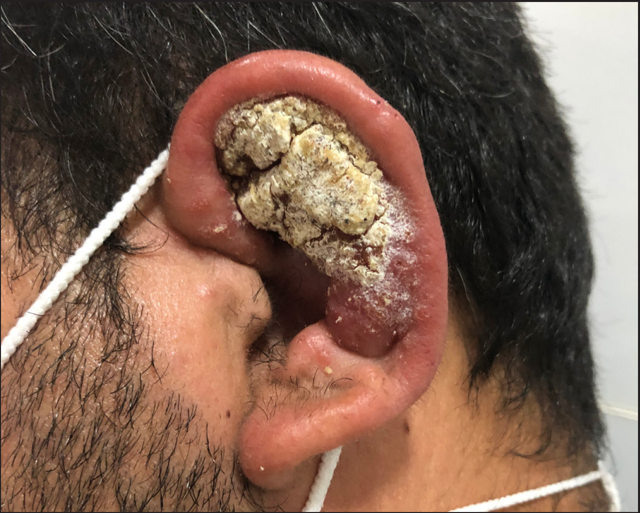
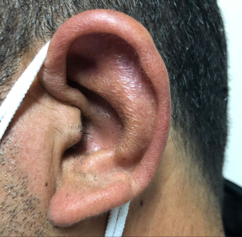
A 41year-old-man was referred to our department from the ear, nose and throat (ENT) department after 12 days of hospitalization with a clinical diagnosis of bacterial perichondritis. The erythematous, edematous inflamed left auricle had not responded to topical and systemic antibiotics over two weeks. The lesion appeared as a red nodule on the concha of the left ear and gradually increased in size over 6 months. The patient denied any trauma to his left ear. A physical examination revealed a verrucous, crusted and mildly tender mass that involved the concha, antihelix and fossa of the left ear. The helix also showed an erythematous swelling, but the other parts of the ear were unaffected [Figure 1]. No cervical lymphadenopathy or organomegaly was detected. Clinically, cutaneous leishmaniasis was suspected, and to confirm the diagnosis, a direct smear from the ear lesion was performed which showed amastigotes on Giemsa staining consistent with a diagnosis of leishmaniasis [Figure 2]. Polymerase chain reaction (PCR) revealed Leishmania major to be the causative agent. All routine laboratory tests and chest X-ray were normal. The patient received meglumine antimoniate (Glucantime, Aventis, Haupt Pharma Livron, France) intralesionally every week. All injections were painless and easy to inject. There was complete resolution without any deformity after 14 injections, and no recurrence or local or systemic complications were seen after 5 months of follow up [Figure 3].

- Auricular cutaneous leishmaniasis: erythematous, edematous verrucous crusted plaque over the left ear

- Giemsa staining showing Leishmania amastigotes inside the histiocytes (Giemsa ×1000)

- Complete cure of the auricular cutaneous leishmaniasis after receiving 14 intralesional injections of meglumine antimoniate
Leishmaniasis is a protozoal infection clinically characterised by a variety of skin lesions that mostly occur on the exposed parts of the body, especially the face and extremities. The diverse morphology of cutaneous lesions depends on the Leishmania species and the host immunity.4 The epidemiology of leishmaniasis is varied in different parts of the world. In the Mediterranean region, in contrast to Mexico, involvement of the ear is reported infrequently.2,3 Leishmaniasis of the ear, commonly seen in Mexico, is known as chiclero’s ulcer.3 The term “chiclero” (derived from Chilce, a material collected by loggers to produce rubber) is used since the disease frequently affects loggers.3 In Mexico, most chiclero ulcers are caused by Leishmania mexicana.3
Leishmaniasis has a long history in Iran. In our case, the causative agent was Leishmania major. Of the 31 provinces, 17, including Khuzestan, located in the Southwest of Iran, are endemic for Leishmania major.1 Youssef et al. previously reported a case of chiclero’s ulcer from Syria in which the causative agent was Leishmania tropica.3 Thus, the presentation of leishmaniasis as chiclero’s ulcer may not be species dependent but possibly may vary depending on the epidemiology variations in different parts of the world. Clinically, a differential diagnosis of chiclero’s ulcer is necessary because sometimes this lesion imitates other entities, such as bacterial perichondritis, angiolymphoid hyperplasia with eosinophilia and squamous cell carcinoma.2,3
Although there are some alternative treatments, pentavalent antimonials are the first-line treatment for all forms of leishmaniasis.5 Our patient received intralesional meglumine antimoniate (glucantime) weekly without any significant pain. The intralesional injection of meglumine antimoniate in the ear lesion, in contrast to the cutaneous leishmaniasis lesions of the other sites, is painless and easy to perform.
Declaration of patient consent
Patient’s consent not required as patients identity is not disclosed or compromised.
Financial support and sponsorship
Nil.
Conflicts of interest
There are no conflicts of interest.
References
- Epidemiological status of leishmaniasis in the Islamic Republic of Iran, 1983–2012. East Mediterr Health J. 2015;21:736-42.
- [CrossRef] [PubMed] [Google Scholar]
- Auricular leishmaniasis mimicking squamous cell carcinoma. J Laryngol Otol. 2009;123:915-8.
- [CrossRef] [PubMed] [Google Scholar]
- Chiclero’s ulcer: An unusual presentation of Leishmania tropica in Syria. Avicenna J Med. 2018;8:117-9.
- [CrossRef] [PubMed] [PubMed Central] [Google Scholar]
- Old word leishmaniasis on the ear lobe: A rare site. Actas Dermosifiliogr. 2014;105:628-30.
- [CrossRef] [PubMed] [Google Scholar]
- Glucantime efficacy in the treatment of zoonotic cutaneous leishmaniasis. Southeast Asian J Trop Med Public Health. 2011;42:502-8.
- [PubMed] [Google Scholar]


