Translate this page into:
Authors' reply
Correspondence Address:
Linda Tognetti
Hospital “S. Maria Alle Scotte,” Viale Bracci 16, 53100, Siena
Italy
| How to cite this article: Tognetti L, Rubegni P. Authors' reply. Indian J Dermatol Venereol Leprol 2018;84:442-443 |
Sir,
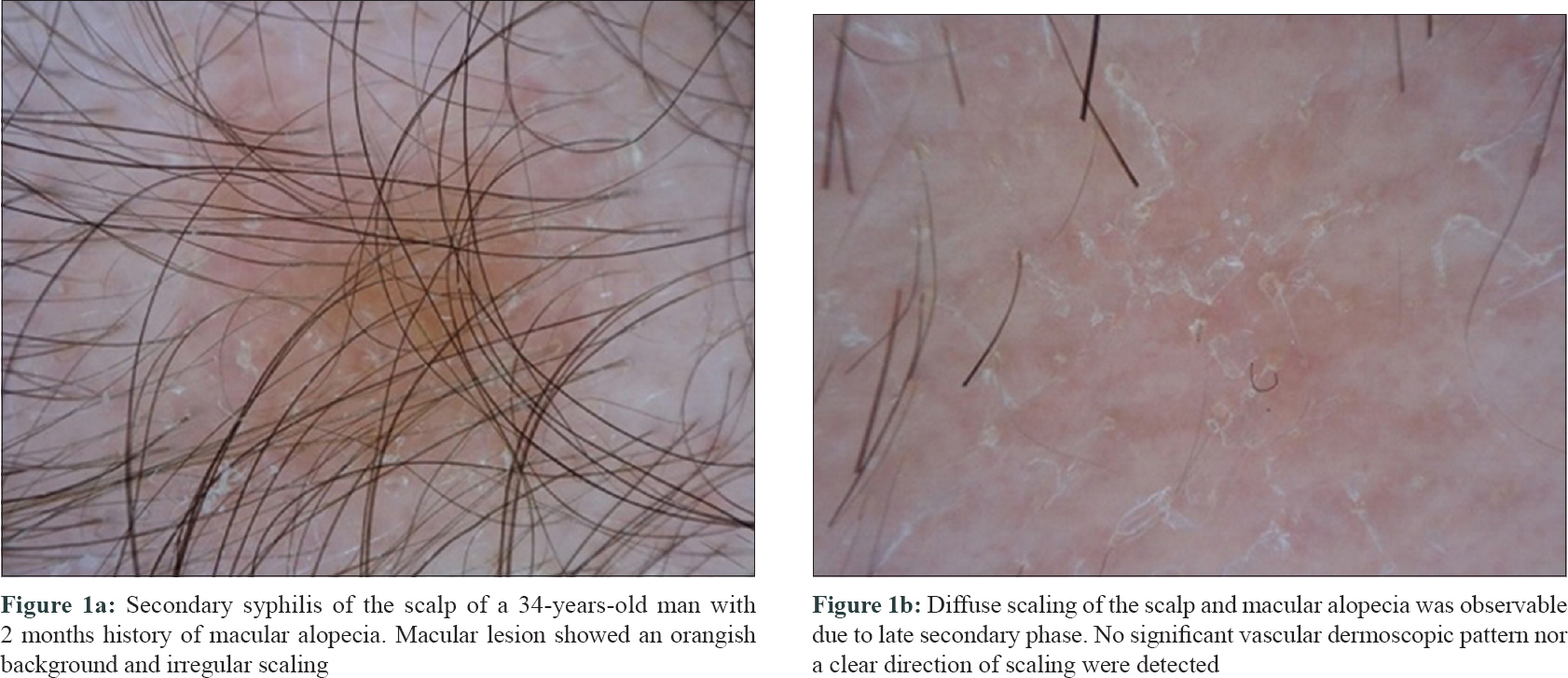
The great variety of clinical presentations of secondary syphilis deserves a careful and continuous attention as well as the collection and comparison of corresponding dermoscopic findings.[1] On the palm of a 28-year-old man with secondary syphilis, we dermoscopically described a classical Biett's sign of a 3-week-old lesion showing monomorphic dotted and glomerular vessels on a diffuse red to orange background within a circular scaling edge outwardly directed, surrounded by an erythematous halo.[2] Errichetti and Stinco reported the same dermoscopic findings, reporting occasional peripheral telangiectatic vessels.[3] In a young adult woman with 1-month history of diffuse macular rash, Streher and Bonamigo recently described the dermoscopic appearance of hyperkeratotic scalp lesions with an inner circular scaling edge inwardly oriented over a diffuse orangish background with monomorphic dotted vessels, suggesting a peculiar appearance of syphilitic Biett's sign. We would add our further observation on the scalp of a man with secondary syphilis-related macular alopecia. Few maculo-papular lesions showed an orangish background and irregular scaling on polarized dermoscopy [Figure - 1]a. No significant vascular dermoscopic pattern was detectable, nor a clear direction of scaling. Due to the timing of observation (i.e. 2-month-old syphilitic alopecia), a diffuse scaling of the scalp was the predominant clinical feature [Figure - 1]b.[4]
 |
| Figure 1: |
Based on these observations, we can speculate that secondary syphilitic maculo-papular lesions observed under polarized dermoscopy show a background ranging from intense to slight orange according to the body site, the thickness of stratum corneum and dermis and the entity of vascular structures. Thus, lesions on the palm can be orange-reddish in background, whereas lesions on the plantar aspect and scalp may appear orange-yellowish. Similarly, the vascular pattern varies according to the lesion age (more or less inflammatory) and body location (i.e., number of capillaries). The direction of scaling is generally less clearly detectable on older lesions, as fragile scales are more susceptible to friction. If a cleared lesion with a slight inflammatory halo (i.e., peripheral dotted vessels) is located on the scalp, where the stratum corneum and the dermis are thicker, scales can also rest attached to the inner edge. Thus, Biett's scaling collarette can also appear inward-directed in certain conditions.
Financial support and sponsorship
Nil.
Conflicts of interest
There are no conflicts of interest.
| 1. |
Balagula Y, Mattei PL, Wisco OJ, Erdag G, Chien AL. The great imitator revisited: The spectrum of atypical cutaneous manifestations of secondary syphilis. Int J Dermatol 2014;53:1434-41.
[Google Scholar]
|
| 2. |
Tognetti L, Sbano P, Fimiani M, Rubegni P. Dermoscopy of Biett's sign and differential diagnosis with annular maculo-papular rashes with scaling. Indian J Dermatol Venereol Leprol 2017;83:270-3.
[Google Scholar]
|
| 3. |
Errichetti E, Stinco G. Dermoscopy in differentiating palmar syphiloderm from palmar papular psoriasis. Int J STD AIDS 2017;28:1461-3.
[Google Scholar]
|
| 4. |
Tognetti L, Cinotti E, Perrot JL, Campoli M, Rubegni P. Syphilitic alopecia: Uncommon trichoscopic findings. Dermatol Pract Concept 2017;7:55-9.
[Google Scholar]
|
Fulltext Views
2,827
PDF downloads
1,518


