Translate this page into:
Capillary malformation associated with multiple accessory tragi
Correspondence Address:
Pravesh Yadav
RZ-97, Phase-III, Prem Nagar, Najafgarh, New Delhi - 110 043
India
| How to cite this article: Yadav P, Mendiratta V, Mittal S, Chander R. Capillary malformation associated with multiple accessory tragi . Indian J Dermatol Venereol Leprol 2014;80:484 |
Sir,
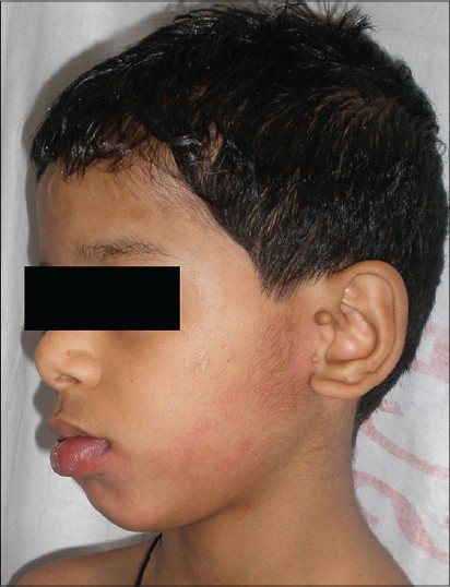
A 5-year-old boy, born of an uneventful pregnancy, was brought to our outpatient department with complaints of reddish discoloration over the left side of face and chin along with two skin-colored swellings in front of the left ear since birth which increased in size in proportion to the body size. The child was developmentally normal and had normal intelligence. There was no history of seizures. There was no family history of capillary malformation or branchial arch anomalies. Examination revealed multiple erythematous, non-blanchable patches present along the mandibular distribution of trigeminal nerve and the lower lip along with a diffuse swelling of the lower lip. There was no thrill or bruit. In addition, there were two skin-colored fleshy papules present just anterior to the left pinna [Figure - 1]. Examination of the oral cavity was unremarkable. Ophthalmic, neurological, and systemic examination was normal. Imaging of the head and neck region, chest radiograph, ultrasound of the abdomen and pelvis, and echocardiograph were unremarkable. Hematological and biochemical investigations were normal. The parents of the child refused biopsy or treatment of both the accessory tragus and capillary malformation.
 |
| Figure 1: Capillary malformation present along the mandibular distribution of trigeminal nerve associated with diffuse swelling of the lower lip and multiple tragi present just anterior to the left pinna |
Capillary malformation is a low-flow vascular malformation commonly affecting the face in the distribution of the trigeminal nerve (V1, V2, and V3). Lesions affecting the face may extend into the mucosal surfaces such as the lips and gingiva. They are sometimes associated with an underlying disorder in the form of various syndromes such as Sturge-Weber syndrome. Immunohistochemical and confocal microscopic studies of capillary malformations reveal a significantly decreased density of perivascular nervous tissue in lesional skin suggesting that inadequate innervation may be responsible for decreased vascular tone and progressive vascular dilatation. [1],[2] A potential role of vascular endothelial growth factor (VEGF)-A and its most active receptor VEGF-R2 expression, [3] as well as RASA1 gene on chromosome 5q encoding a protein (p120-rasGAP) [4] has been elucidated probably by inducing vessel proliferation and/or vasodilatation.
A normal tragus is derived from the dorsal portion of the first branchial arch as it grows ventrally to join in the midline during embryonic life. Isolated accessory auricles may be inherited as an autosomal dominant trait, mapped to chromosome 14q11.2-12. [5] Accessory tragi may also occur with other malformations and syndromes of the first branchial arch, such as Treacher Collins, Goldenhar, Nager′s acrofacial dysostosis, Wolf-Hirschhorn syndrome (4p-), oculocerebrocutaneous syndrome, Townes′ syndrome, and Down′s syndrome.
The mandibular and maxillary branches of the trigeminal nerve are also derived from the first branchial arch and innervate structures derived from the first arch. A defect in the first branchial arch possibly associated with a linked mutation potentially offers an explanation for this concurrence. The associated mutation may lead to defective trigeminal nerve innervation of blood vessels in the mandibular region leading to progressive vascular dilatation manifesting as a capillary malformation and the first branchial defect may lead to defective closure ventrally leading to accessory tragi. Also, both capillary malformation and accessory tragi have been known to be autosomal dominantly inherited.
We were unable to find previous reports of an association of capillary malformation with accessory tragus. The association in our patient may be conicidental, but such associations provide an opportunity to explore possible genetic or pathogenetic links between the two conditions. Unfortunately, we were unable to carry out genetic studies in our patient. Though our patient did not have any systemic involvement, patients with these pathologies should be screened for other local and systemic abnormalities.
| 1. |
Smoller BR, Rosen S. Port-wine stains. A disease of altered neural modulation of blood vessels? Arch Dermatol 1986;122:177-9.
[Google Scholar]
|
| 2. |
Selim MM, Kelly KM, Nelson JS, Wendelschafer-Crabb G, Kennedy WR, Zelickson BD. Confocal microscopy study of nerves and blood vessels in untreated and treated port wine stains: Preliminary observations. Dermatol Surg 2004;30:892-7.
[Google Scholar]
|
| 3. |
Vural E, Ramakrishnan J, Cetin N, Buckmiller L, Suen JY, Fan CY. The expression of vascular endothelial growth factor and its receptors in port-wine stains. Otolaryngol Head Neck Surg 2008;139:560-4.
[Google Scholar]
|
| 4. |
Eerola I, Boon LM, Mulliken JB, Burrows PE, Dompmartin A, Watanabe S, et al. Capillary malformation-arteriovenous malformation, a new clinical and genetic disorder caused by RASA1 mutations. Am J Hum Genet 2003;73:1240-9.
[Google Scholar]
|
| 5. |
Yang Y, Guo J, Liu Z, Tang S, Li N, Yang M, et al. A locus for autosomal dominant accessory auricular anomaly maps to 14q11.2-q12. Hum Genet 2006;120:144-7.
[Google Scholar]
|
Fulltext Views
2,203
PDF downloads
2,232


