
Translate this page into:
Cerebriform congenital melanocytic naevus on the sole

Corresponding author: Dr. Sheetanshu Kumar, Department of Dermatology & Sexually Transmitted Diseases, Jawaharlal Institute of Postgraduate Medical Education and Research, Puducherry, India. kumar.sheetanshu@gmail.com
-
Received: ,
Accepted: ,
How to cite this article: Meena A, Somasundaram A, L K, Kumar S, Toi PC. Cerebriform congenital melanocytic naevus on the sole. Indian J Dermatol Venereol Leprol. doi: 10.25259/IJDVL_1518_2024
A 24-years old woman presented with asymptomatic, raised, black, skin lesions on the sole of her right foot since birth. Examination revealed three, hyperpigmented nodulo-plaques on the right foot distal plantar aspect proximal to the second toe. [Figure 1a]. Dermoscopy showed cerebriform appearance with sulci and gyri [Figure 1b]. Histopathology showed basaloid naevus cells with melanin in the upper dermis and naevus cells interspersed within the dense collagen bundles and extended around the adnexal structures. A diagnosis of cerebriform congenital melanocytic naevus (CMN) was made, which is a rare morphological variant of CMN reported previously on the scalp. The index case is the first report of cerebriform CMN on the sole of the foot.
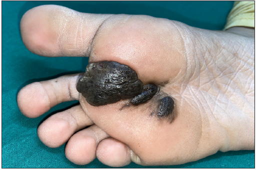
- Three well-defined hyperpigmented plaques on the sole of the right foot.
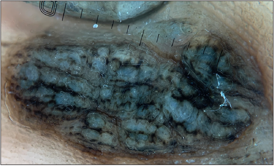
- Dermoscopy (Dermlite, 10x, polarised mode) showing prominent sulci and gyri, giving a cerebriform appearance of the naevus with accentuation of pigmentation in the sulci.
Declaration of patient consent
The authors certify that they have obtained all appropriate patient consent.
Financial support and sponsorship
Nil.
Conflicts of interest
There are no conflicts of interest.
Use of artificial intelligence (AI)-assisted technology for manuscript preparation
The authors confirm that there was no use of artificial intelligence (AI)-assisted technology for assisting in the writing or editing of the manuscript and no images were manipulated using AI.


