Translate this page into:
CHILD syndrome
Correspondence Address:
Ram M Kukreja
Department of Dermatology and STD, Dr. D.Y. Patil Medical College and Hospital, Room No. 207, Kadamwadi, Kolhapur - 416 006, Maharashtra
India
| How to cite this article: Heda G D, Valivade V, Sanghavi P, Kukreja RM, Phulari YJ. CHILD syndrome . Indian J Dermatol Venereol Leprol 2014;80:483 |
Sir,
Congenital hemidysplasia with icthyosiform erythroderma and limb defects (CHILD) syndrome is a rare congenital disorder characterized by hemidysplasia and ipsilateral erythroderma with ipsilateral limb and organ defects. It is caused by an X-linked dominant mutation in the NSDHL gene. [1] In most of the cases, mutations in NSDHL (NAD (P) H steroid dehydrogenase-like protein) at Xq28 were identified to be the cause of this syndrome. [2] Four additional NSDHL mutations were subsequently reported in individuals with CHILD syndrome. A shortage of this enzyme may also allow potentially toxic byproducts of cholesterol production to build up in the tissues. Low cholesterol levels and/or an accumulation of other substances disrupt the growth and development of many parts of the body. It has been found that cholesterol interacts with proteins controlling embryonic development through the hedgehog protein signaling pathway. [3]
Multiple ipsilateral anomalies of the viscera and central nervous system are observed in individuals with CHILD syndrome. These anomalies include cardiac malformations and ipsilateral hypoplasia of the brain, lungs, thyroid and reproductive tract. The severity of the limb defects may vary from hypoplasia of some metacarpals or phalanges to complete absence of an extremity. Non-specific punctuate calcification of cartilage may be observed. At times, the patient may present with bilateral epidermal nevus. [4] The distinct unilateral pattern of lesions may be diffuse and/or linear, with streaks following the lines of Blaschko. Ptychotropism, or affinity for body folds is common. [5]
The management of ichthyosiform erythroderma includes oral and/or topical steroids along with emollients. The use of lovastatin topically led to complete healing of the inflammatory CHILD nevus in a few cases, whereas cholesterol application alone had no satisfactory effect. [6] Approximately two-thirds of reported CHILD patients have right-sided involvement. It appears that the localization of lesions to the left side is often associated with more severe internal anomalies and early death.
A 1½-year-old girl, the second child of a non-consanguineous marriage, presented with a deformity of the right arm and shortening of right leg [Figure - 1] since birth along with redness and scaling over the body since 2 months of age. There was no history suggestive of systemic involvement. The child was born by emergency lower segment cesarean section due to failure in progression of labor. The baby cried immediately at birth and birth weight was 2.5 kg. The mother had no history of miscarriage and the elder sister was normal.
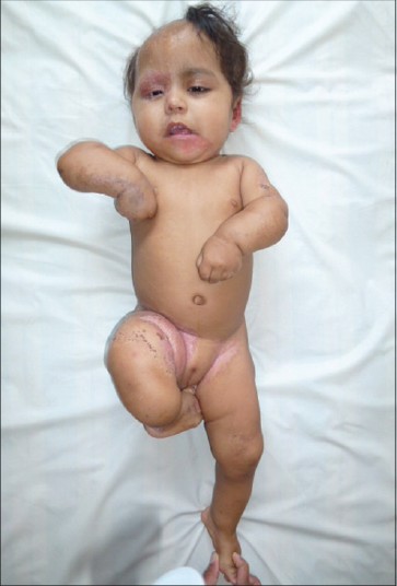 |
| Figure 1: Ipsilateral limb shortening |
Cutaneous examination revealed multiple erythematous scaly plaques on forehead, right upper eyelid, ears, axillae, forearms, buttocks, groins, thighs and dorsum of right foot. There was a sharp demarcation and similar ichthyotic plaques arranged in linear streaks were noted on ears, chin, groins and buttocks [Figure - 2]. Ptychotropism in the form of involvement of the flexural creases of the axillae and groins was conspicuous [Figure - 3]. Scarring alopecia involving right frontal and temporal area was also noted [Figure - 4]. Nails of right 4 th and 5 th toe were dystrophic. Mucosal involvement was absent.
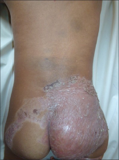 |
| Figure 2: Sharply demarcated ichthyosiform plaques |
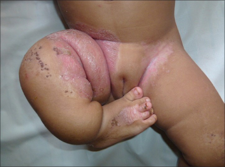 |
| Figure 3: Ptychotropism |
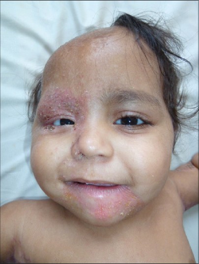 |
| Figure 4: Erythematous plaques over right eyelids and chin with scarring alopecia |
Laboratory investigations revealed anemia and thrombocytosis. All the other investigations were within normal limits.
Lower limbs X-ray showed shortening of right femur, tibia and fibula [Figure - 5] Further examination did not show any involvement of other organs such as the eyes, brain, heart, lungs, or kidneys.
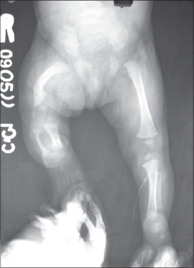 |
| Figure 5: X-ray: Shortening of right femur, tibia and fibula |
Histological examination revealed hyperkeratosis with focal parakeratosis and mild hyperplasia. Granular layer was almost absent. Rudimentary keratotic plugs were seen. The superficial and mid-dermis exhibited sparse perivascular and periadnexal lymphocytic inflammatory infiltrate. The biopsy was suggestive of an ichthyosiform dermatosis.
Based upon the clinical and radiological findings, a diagnosis of CHILD syndrome was made. The patient was managed conservatively with topical and oral steroids along with emollients for ichthyosiform erythroderma. The patient was being followed-up for a period of 6 months and she responded well to treatment.
| 1. |
Bornholdt D, König A, Happle R, Leveleki L, Bittar M, Danarti R, et al. Mutational spectrum of NSDHL in CHILD syndrome. J Med Genet 2005;42:e17.
[Google Scholar]
|
| 2. |
König A, Happle R, Bornholdt D, Engel H, Grzeschik KH. Mutations in the NSDHL gene, encoding a 3beta-hydroxysteroid dehydrogenase, cause CHILD syndrome. Am J Med Genet 2000;90:339-46.
[Google Scholar]
|
| 3. |
Grzeschik KH. Human limb malformations; an approach to the molecular basis of development. Int J Dev Biol 2002;46:983-91.
[Google Scholar]
|
| 4. |
Fink-Puches R, Soyer HP, Pierer G, Kerl H, Happle R. Systematized inflammatory epidermal nevus with symmetrical involvement: An unusual case of CHILD syndrome? J Am Acad Dermatol 1997;36:823-6.
[Google Scholar]
|
| 5. |
Happle R. Ptychotropism as a cutaneous feature of the CHILD syndrome. J Am Acad Dermatol 1990;23:763-6.
[Google Scholar]
|
| 6. |
Merino De Paz N, Rodriguez-Martin M, Contreras-Ferrer P, Garcia Bustinduy M, Gonzalez Perera I, Virgos Aller T, et al. Topical treatment of CHILD nevus and Sjögren-Larsson Syndrome with combined lovastatin and cholesterol. Eur J Dermatol 2011;21:1026-7.
[Google Scholar]
|
Fulltext Views
6,341
PDF downloads
3,497


