Translate this page into:
Chronic zosteriform cutaneous leishmaniasis
Correspondence Address:
M Omidian
Department of Dermatology, Imam Hospital, Ahwaz Jundishapour University of Medical Sciences, Ahwaz
Iran
| How to cite this article: Omidian M, Mapar M A. Chronic zosteriform cutaneous leishmaniasis. Indian J Dermatol Venereol Leprol 2006;72:41-42 |
Abstract
Cutaneous leishmanasis (CL) may present with unusual clinical variants such as acute paronychial, annular, palmoplantar, zosteriform, erysipeloid, and sporotrichoid. The zosteriform variant has rarely been reported. Unusual lesions may be morphologically attributed to an altered host response or owing to an atypical strain of parasites in these lesions. We report a patient with CL in a multidermatomal pattern on the back and buttock of a man in Khozestan province in the south of Iran. To our knowledge, this is the first reported case of multidermatomal zosteriform CL. It was resistant to conventional treatment but responded well to a combination of meglumine antimoniate, allopurinol, and cryotherapy. |
 |
 |
 |
INTRODUCTION
Cutaneous leishmaniasis (CL) is a common protozoal disease, caused by several species of Leishmania . CL, owing to L. major and L. tropica , is an important public-health problem in Iran.[1] The common clinical types are the nodular and papular; rare variants are erysipeloid, annular, paronychial, palmoplantar, sporotrichoid, and genital forms. [2],[3],[4],[5] We present a patient with a very rare and chronic variant-multidermatomal zosteriform CL. The patient was from the Khozestan province, which is in the south of Iran.
CASE REPORT
A 60-year-old man was referred to us with a large erythematous plaque, 20 x 30 cm[2] in size, studded with papules, pseudovesicles, and small nodules on the right lower portion of the back and buttock since 3 years [Figure - 1]. There was also some crusting and slight oozing. He had been treated with many therapeutic modalities, including short courses of meglumine antimoniate and acyclovir (because of misdiagnosis as herpes zoster).
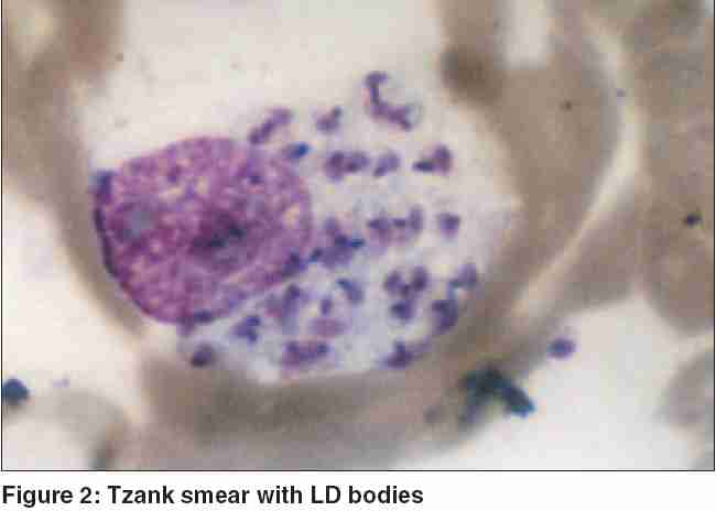
A Tzanck smear from the lesion did not show any feature of herpes zoster but numerous Leishman Donovan (LD) bodies were seen [Figure - 2]. A biopsy specimen showed a granulomatous infiltrate in the dermis. Culture on Novy-Nicolle-MacNeal medium was positive for Leishmania .
The patient′s electrocardiogram (ECG), complete blood picture, chest X-ray, and serum urea, creatinine and electrolytes were within normal limits. He was given a combination of meglumine antimoniate in a dose of 20 mg/kg body weight intramuscularly daily for 20 days, 20 mg/kg body weight of allopurinol daily for 30 days (with monitoring of the ECG and routine hematological parameters), and liquid-nitrogen cryotherapy every 2 weeks. After 2 months almost all the lesions had cleared.
DISCUSSION
CL is a disease with different clinical features. [2],[3],[4],[5] Many of the lesions are typical and present no diagnostic difficulties. However, a zosteriform presentation is rare, especially with multidermatomal involvement. The most commonly involved sites are exposed areas.[5] Our patient presented with an unusual lesion on a covered area. The lesion was zosteriform and multidermatomal, on the right lower part of the back, flank, and buttock with nodules, papules, and pseudovesicular lesions on an erythematous background.
Zosteriform CL has rarely been reported,[3],[5] but a multidermatomal zosteriform pattern has not been reported. Our patient had been mistakenly treated for herpes zoster on several occasions. A Tzanck smear from lesions showed numerous LD bodies. The mechanism of multidermatomal involvement is not clear, but altered host immunity may be involved. The clinical pattern of the disease is determined by the interaction between the host immune responses and the strain of the parasite involved and, to some extent, on the site of involvement.[3],[4] In an endemic area it is necessary for the physician to be aware that any atypical lesion should be investigated for CL.
| 1. |
Momeni AZ, Aminjvahery M. Successful treatment of cutaneous leishmaniasis, using a combination of meglumine antimoniate plus allopurinol. Eur J Dermatol 2003;13:40-3.
[Google Scholar]
|
| 2. |
Faber WR, Oskem L, van Gool T, Kroon NC, Knegt-Junk KJ, Hofwegen H, et al . Value of diagnostic techniques for cutaneous leishmanaisis. J Am Acad Dermatol 2003;49:70-4.
[Google Scholar]
|
| 3. |
Raja KM, Kaan AA, Hameed A, Rahman SB. Unusual clinical variants of cutaneous leishmaniasis in Pakistan. Br J Dermatol 1998;139:111-3.
[Google Scholar]
|
| 4. |
Iftikhar N, Bari I, Ejaz A. Rare variants of cutaneous leishmaniasis: whitlow, paronychia, and sporotrichoid. Int J Dermatol 2003;42:807-9.
[Google Scholar]
|
| 5. |
Momeni AZ, Aminjavahery M. Clinical picture of cutaneous leishmaniasis in Isfahan, Iran. Int J Dermatol 1994;33:260-5.
[Google Scholar]
|
Fulltext Views
2,305
PDF downloads
1,026


