Translate this page into:
Clear zone phenomenon: A rare phenomenon in ichthyosis with co-existing superficial fungal infection
Corresponding author: Dr. Pravesh Yadav, Department of Dermatology and STD, Lady Hardinge Medical College and SSKH, KSCH, Delhi - 110 001, India. rao.pravesh@gmail.com
-
Received: ,
Accepted: ,
How to cite this article: Agrawal M, Yadav P, Yadav J, Chander R. Clear zone phenomenon: A rare phenomenon in ichthyosis with co-existing superficial fungal infection. Indian J Dermatol Venereol Leprol 2021;87:103-105.
Sir,
Ichthyosis is a common disorder of keratinization, manifesting as noninflammatory scaling, which may range in severity from minimal involvement of a few sites to generalized scaling. Several types of ichthyosis have been classified according to the inheritance, clinical appearance, pathological features and systemic involvement.
Cutaneous dermatophytosis is a common condition with a global prevalence of nearly 20%–25%.1 The most common species implicated for dermatophytosis in India is Trichophyton rubrum.2 There are a handful of reports describing the coexistence of dermatophyte infections in patients with ichthyoses. We hereby report two cases of unusual presentations of dermatophyte infections showing clearing of ichthyotic scaling in patients with congenital ichthyosis.
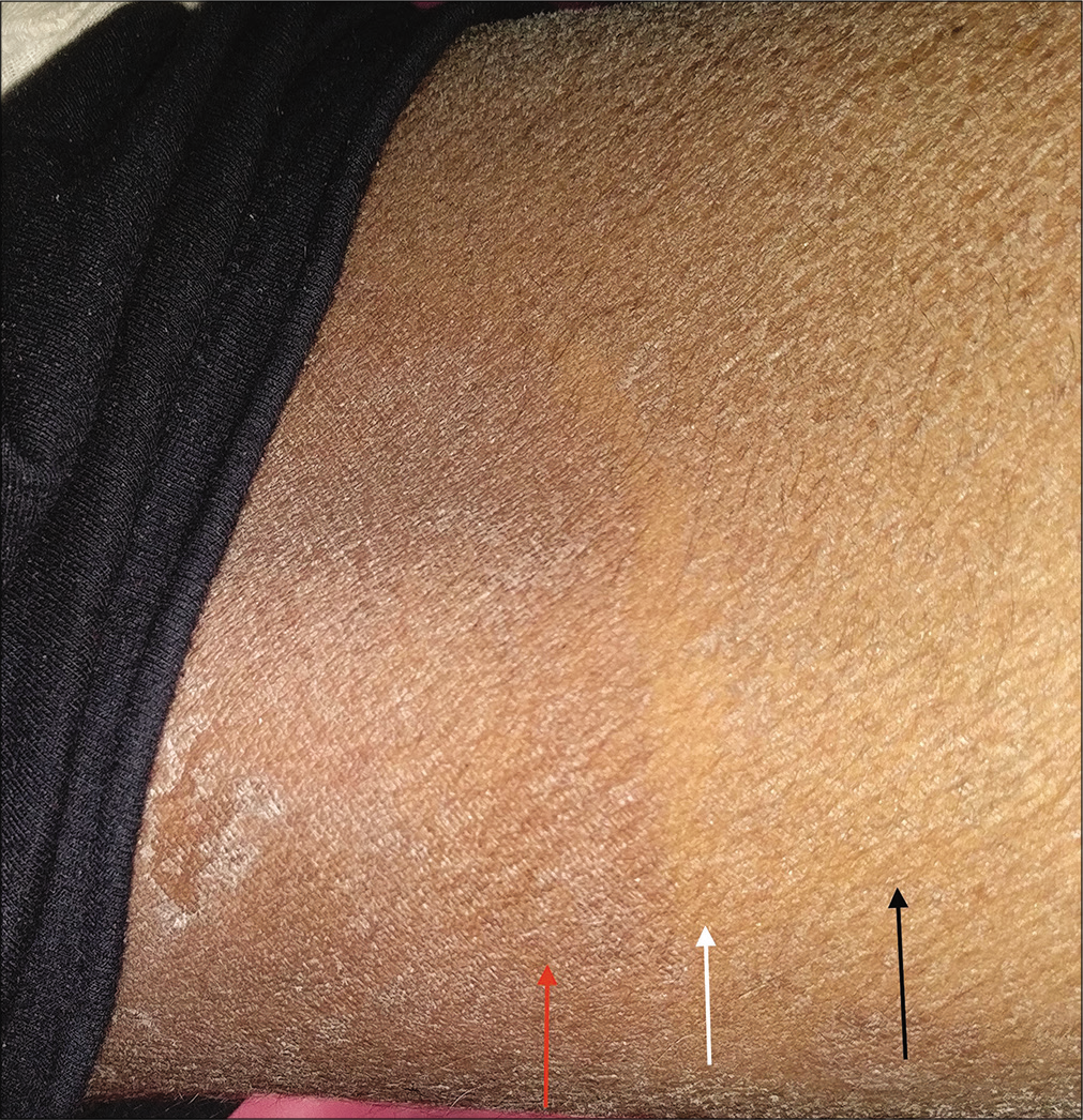
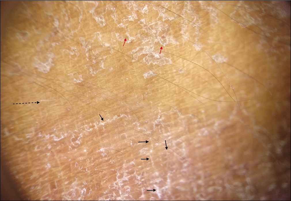
A 16-year-old Indian male, known case of X-linked recessive ichthyosis (XLRI), presented with itchy lesions in the groin of 20 days duration. Examination revealed generalized light-brown coloured adherent polygonal ichthyotic scales with upturned edges. There were well-defined erythematous scaly plaques in bilateral groins extending to inner thighs suggestive of tinea cruris with a peculiar finding of clear uninvolved zone between the erythematous plaque and ichthyotic skin [Figure 1]. Dermoscopy showed erythema and thicker white scales following the skin creases in the erythematous plaque (probably due to affinity of fungus towards creases due to increased moisture content), surrounded by a clear zone showing neither erythema nor scaling and the adjacent ichthyotic skin showed fine white scales irregularly arranged not respecting the skin creases [Figure 2]. Potassium hydroxide (KOH) mounts and cultures were done from the erythematous plaque, adjacent clear zone and the margin of clear zone with ichthyotic skin. Septate fungal hyphae were demonstrated only from the erythematous plaque. Culture from the erythematous plaque was positive for Trichophyton rubrum while the other two areas were negative. A diagnosis of XLRI with tinea cruris was made.

- Case 1 - Well-defined erythematous scaly plaques in the groins extending to inner thigh (red arrow), adjacent clear uninvolved zone devoid of scaling (white arrow) and generalized ichthyotic fine scales (black arrow)

- Polarized ×10 dermoscopic image from case 1 showing erythema and thick white scales following the skin creases (short black arrows) with adjacent area devoid of erythema or scaling (long black dashed arrow) and ichthyotic skin showing finer white scales irregularly arranged not respecting the skin creases (red arrows) (Dermlite DL4)
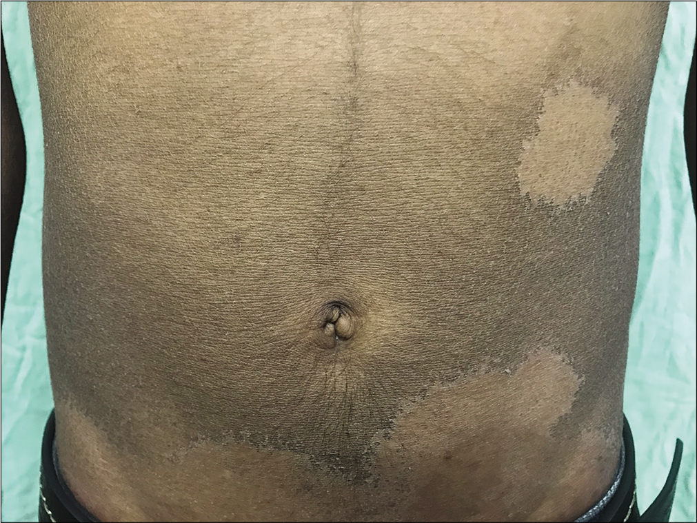
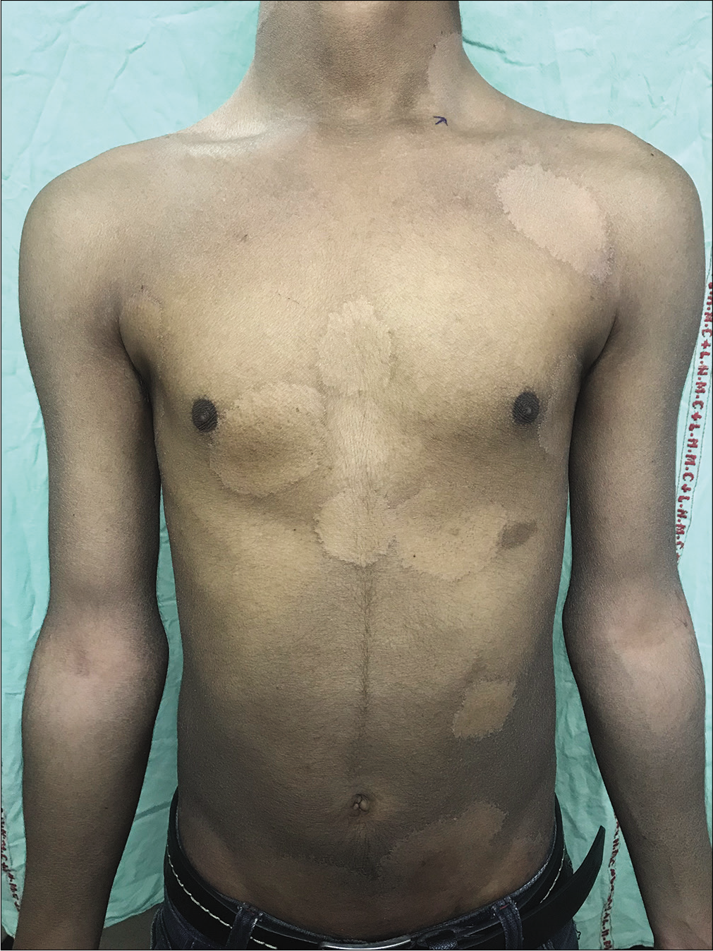
Another 19-year-old male having XLRI presented with complaints of round itchy lesions over the chest, abdomen and back of 1 month duration. Examination showed diffuse light-brown scaling all over the body with well-defined discrete as well as coalescing round zones of complete clearance of ichthyotic scales on the trunk, neck, and arms [Figure 3a and b]. KOH examination from the margins of clearing showed fungal elements with culture positive for Trichophyton rubrum.

- Case 2 - Well-defined discrete as well as coalescing round zones of complete clearance of ichthyotic scales on abdomen

- Case 2 - generalized distribution of similar clear areas on neck, chest and abdomen
Both patients were treated with oral terbinafine 250mg and topical 1% luliconazole cream once daily along with antihistaminics, emollients and general care for ichthyotic skin. Both of them showed complete clearance of tinea lesions and marked improvement of ichthyosis at 8 weeks follow-up.
Dermatophyte infections may be difficult to suspect in ichthyosis patients or may have unusual presentations.3 There are a number of ways dermatophytosis and the ichthyotic background may interact with each other. Excessive keratin in ichthyosis provides a favorable habitat for fungal growth. Moreover, ichthyosis vulgaris is associated with atopy, which in turn is a risk factor for tinea infections (due to shift from T-helper (Th) 1 to a Th2 response in atopy).4 “Anatopic phenomenon” is a term used to describe the modulation of inflammatory response of one dermatoses by another unrelated cutaneous infection at the same site. 5 The clearance of ichthyotic scaling seen in our cases could be attributed to the arsenal of proteases secreted by dermatophytes aimed at the digestion of the keratin network into assimilable oligopeptides or amino acids. They secrete multiple serine-subtilisins and metallo-endoproteases (fungalysins) formerly called keratinases.6 Production of these enzymes may have led to disintegration of ichthyotic scaling in our cases producing such clear zones. Furthermore, in T. rubrum, the mannans in the cell wall are known to decrease epidermal proliferation.6 This mechanism may also explain reduced scaling observed in our cases. It is difficult to postulate as to why only a clear margin was observed in first case as opposed to complete round clear zones in the latter: probably the extensive involvement in the second case could have led to complete clearing.
In a previously reported case similar to our second case, the authors concluded that the fungus can alter intercellular lipid layers, causing normalization of the cholesterol sulphate-cholesterol ratio correcting the pathology in X linked recessive ichthyosis thereby resolving ichthyotic scaling.3
If further molecular studies are undertaken and the cause is delineated, then extrapolation of clear zone phenomenon can potentially lead to novel treatment modalities for the treatment of congenital ichthyosis.
To conclude, dermatophytic infections must be suspected if cases of ichthyoses present with localized areas of erythema, clear zones and/or increased pruritis.Further research into the underlying pathomechanisms responsible for this clear zone phenomenon may lead to newer therapeutic modalities for ichthyosis.
Declaration of patient consent
The authors certify that they have obtained all appropriate patient consent forms. In the form the patients have given their consent for their images and other clinical information to be reported in the journal. The patients understand that their names and initials will not be published and due efforts will be made to conceal their identity, but anonymity cannot be guaranteed.
Financial support and sponsorship
Nil.
Conflicts of interest
There are no conflicts of interest.
References
- Prevalence of dermatophytic infection and the spectrum of dermatophytes in patients attending a tertiary hospital in Addis Ababa, Ethiopia. Int J Microbiol. 2015;2015:653419.
- [CrossRef] [PubMed] [Google Scholar]
- Management of tinea corporis, tinea cruris, and tinea pedis: A comprehensive review. Indian Dermatol Online J. 2016;7:77-86.
- [CrossRef] [PubMed] [Google Scholar]
- Tinea corporis due to Trichophyton verrucosum in recessive, X-linked ichthyosis. Mycoses. 1993;36:319-20.
- [CrossRef] [PubMed] [Google Scholar]
- Chronic dermatophytosis in lamellar ichthyosis: Relevance of a T-helper 2-type immune response to Trichophyton rubrum. Br J Dermatol. 2001;145:518-21.
- [CrossRef] [PubMed] [Google Scholar]
- Sparing phenomena in dermatology. Indian J Dermatol Venereol Leprol. 2013;79:545-50.
- [CrossRef] [PubMed] [Google Scholar]
- Dermatophytosis and the immune response. J Am Acad Dermatol. 1994;31:S34-41.
- [CrossRef] [Google Scholar]


