
Translate this page into:
Clinical characteristics and management outcomes in isolated nail lichen planus: A retrospective case series

Corresponding author: Dr Archana Singal, Dermatology and STD, University College of Medical Sciences and GTB Hospital, New Delhi, India. archanasingal@gmail.com
-
Received: ,
Accepted: ,
How to cite this article: Singal A, Gaurav V, Kaur I. Clinical characteristics and management outcomes in isolated nail lichen planus: A retrospective case series. Indian J Dermatol Venereol Leprol. 2024;90:329-35. doi: 10.25259/IJDVL_449_2023
Abstract
Background
Isolated nail lichen planus (NLP) without mucocutaneous involvement is rare. Literature about the clinical profile and management is scarce.
Aims/Objective
We attempted to characterize the clinico-demographic profile and analyze the management outcome of patients with isolated nail lichen planus.
Methods
Records of 15 patients were evaluated, and their demographic profile, clinical features of the nail matrix and nail bed disease, onychoscopy findings, histopathological features, treatment given, and follow-up progress were analysed.
Results
Data from 15 patients were collected. The mean age of the patients was 28.6 ± 19.0 years (range 3.5 years to 56 years). The gender ratio was 0.8 with 7 males and 8 females. The average disease duration at presentation was 2.8 ± 2.6 years (6 months–9 years). The average number of nails involved was 16.4 ± 4.6 (8-20 nails). All 20 nails were involved in 9 (60%) patients. Nail matrix involvement was seen in all, with onychorrhexis being the most common manifestation, which was seen in 11 (73.3%) patients. Nail bed involvement was seen in 11 (73.3%) patients, with onycholysis being the most common presentation. Severe nail disease was seen in 7 (46.7%) patients, and 5 (33.3%) had pterygium involving an average of 3.4 nails. Moderate to good improvement was observed in 9 (60%) patients after an average of 6.1 ± 2.4 (3-9) treatment sessions with intramuscular and intramatricial triamcinolone acetonide injection. Of these, 2/9 (22.2%) developed disease recurrence in a few nails after an interval of 1 and 1.5 years, respectively. Two patients achieved complete clinical cures that persisted beyond 2 years of follow-up.
Limitations
Retrospective nature of the series and the small sample size are the major limitations.
Conclusion
The risk of permanent disfigurement is high in NLP and calls for an early diagnosis and prompt treatment. Intralesional and intramuscular steroids are first-line therapeutic options depending upon the number of nails involved.
Keywords
Lichen planus
nail lichen planus
isolated nail lichen planus
intramuscular triamcinolone acetonide
intramatricial triamcinolone acetonide.

Plain Language Summary
Isolated nail lichen planus is a rare entity. Early diagnosis is imperative for early intervention, as delayed treatment may lead to permanent nail dystrophy. Literature on clinico-epidemiological aspects and management is scarce. In the present study, the authors describe the various clinical manifestations of isolated nail lichen planus in 15 patients. Nail matrix involvement was seen in all, with onychorrhexis being the most common feature, seen in 11 (73.3%) patients. Nail bed involvement was seen in 11 (73.3%) patients with onycholysis being the most common presentation. Severe nail disease was present in 7 (46.7%) patients, and 5 (33.3%) patients had pterygium involving an average of 3.4 nails. Moderate to good improvement was observed in 9 (60%) patients after an average of 6.1 ± 2.4 (3-9) treatment sessions with intramuscular and intramatricial triamcinolone acetonide given 4-weekly. Of these, 2/9 (22.2%) patients developed disease recurrence in a few nails after an interval of 1 and 1.5 years, respectively. Two patients achieved complete clinical cures that persisted beyond 2 years of follow-up. The study also highlights the efficacy of intramatricial and intramuscular steroids. The authors concluded that intralesional and intramuscular steroids may be used as first-line therapeutic options depending on the number of nails involved.
Introduction
Lichen Planus (LP) is a T-cell mediated chronic, inflammatory autoimmune dermatosis involving the skin, mucosae, hair, and nails.1 Nail involvement can occur in up to 10% of mucocutaneous LP.2 Isolated nail lichen planus (NLP) is a rare and severely disabling disease. Clinical manifestations depend upon the involvement of the nail matrix, nail bed, or both. Thus, varied presentations of NLP may be seen, ranging from nail plate thinning, onychorrhexis, trachyonychia, longitudinal melanonychia, pterygium formation, and even anonychia in severe cases.3 These clinical features of NLP may overlap with other nail disorders, so confirmation by onychoscopy and histology is essential to formulate a treatment plan and for prognostication. The published literature about NLP is limited to small case series, except for two retrospective studies: a 20-year study from Italy and a 12-year study from France, comprising 75 and 67 patients, respectively.1–5 We present a retrospective case series of 15 patients with isolated NLP to highlight the clinico-epidemiological profile and management outcomes.
Materials and Methods
This case series is a retrospective analysis of 15 patients with isolated nail lichen planus (NLP) attending dermatology outpatients of a tertiary care hospital in North India from January 2016 to December 2019 with at least a 2-year follow-up. We followed the principles of the Declaration of Helsinki. Informed consent was obtained from all adult patients or parents in the case of children. A detailed history of the onset and progression of nail lesions, drug intake, and family history of lichen planus was elicited in each patient, followed by a physical and mucocutaneous examination to look for co-existent lesions of LP involving the skin, orogenital mucosae, and scalp. The diagnosis was based on characteristic clinical nail lesions involving matrix and nail bed [Table 1] [Figures 1–3]. The disease severity was classified into mild, moderate, and severe based on the clinical findings [Table 2].6 The clinical diagnosis was supplemented by onychoscopy and histology. Onychoscopy was performed with a hand-held dermoscope (DermLite DL3 Gen, San Jan Capistrano, CA, USA). Observed onychoscopic features were similar to clinical features. Nail biopsy was performed in 10/15 patients. Depending upon the predominant clinical manifestations, a biopsy was taken from the distal nail matrix in 6 patients, while the nail bed was biopsied in 4 patients. Histopathologic diagnosis was based on the criteria proposed by Hanno et al.7 Hypergranulosis, acanthosis, and a band-like lymphocytic infiltrate involving the nail matrix and bed, in varying combinations, was seen on histology. All patients underwent routine hematological and biochemical investigations.
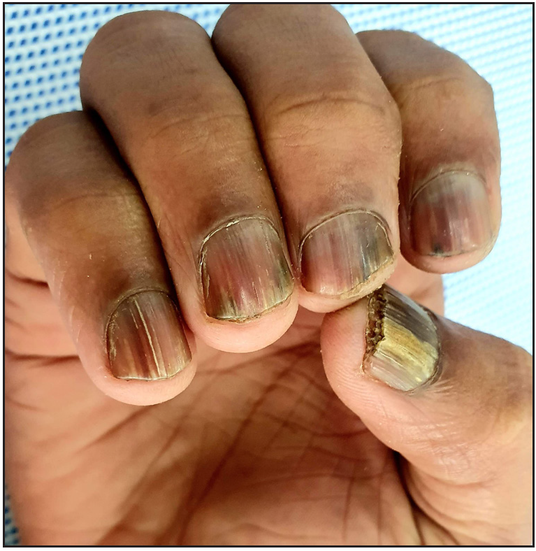
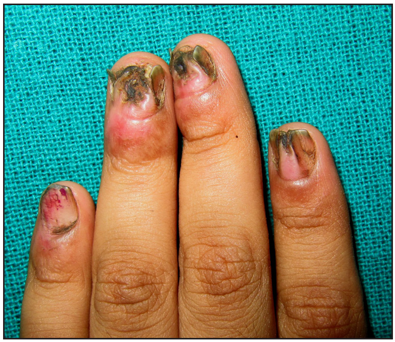
- Nail lichen planus presenting as melanonychia, onychorrhexis, and longitudinal ridging involving right fingernails.
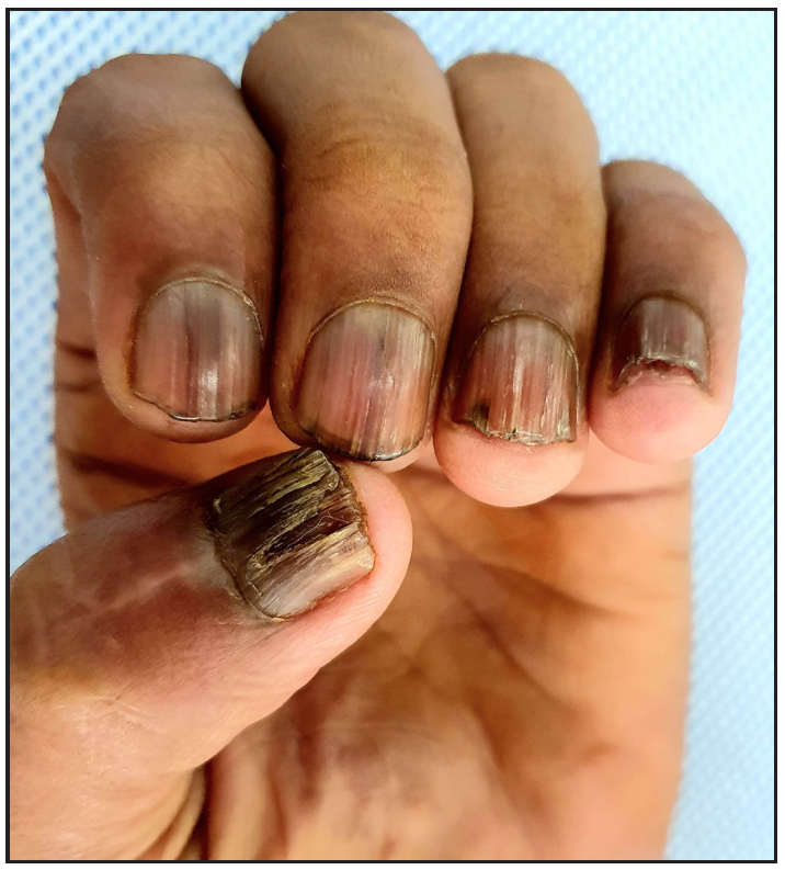
- Nail lichen planus presenting as melanonychia, onychorrhexis, and longitudinal ridging involving left fingernails.
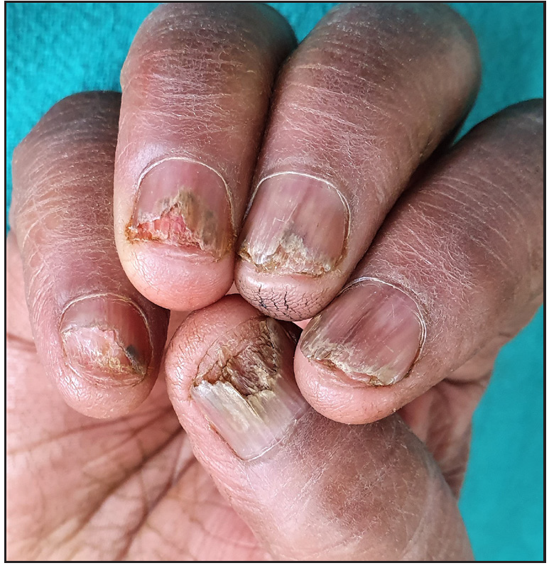
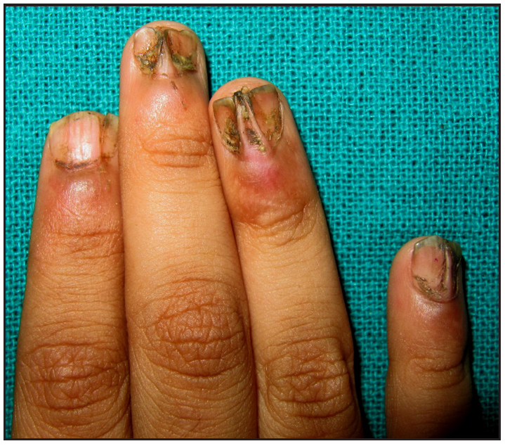
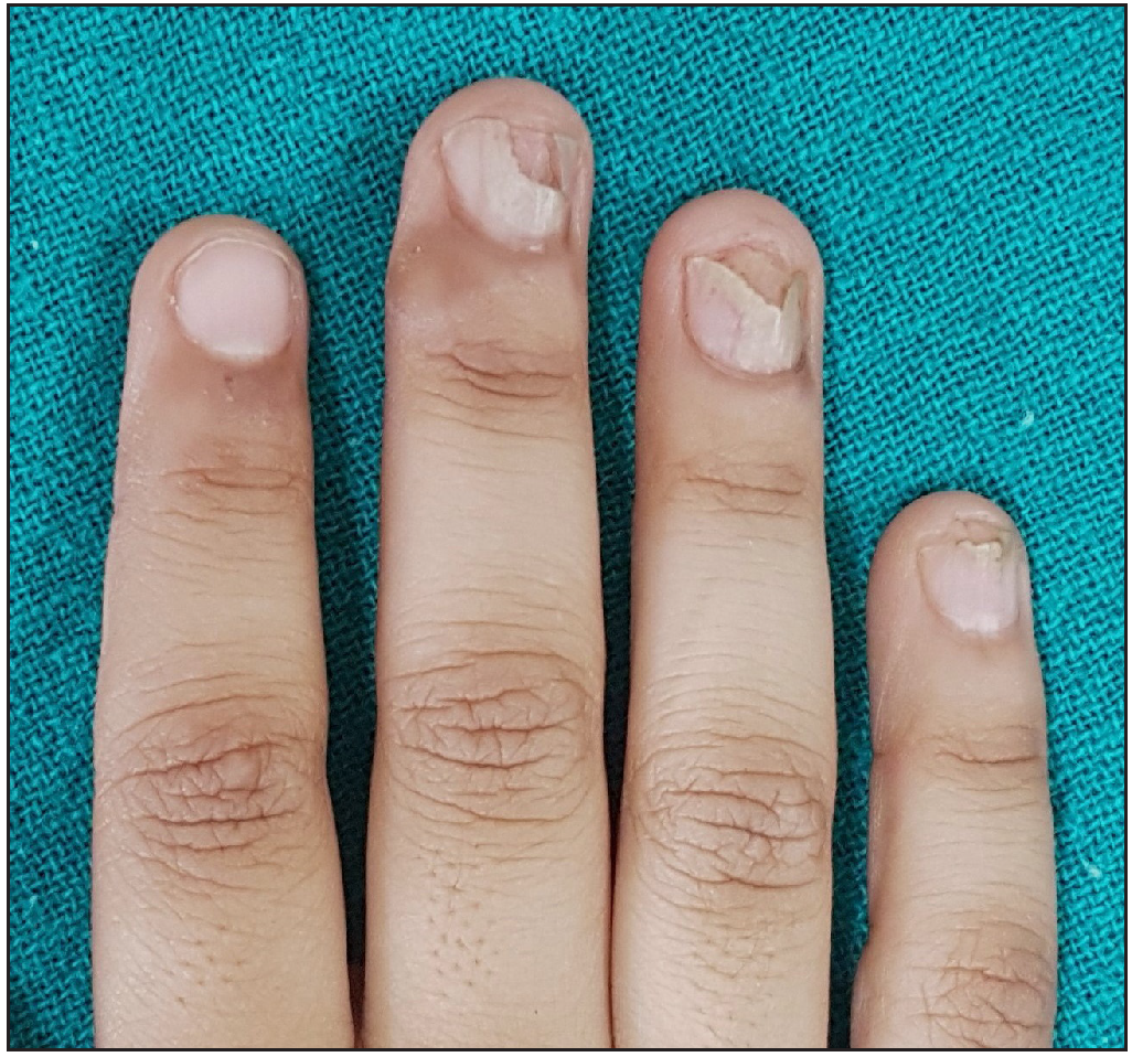
- Nail lichen planus presenting as onycholysis, subungual hyperkeratosis, splinter hemorrhage, and nail bed erythema with longitudinal ridging and distal splitting involving right fingernails.
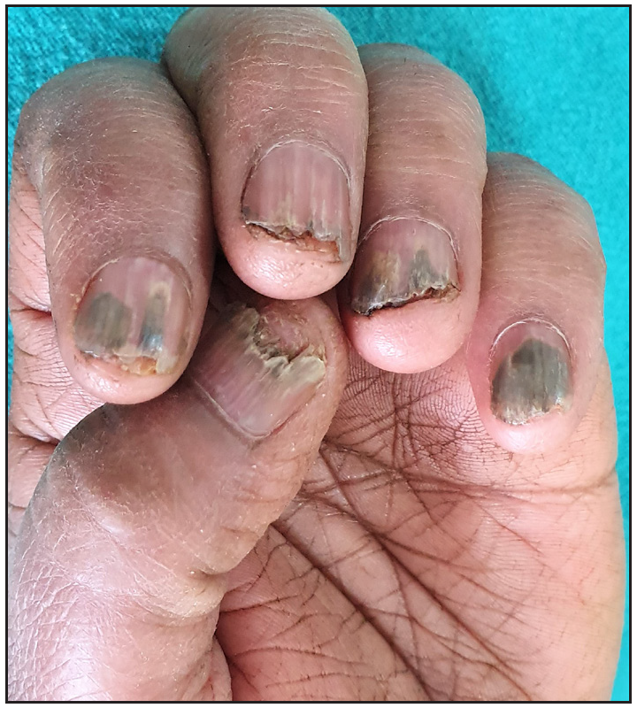
- Nail lichen planus presenting as onycholysis, subungual hyperkeratosis, splinter hemorrhage, and nail bed erythema with longitudinal ridging and distal splitting involving left fingernails.
- Pterygium involving left index, middle, and ring fingernails.
- Pterygium involving right middle, index, and little fingernails.
| Site of involvement and clinical presentation | |
|---|---|
| Nail Matrix | |
| • Nail plate thinning | 12 (80%) |
| • Onychorrhexis | 11 (73.3%) |
| • Longitudinal ridging | 10 (66.7%) |
| • Longitudinal splitting | 10 (66.7%) |
| • Melanonychia | 8 (53.3%) |
| • Mottled/ Red/ distorted lunula | 9 (60%) |
| • Onychoschizia | 8 (53.3%) |
| • Trachyonychia | 5 (33.3%) |
| • Dorsal pterygium | 5 (33.3%) |
| • Anonychia | 4 (26.7%) |
| • Beau‘s line | 1 (6.7%) |
| Nail Bed | |
| • Onycholysis | 11 (73.3%) |
| • Subungual hyperkeratosis | 6 (40%) |
| • Splinter hemorrhage | 2 (13.3%) |
| • Nail bed erythema | 10 (66.7%) |
| • Pup-tent appearance of nail plate | 5 (33.3%) |
Severity |
Clinical features |
Number of patients in each group |
|---|---|---|
| Mild | Nail thinning, longitudinal ridging, distal splitting of <3 mm in length, onycholysis <25%, and no nail bed hyperkeratosis | 2 (13.3%) |
| Moderate | Partial fissuring, longitudinal grooves, distal splitting of 3 to 5mm in length, onycholysis between 25% and 50%, mottled erythema of the lunula, and subungual hyperkeratosis | 6 (40%) |
| Severe | Complete fissuring, deep grooves, splitting of >5mm in length, onycholysis >50%, diffuse erythema of the lunula, pterygium, and anonychia | 7 (46.7%) |
Treatment: Patients were counseled to follow general precautions, like avoidance of trauma and liberal use of emollients. Two patients with mild disease received clobetasol propionate 0.05% ointment and topical tacrolimus 0.1% ointment, each for once daily application not exceeding 3 months. Eleven patients with moderate (n = 6) and severe (n = 5) disease received intramuscular triamcinolone acetonide (TA) (40 mg for adults and 0.5-1 mg/kg for children) injections every 4 weeks with topical tacrolimus (0.1%) ointment. Two patients (an 11-year-old boy and a 54-years man with multiple co-morbidities) with extensive dorsal pterygium and anonychia involving multiple digits, were counseled regarding the nature of the disease and prognosis, and treated with topical emollients and tacrolimus ointment. Patients were followed up every 4-6 weeks for clinical evaluation and photographic documentation. Intramatricial injection of TA (5mg/ml) at monthly intervals was given only for the nails that showed evidence of disease even after six intramuscular injections and was delivered in 17 nails of 4 patients.
The treatment outcome was assessed as no improvement/worsening, minimal improvement (25% improvement in the nail plate abnormality), mild improvement (26% to 50%), moderate improvement (51% to 75%) and significant improvement (76% to 99%) based on the photographic comparison and investigator‘s global assessment. Clinical cure was defined as 100% disease resolution or a normal nail appearance.
Statistics: All the demographic and clinical data analysis was performed using SPSS version 20 (SPSS, Inc., Chicago, IL). Frequencies and percentages were used for categorical variables.
Results
Demographic profile: The demographic profile and baseline disease characteristics have been detailed in Table 3. Of the 15 patients, 7 were females, and 8 were males, the youngest being 3.5 years. Childhood nail LP (≤ 12 years) was diagnosed in 4 (26.7%) patients:3 boys and 1 girl child. Co-morbidities were present in 6 (40%) patients; alopecia areata (concurrent in 1 and past history in 2) in 3 (20%), while metabolic syndrome with hypothyroidism, dyslipidemia, and history of myocardial infection was present in one patient each.
| Features | Present study | Tosti et al1, 1993 | Tosti et al9, 2001 | Piraccini et al2, 2010 | Goettman et al3, 2012 | Chiheb et al4, 2015 | Wechsuruk et al5, 2021 |
|---|---|---|---|---|---|---|---|
| Epidemiological | |||||||
| Place | India | Italy | Italy | Italy | France | Morocco | Thailand |
| Study population | Adults and children | Adults and children | Children | Adults and children | Adults and children | Adults and children | Adults |
| Number of patients | 15 | 24 | 15 | 75 | 67 | 20 | 14 |
| Age (in years) | 28.6 ± 19.0 years (3.5-56 years) | 49.5 ± 14.7 years (11 to 68 years | 9.6 ± 2.2 years (6-12 years) | 54 years (7 to 80 years) | 47 years ± 20.2 (6 to 78 years) | 35 years (9 to 56 years) | 53.3 ± 13.9 years |
| Sex ratio (Male: Female) | 0.8 (M = 7, F = 8) | 1.2:1 (M = 13, F = 11) | 2.75:1 (M = 11, F = 4) | 1.1:1 (M = 39, F = 36) | 1.8:1 (M = 43, F = 24) | 1:1 (M = 10, F = 10) | 0.6:1 (M = 6; F = 9) |
| Childhood NLP (0-12 years) | 4 (30.7%) | 1 (4.1%) | 15 (100%) | 4 (5.3%) | 7 (10.4%) | 8 (40%) | - |
| Clinical | |||||||
| Duration of disease (Mean ± SD) | 2.8 ± 2.6 years (6 months – 9 years) | 2 ± 5 years (0 – 25 years) | 2.5 ± 2.9 years (2 months to 9 years) | 1.3 years (6 months -51 months) | 3.2 years ± 64.4 (2m–30y) 20 (30.0%) | 4 years (2 months to 10 years | 3.5 years (2 months to 30 years) |
| Number of nails involved | |||||||
| 1. Total | 16.4 ± 4.6 (8-20 nails) | 12.3 ± 7.2 | 15.3 ± 5.5 | 12.7 ± 6.4 (1- 20 nails) | 9.3 ± 5.6 (1–20 nails) | - | - |
| 2. Fingernail | 8.7 ± 3.0 (3-10 nails) | 8 ± 3.3 | 9.5 ± 1.8 | 8.7 ± 2.7 (1-10 nails) | 7 ± 4 (1–10 nails) | - | - |
| 3. Toenail | 8.7 ± 2.8 (2-10 nails) | 4.3 ± 4.9 | 5.7 ± 4.8 | 4.3 ± 4.8 (2-10) nails | 3 ± 3.6 (0–10 nails) | 11 | - |
| Number of patients with isolated fingernail/toenail/twenty nail involvement | |||||||
| 1. Isolated fingernail involvement | 2 (13.3%) | 12 (50%) | 3 (20%) | 41 (54.7%) | 8 (11.9%) | - | - |
| 2. Isolated toenail involvement | 1 (6.6%) | - | - | - | 4 (6.0%) | - | - |
| 3. Total 20nail involvement | 9 (60%) | 10 (41.6%) | 8 (53.3%) | 29 (38.7%) | 7 (10.44%) | - | - |
| Severity of NLP | |||||||
| 1. Mild | 2 (13.3%) | - | 3 (20%) | - | 7 (10.44%) | - | 0 |
| 2. Moderate | 6 (40%) | - | 3 (20%) | - | 35 (49.25%) | - | 4 (28.6%) |
| 3. Severe | 7 (46%) | - | 9 (60%) | - | 27 (40.29%) | - | 10 (71.4%) |
| Characteristics | |||||||
| Nail matrix involvement | 13 (100%) | 22 (91.6%) | 15 (100%) | 66 (88%) | 61 (91%) | 17 (85%) | 14 (100%) |
| Melanonychia | 8 (53.3%) | 1 (4.1%) | 9 (60%) | - | - | - | 12 (85.7%) |
| Longitudinal ridging and splitting | 10 (66.7%) | 17 (70.8%) | 1 (6.7%) | - | 60 (89.5%) | 8 (40%) | 9 (64.3%) |
| Red lunula | 9 (60%) | 6 (25%) | 2 (13.3%) | - | - | - | - |
| Trachyonychia | 5 (33.3%) | - | - | - | 21 (31.3%) | - | 7 (50%) |
| Anonychia | 4 (26.7%) | 4 (16.6%) | 4 (26.6%) | - | 15 (22.4%) | 4 (20%) | 7 (50%) |
| Pterygium | 5 (33.3%) | 12 (50%) | 5 (33.3%) | - | 24 (35.8%) | 2 (10%) | 4 (28.6%) |
| Nail plate thinning | 12 (80%) | 14 (58.3%) | 6 (40%) | - | 12 (17.9%) | 3 (15%) | 6 (42.9%) |
| Nail bed involvement | 11 (73.3%) | 10 (41.6%) | 6 (40%) | - | 29 (43.3%) | 11 (55%) | 8 (57.1%) |
| Onycholysis | 11 (73.3%) | 9 (37.5%) | - | 4 (5.3%) | 29 (43.3%) | 7 (35%) | 5 (35.7%) |
| Subungual hyperkeratosis | 6 (40%) | 1 (4.1%) | - | - | 16 (23.8%) | 4 (20%) | 4 (28.6%) |
| Splinter hemorrhage | 2 (13.3%) | - | - | 27 (36%) | - | - | 2 (14.3%) |
| Treatment | |||||||
| Systemic corticosteroids | 10 (66.7%) | 17 (70.8%) | 10 (66.7%) | 67 (89.3%) | 46 (68.6%) | 10 (50%) | 9 (64.2%) |
| Oral prednisone | - | 15 (62.5%) | - | - | 10 (14.9%) | - | |
| Intramuscular triamcinolone acetonide | 10 (66.7%) | 2 (8.3%) | 10 (66.7%) | 67 (89.3%) | 36 (53.7%) | 10 (50%) | - |
| Intralesional triamcinolone acetonide | 3 (20.0%) | 4 (16.6%) | - | 8 (10.6%) | 21 (31.3%) | - | - |
| No of sessions | 3-9 | 2-4 | 3-6 | 4-7 | 6 | 3-6 | |
| Improvement | 10 (66.6%) | 17 (67%) | 9 (90%) | 51 (68%) | 56 (83.6%) | 10 (100%) | 9 (100%) |
| Duration of follow-up | 2-5 years | 1 to 4 years | 2 years | - | - | 2 years | 5 years |
| No response | 3 (23.0%) | - | 2 (20%) | 9 (33.3%) | 11 (16.4%) | - | - |
| Relapse | 2 (20%) | 8 (33%) | 2 years | 11 (44.7%) | 37 (55.2%) | - | - |
| Relapse free interval | 1 – 1.5 years | 1 to 4 years | - | 5 years | 18 ± 2.85 months | - | - |
Clinical Presentation: Clinical features of the nail matrix and nail bed involvement were recorded as shown in Table 1, and severity was graded as shown in Table 2.
Among patients with severe nail disease (7 cases), 5 (33.3%) had pterygium involving an average of 3.4 nails. The presence of pterygium correlated with the disease duration in years (r = 0.87). The average disease duration was higher in patients with pterygium formation than those without it (2.2 years vs. 1.3 years).
Treatment outcome:
Two patients (13.3%) with extensive pterygium and anonychia treated with topical tacrolimus and emollients showed no improvement. Two (13.3%) patients with severe disease treated with monthly intramuscular TA injections showed minimal improvement at 2 months and 5 months, respectively. Mild improvement was seen in one patient. Moderate to good improvement was observed in 10 (66.6%) patients after an average of 6.1 ± 2.4 (3-9) treatment sessions (including up to 6 sessions of intramuscular TA injections and up to 3 sessions of intramatricial TA injections, depending upon number and severity of nail involvement) [Figures 4a-4d], and of these, 2/10 (20%) developed disease recurrence in a few nails after an interval of 1 and 1.5 years, respectively. These nails were treated successfully with intramatricial TA injections. Two patients achieved clinical cures that persisted beyond 2 years of follow-up.
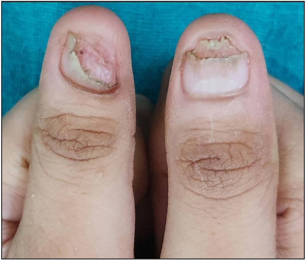
- Nail lichen planus of the thumbnails before treatment with intramuscular triamcinolone acetonide, and fingernails.
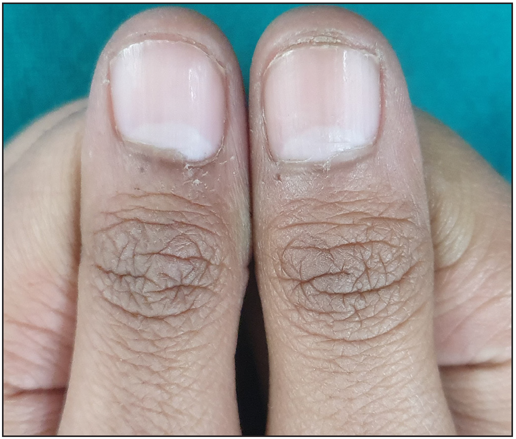
- Nail lichen planus of the thumbnails 6 months after treatment with intramuscular triamcinolone acetonide.
- Nail lichen planus of the fingernails before treatment.
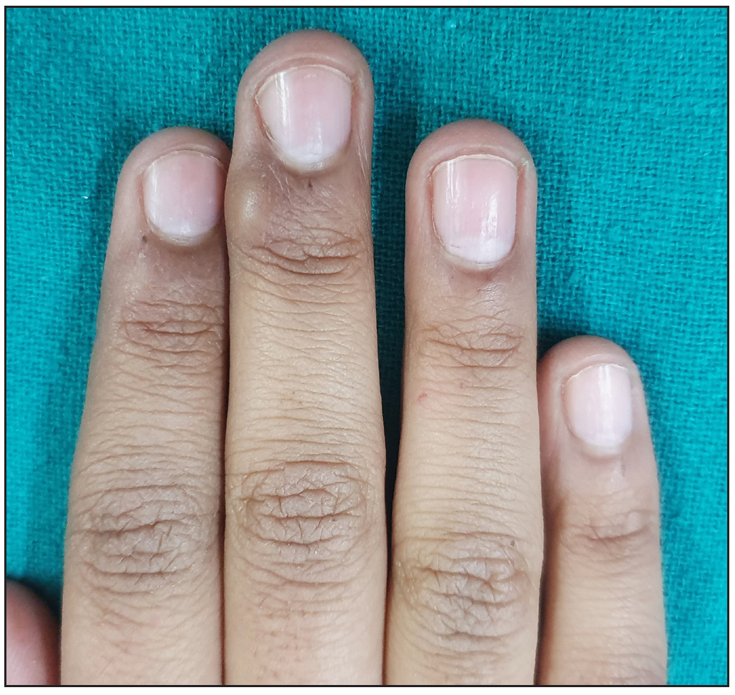
- Nail lichen planus of the fingernails 6 months after treatment.
Discussion
Isolated nail lichen planus (NLP) is rare; its incidence and prevalence are unknown. NLP occurs with increased frequency in adults (especially in the 5th-6th decade) with a male preponderance.2 Fingernails are reportedly more commonly involved than toenails.3 Early diagnosis and treatment are necessary to prevent permanent scarring, nail disfigurement, and anonychia, which leads to severe psychological and functional impairment. Demographic and clinical characteristics and management outcomes of the present case series and their comparison with the previously published series have been summarised in Table 3.
The average age at presentation in this case series (28.6 ± 19.0 years) was significantly lower than most previous studies (5th to 6th decade).1–3,5 However, similar age has been reported from Morocco and India.4,8 Increasing disease awareness among patients, early clinical diagnosis, and geographical variations may account for this deviation. The proportion of childhood NLP was also higher, except in the study by Tosti et al. on childhood NLP and Chiheb et al. from Morocco.4,9 No gender predilection was noted in this series similar to many studies.1-4 However, some authors have reported a male preponderance.9,3 Extreme visibility of nails probably invites similar attention from both genders. The average number of involved nails is higher than previously reported cases, implying that Indians may be predisposed to extensive disease.1-5 Toe nails and 20 nail involvement were also higher in this case series. This could be due to nail trauma from barefoot walking, a typical Indian practice. Similar to the previous studies, most patients had moderate to severe disease, probably resulting from delayed diagnosis.
Nail matrix involvement was seen in all 15 patients; as reported by previous studies, nail plate thinning was the commonest feature, which was seen in 12 (80%) patients, followed by onychorrhexis which was seen in 11 (73.3%) patients. This is in contrast to previous studies where melanonychia, and longitudinal ridging and splitting were the commonest presentations.5,9 Nail bed involvement (onycholysis) occurred in 11(73.3%) patients, which is significantly higher than previously reported. This also points to severe nail disease in our series with a higher frequency of nail plate and bed involvement. Like other studies, dorsal pterygium was seen in 33.3% of the patients.3,5,9 While describing the most severe signs in NLP, Ceccarelli et al. concluded that anonychia was associated with severe nail plate thinning, retraction of the nail plate and nail bed, loss of proximal nail fold limits, and onychoatrophy. However, dorsal pterygium was associated with loss of proximal nail fold limits, onychoatrophy, and distal splitting greater than 50%.10 Similar observations were made by us. [Figures 3 and 5].
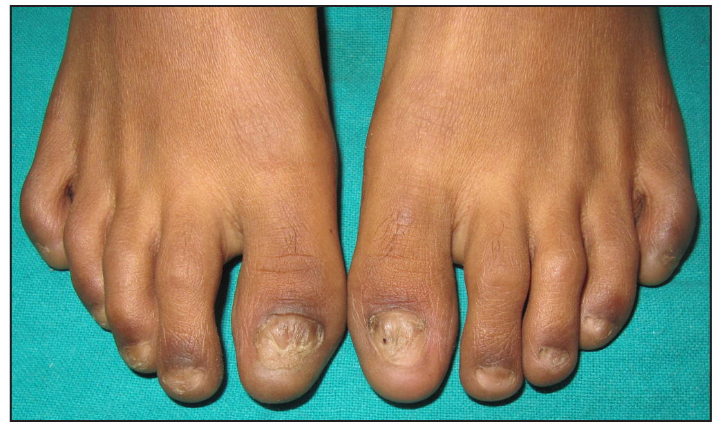
- Anonychia associated with nail bed retraction involving all toenails.
Treatment modalities for NLP are limited, and large-scale studies with long-term follow-up are scarce. Therapeutic options include topical preparations (0.05% clobetasol propionate, 0.1% tacrolimus, 0.1% tazarotene), intramatricial triamcinolone acetonide, and systemic steroids (oral and intramuscular). Topical treatment has been proven to be less efficacious due to limited absorption and there is risk of adverse effects from prolonged use.6 First-line therapy includes intralesional or intramuscular steroids. Oral steroids, cyclosporine, and retinoids may be considered in resistant cases. Tosti et al. in 1993 reported oral, intramuscular, and intralesional steroids to be effective treatment options with relapse in 8/24 (33%) patients after a disease-free interval of 1-4 years.1 Piraccini et al. and Goetterman et al. found intramuscular and intralesional triamcinolone acetonide to be efficacious with relapse observed in a few nails after a disease-free period of 2-5 years [Table 3].2,3 A recent study by Wechsuruk et al. concluded that systemic steroids (oral and intramuscular) can provide better outcomes than topical treatment.5 However, an expert consensus by Iorizzo et al. has cautioned about the use of systemic steroids due to the risk of adverse effects on long-term treatment. Recently, JAK inhibitors have been effective in nail lichen planus. In isolated case reports, both tofacitinib and baricitinib have been used successfully for nail lichen planus.11,12
With a comparable efficacy between intralesional and intramuscular steroids, we suggest intralesional steroids as a first-line approach for mild to moderate NLP involving few nails. In contrast, IM steroids can be reserved for severe, extensive, or recalcitrant cases. None of the patients developed side effects of systemic corticosteroid administration.
Limitation- The major limitations of this case series are the small patient number and retrospective data analysis.
Conclusion
Isolated NLP requires a high degree of suspicion for diagnosis. Clinical diagnosis should be confirmed on onychoscopy and histology. NLP is a potentially scarring disease, and treatment remains ineffective when pterygium sets in, making early diagnosis and prompt treatment mandatory. Intramatricial injections for limited nail involvement and intramuscular steroids for extensive disease are efficacious therapeutic modalities.
Declaration of patient consent
The authors certify that they have obtained all appropriate patient consent.
Financial support and sponsorship
Nil.
Conflicts of interest
There are no conflicts of interest.
Use of Artificial Intelligence (AI)-Assisted Technology for manuscript preparation
The authors confirm that there was no use of Artificial Intelligence (AI)-Assisted Technology for assisting in the writing or editing of the manuscript and no images were manipulated using the AI.
References
- Nail lichen planus: Clinical and pathologic study of twenty-four patients. J Am Acad Dermatol.. 1993;28:724-30.
- [CrossRef] [PubMed] [Google Scholar]
- Nail lichen planus: Response to treatment and long-term follow-up. Eur J Dermatol.. 2010;20:489-96.
- [CrossRef] [PubMed] [Google Scholar]
- Nail lichen planus: Epidemiological, clinical, pathological, therapeutic and prognosis study of 67 cases. J EurAcad Dermatol Venereol.. 2012;26:1304-9.
- [Google Scholar]
- Caractéristiquescliniques et évolutives du lichen plan unguéal : étude descriptive de 20 patients [Clinical characteristics of nail lichen planus and follow-up: A descriptive study of 20 patients] Ann Dermatol Venereol.. 2015;142:21-5.
- [CrossRef] [PubMed] [Google Scholar]
- Clinical features and treatment outcomes of nail lichen planus: A retrospective study. JAAD Case Rep.. 2021;22;17:43-8.
- [PubMed] [Google Scholar]
- Isolated nail lichen planus: An expert consensus on treatment of the classical form. J Am Acad Dermatol.. 2020;83:1717-23.
- [CrossRef] [PubMed] [Google Scholar]
- Longitudinal nail biopsy in evaluation of acquired nail dystrophies. J Am Acad Dermatol.. 1986;14:803-9.
- [CrossRef] [PubMed] [Google Scholar]
- Histopathological evaluation of nail lichen planus: A cross-sectional study. J CutanPathol.. 2021;48:11-7.
- [Google Scholar]
- Nail lichen planus in children: Clinical features, response to treatment, and long-term follow-up. Arch Dermatol.. 2001;137:1027-32.
- [PubMed] [Google Scholar]
- Description of the most severe signs in nail lichen planus: A strategy to contribute to the diagnosis of the severe stage. Int J Dermatol.. 2022;61:1124-30.
- [CrossRef] [PubMed] [Google Scholar]
- Tofacitinib as treatment for nail lichen planus associated with alopecia universalis. JAMA Dermatol.. 2021;157:352-3.
- [CrossRef] [PubMed] [Google Scholar]
- Treatment of severe nail lichen planus with baricitinib. JAMA Dermatol.. 2022;158:107-8.
- [CrossRef] [PubMed] [Google Scholar]


