Generic selectors
Exact matches only
Search in title
Search in content
Post Type Selectors
Search in posts
Search in pages
Filter by Categories
15th National Conference of the IAOMFP, Chennai, 2006
Abstract
Abstracts from current literature
Acne in India: Guidelines for management - IAA Consensus Document
Addendum
Announcement
Art & Psychiatry
Article
Articles
Association Activities
Association Notes
Award Article
Book Review
Brief Report
Case Analysis
Case Letter
Case Letters
Case Notes
Case Report
Case Reports
Clinical and Laboratory Investigations
Clinical Article
Clinical Studies
Clinical Study
Commentary
Conference Oration
Conference Summary
Continuing Medical Education
Correspondence
Corrigendum
Cosmetic Dermatology
Cosmetology
Current Best Evidence
Current Issue
Current View
Derma Quest
Dermato Surgery
Dermatopathology
Dermatosurgery Specials
Dispensing Pearl
Do you know?
Drug Dialogues
e-IJDVL
Editor Speaks
Editorial
Editorial Remarks
Editorial Report
Editorial Report - 2007
Editorial report for 2004-2005
Errata
Erratum
Focus
Fourth All India Conference Programme
From Our Book Shelf
From the Desk of Chief Editor
General
Get Set for Net
Get set for the net
Guest Article
Guest Editorial
History
How I Manage?
IADVL Announcement
IADVL Announcements
IJDVL Awards
IJDVL AWARDS 2015
IJDVL Awards 2018
IJDVL Awards 2019
IJDVL Awards 2020
IJDVL International Awards 2018
Images in Clinical Practice
In Memorium
Inaugural Address
Index
Knowledge From World Contemporaries
Leprosy Section
Letter in Response to Previous Publication
Letter to Editor
Letter to the Editor
Letter to the Editor - Case Letter
Letter to the Editor - Letter in Response to Published Article
LETTER TO THE EDITOR - LETTERS IN RESPONSE TO PUBLISHED ARTICLES
Letter to the Editor - Observation Letter
Letter to the Editor - Study Letter
Letter to the Editor - Therapy Letter
Letter to the Editor: Articles in Response to Previously Published Articles
Letters in Response to Previous Publication
Letters to the Editor
Letters to the Editor - Letter in Response to Previously Published Articles
Letters to the Editor: Case Letters
Letters to the Editor: Letters in Response to Previously Published Articles
Medicolegal Window
Messages
Miscellaneous Letter
Musings
Net Case
Net case report
Net Image
Net Images
Net Letter
Net Quiz
Net Study
New Preparations
News
News & Views
Obituary
Observation Letter
Observation Letters
Oration
Original Article
ORIGINAL CONTRIBUTION
Original Contributions
Pattern of Skin Diseases
Pearls
Pediatric Dermatology
Pediatric Rounds
Perspective
Presedential Address
Presidential Address
Presidents Remarks
Quiz
Recommendations
Regret
Report
Report of chief editor
Report of Hon : Treasurer IADVL
Report of Hon. General Secretary IADVL
Research Methdology
Research Methodology
Resident page
Resident's Page
Resident’s Page
Residents' Corner
Residents' Corner
Residents' Page
Retraction
Review
Review Article
Review Articles
Reviewers 2022
Revision Corner
Self Assessment Programme
SEMINAR
Seminar: Chronic Arsenicosis in India
Seminar: HIV Infection
Short Communication
Short Communications
Short Report
Snippets
Special Article
Specialty Interface
Studies
Study Letter
Study Letters
Supplement-Photoprotection
Supplement-Psoriasis
Symposium - Contact Dermatitis
Symposium - Lasers
Symposium - Pediatric Dermatoses
Symposium - Psoriasis
Symposium - Vesicobullous Disorders
SYMPOSIUM - VITILIGO
Symposium Aesthetic Surgery
Symposium Dermatopathology
Symposium-Hair Disorders
Symposium-Nails Part I
Symposium-Nails-Part II
Systematic Review and Meta-Analysis
Systematic Reviews and Meta-analyses
Systematic Reviews and Meta-analysis
Tables
Technology
Therapeutic Guideline-IADVL
Therapeutic Guidelines
Therapeutic Guidelines - IADVL
Therapeutics
Therapy
Therapy Letter
Therapy Letters
View Point
Viewpoint
What’s new in Dermatology
View/Download PDF

Translate this page into:

Observation Letter
87 (
3
); 399-401
doi:
10.25259/IJDVL_399_19
pmid:
33666036

Clinical, dermoscopic and histopathological features of solitary nevus lipomatosus cutaneous superficialis

Department of Specialized, Experimental and Diagnostic Medicine, University of Bologna, Bologna, Italy
Corresponding author: Dr. Carlotta Baraldi, Via Massarenti 1, 40138 Bologna, Italy. carlotta.baraldi3@unibo.it
Licence
This is an open-access article distributed under the terms of the Creative Commons Attribution-Non Commercial-Share Alike 4.0 License, which allows others to remix, tweak, and build upon the work non-commercially, as long as the author is credited and the new creations are licensed under the identical terms.

How to cite this article: Baraldi C, Barisani A, Fanti PA, Patrizi A. Clinical, dermoscopic and histopathological features of solitary nevus lipomatosus cutaneous superficialis. Indian J Dermatol Venereol Leprol 2021;87:399-401.

Sir,

Nevus lipomatosus cutaneous superficialis is a rare congenital or acquired hamartoma, characterized by the presence of mature adipose tissue, localized within the dermis without continuity with the hypodermal adipose tissue. Two clinical variants of nevus lipomatosus cutaneous superficialis have been described: The multiple (the classic Hoffmann-Zurhelle form) and the solitary type. The classic type usually occurs at birth or during the first three decades of life and consists of multiple, soft skin-colored or yellowish papules or nodules, covered with smooth or wrinkled skin, sometimes with a cerebriform appearance.1 The lesions are mainly located in the lower back, buttocks and thighs. The classic form is usually unilateral and may have a linear or zosteriform configuration or can be grouped into a plaque. Although the classic type of nevus lipomatosus cutaneous superficialis is not typically associated with other malformations, there are reports of its coexistence with caféau-lait spots, solitary angiokeratoma of Fordyce and cherry angioma. On the other hand, the solitary form is expressed as a skin-colored, single nodule or papule without predilection for a specific age group or a typical location.2,3

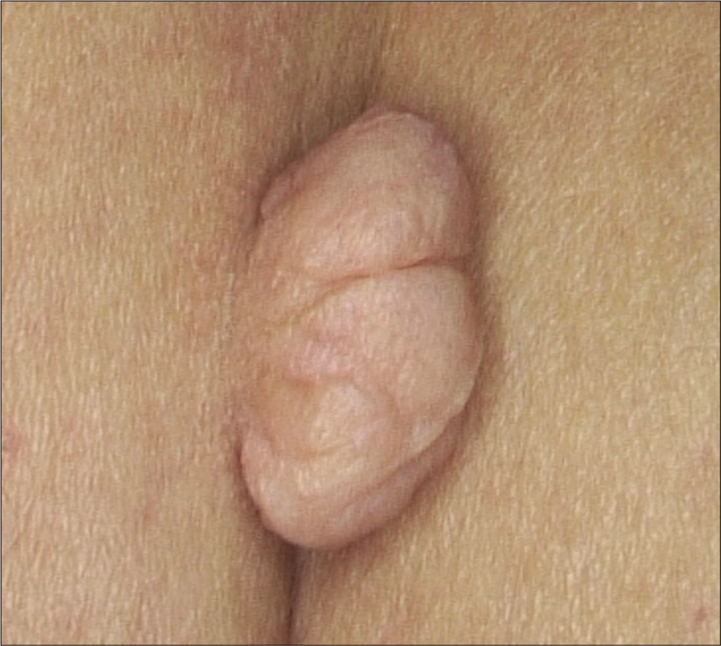
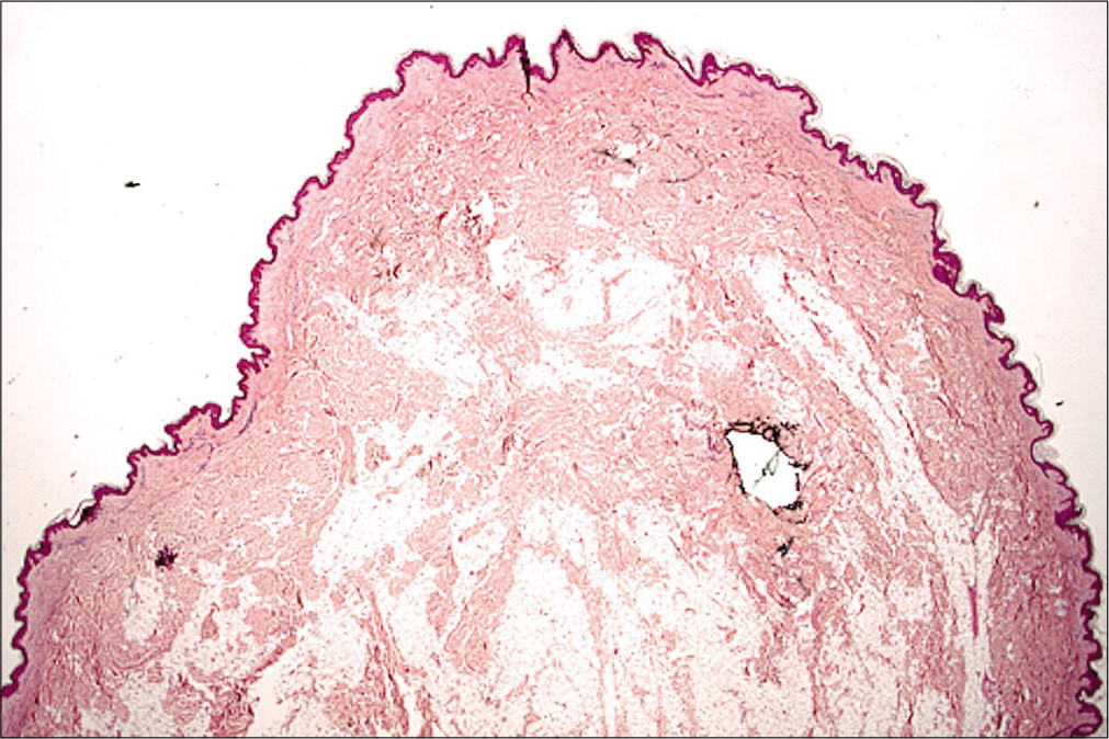
We report here a series of 34 cases of solitary nevus lipomatosus cutaneous superficialis to assess the epidemiological, clinical, dermoscopic and pathological features. We identified 34 Caucasian patients with histopathologically confirmed solitary nevus lipomatosus cutaneous superficialis (dermatopathology service, University of Bologna) between January 2005 and December 2017. There were 22 (64.7%) women and 12 (35.3%) men, with a female to male ratio of 1.83:1. The average age at diagnosis was 51.8 years (range 3282 years). The legs were mostly affected across all genders (n = 10, 29.4%) followed by the trunk (n = 8, 23.5%), the buttocks (n = 7, 20.5%), the groin, the shoulders and the arms. We observed two different aspects of the solitary type. The first one was a skin-colored solitary pedunculated papule or nodule with a smooth skin-colored surface (n =19, 55.8%) [Figure 1a] and the second type was a skin-colored single, pedunculated nodule with a cerebriform, pinkish surface (n =15, 44.8%) [Figure 1b]. The size ranged from 0.3 mm to 2.5 mm. No associated comedones or dilated follicular ostia were observed. There was no association with any other skin lesion or malformation [Table 1]. All the lesions were examined by dermoscopy at different magnification. The main dermoscopic features of nevus lipomatosus cutaneous superficialis were pinkish background and linear whitish structures. At high magnification, the presence of linear-coiled and/or linear-loop-like vessels was evident. Dermoscopy of smooth-surfaced nevus lipomatosus cutaneous superficialis showed linear whitish structures with linear-coiled vessels and dermoscopy of nevus lipomatosus cutaneous superficialis with cerebriform surface showed brownish pigmented areas with irregularly distributed linear-coiled vessels and linear-loop-like vessels [Figures 2a and b]. Histopathology showed polypoid or papillomatous proliferations, hyperkeratosis of the epidermis and mature adipocytes embedded between the collagen bundles. A proliferation of dilated blood vessels with venular features, lined by regular single linear endothelium was evident in the upper dermis in all the nevus lipomatosus cutaneous superficialis. In the case of nevus lipomatosus cutaneous superficialis with a clinically smooth surface, adipocytes were present between the collagen bundles only in the reticular and adipocytes are present both in reticular and papillary dermis in the cerebriform type [Figures 3a and b].

Figure 1a:
“Smooth surface” nevus lipomatosus cutaneous superficialis clinical presentation: skin-colored pedunculated nodule
Figure 1b:
“Cerebriform surface” nevus lipomatosus cutaneous superficialis clinical presentation: pedunculated pinkish nodule with cerebriform surface
Figure 2a:
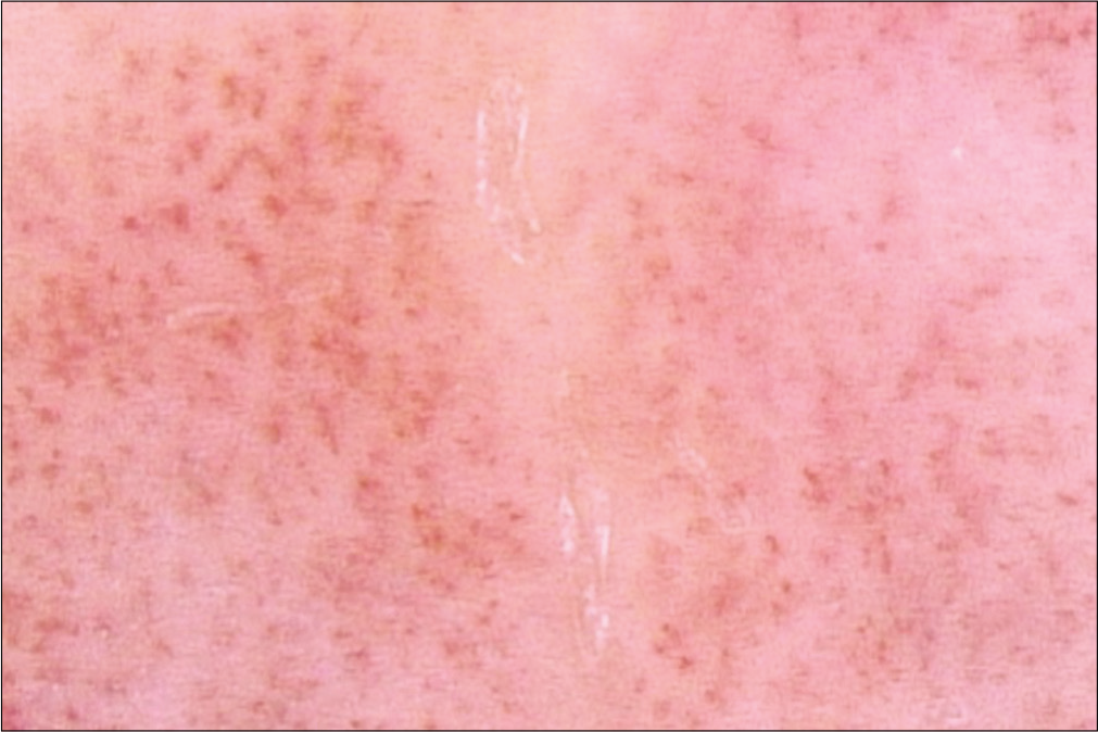
“Smooth surface” nevus lipomatosus cutaneous superficialis’ dermoscopic features: linear-coiled vessels with whitish structures
Figure 2b:
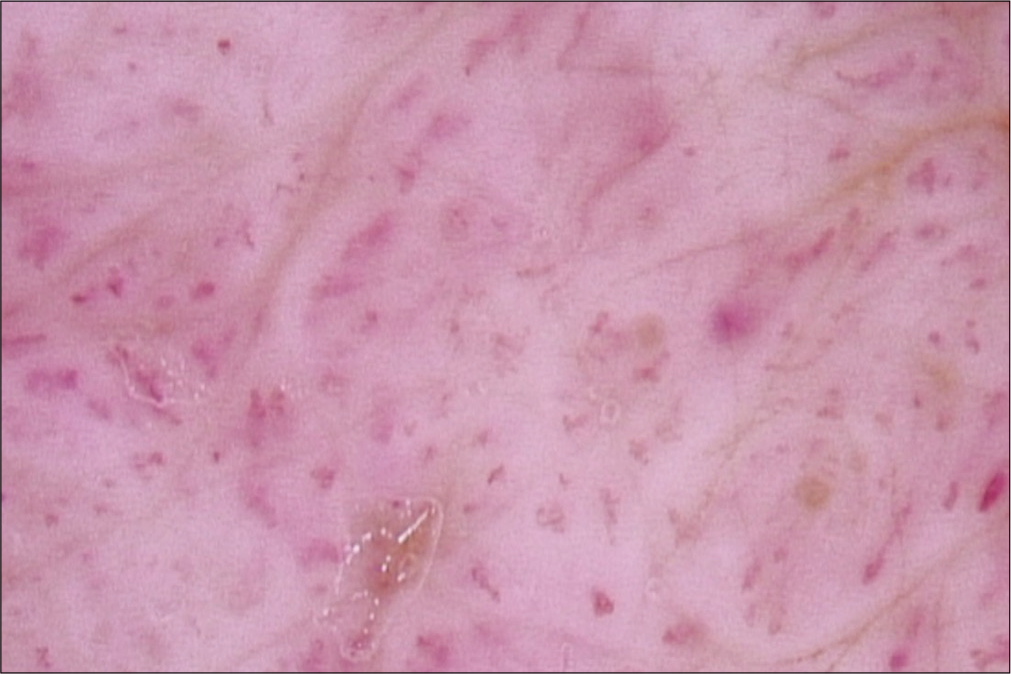
“Cerebriform” nevus lipomatosus cutaneous superficialis’ dermoscopic features: irregular distributed linear-coiled and linear-loop-like vessels and brownish pigmented areas
Figure 3a:
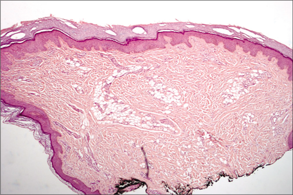
Histological features of “smooth surface” nevus lipomatosus cutaneous superficialis (H and E, ×400)
Figure 3b:
Histological features of “Cerebriform surface” nevus lipomatosus cutaneous superficialis (H and E,×200)
Table 1: Clinical features of nevus lipomatosus cutaneous superficialis
Clinical features Numbers of cases
Solitary type of nevus lipomatosus cutaneous superficialis 34
Multiple types of nevus lipomatosus cutaneous superficialis 0
Smooth surface 19
Distribution
Legs 7
Trunk 4
Shoulders 2
Buttock 3
Arms 0
Groin 3
Size (cm)
Average 0.9
Range 0.3-2
Cerebriform surface 15
Distribution
Legs 3
Trunk 4
Shoulders 1
Buttock 4
Arms 1
Groin 2
Size (cm)
Average 1.2
Range 2.5

Our report of 34 cases of solitary nevus lipomatosus cutaneous superficialis is the largest series to date and could help to improve our familiarity with this rare hamartoma. In accordance with the published literature, we evidence that solitary form of nevus lipomatosus cutaneous superficialis is a tumor of adults, with a mean age of onset at 51.8 years and a female predominance.4Our study may give additional data about the clinical pattern of this rare hamartoma. We identified two clinical aspects of nevus lipomatosus cutaneous superficialis solitary type: One with a smooth surface and the other with the cerebriform surface, without any correlation with the distribution of the lesions. Indeed, in our study, any type of nevus lipomatosus cutaneous superficialis frequently arises on the legs. Histopathologically, the features of the single type nevus lipomatosus cutaneous superficialis are similar, but adipocytes are present only in the reticular dermis in the smooth-surfaced type; adipocytes are present both in reticular and papillary dermis in the cerebriform type. A proliferation of dilated blood vessels lined by regular single linear endothelium was evident in the upper dermis in all the nevus lipomatosus cutaneous superficialis. This is the first study to describe dermoscopic features of solitary nevus lipomatosus cutaneous superficialis. We found only one study in 2017, in which the authors describe the dermoscopic features of three classical nevus lipomatosus cutaneous superficialis but they did not identify any distinctive dermoscopic features of classical nevus lipomatosus cutaneous superficialis.5 In our opinion, the presence of linear-coiled or linear-loop-like vessels in the papillomatous solitary skin-colored lesion may be a clue for the dermoscopic diagnosis of a single nevus lipomatosus cutaneous superficialis.

Declaration of patient consent

The authors certify that they have obtained all appropriate patient consent.

Financial support and sponsorship

The authors did not receive any financial support or sponsorship.

Conflicts of interest

There are no conflicts of interest.

References

  1. , . UBER einennaevuslipomatodes cutaneous superficialis der linkenglutaalgegend. Arch Dermatol Syphilol. 1921;130:327-33.
    [CrossRef] [Google Scholar]
  2. , , . Naevussuperficialislipomatosus. A clinicopathological report of twenty cases. Br J Dermatol1975;. ;93:121-33.
    [CrossRef] [Google Scholar]
  3. , . Giant nevus lipomatosussuperficialis with multiple folliculo sebaceous cystic hamartomas and dermoid cysts. J Eur Acad Dermatol Venereol. 2005;19:84-6.
    [CrossRef] [PubMed] [Google Scholar]
  4. , , , , , . Colocalization of lipedematous scalp and nevus lipomatosussuperficialis: A case report. J CutanPathol2007;. ;34:342-5.
    [CrossRef] [PubMed] [Google Scholar]
  5. , , , . Dermatoscopic evaluation of three cases of nevus lipomatosus cutaneous superficialis. Indian J Dermatol Venereol Leprol. 2017;83:383-6.
    [CrossRef] [PubMed] [Google Scholar]

Fulltext Views
5,488

PDF downloads
2,095
View/Download PDF
Download Citations
BibTeX
RIS
Show Sections