Generic selectors
Exact matches only
Search in title
Search in content
Post Type Selectors
Search in posts
Search in pages
Filter by Categories
15th National Conference of the IAOMFP, Chennai, 2006
Abstract
Abstracts from current literature
Acne in India: Guidelines for management - IAA Consensus Document
Addendum
Announcement
Art & Psychiatry
Article
Articles
Association Activities
Association Notes
Award Article
Book Review
Brief Report
Case Analysis
Case Letter
Case Letters
Case Notes
Case Report
Case Reports
Clinical and Laboratory Investigations
Clinical Article
Clinical Studies
Clinical Study
Commentary
Conference Oration
Conference Summary
Continuing Medical Education
Correspondence
Corrigendum
Cosmetic Dermatology
Cosmetology
Current Best Evidence
Current Issue
Current View
Derma Quest
Dermato Surgery
Dermatopathology
Dermatosurgery Specials
Dispensing Pearl
Do you know?
Drug Dialogues
e-IJDVL
Editor Speaks
Editorial
Editorial Remarks
Editorial Report
Editorial Report - 2007
Editorial report for 2004-2005
Errata
Erratum
Focus
Fourth All India Conference Programme
From Our Book Shelf
From the Desk of Chief Editor
General
Get Set for Net
Get set for the net
Guest Article
Guest Editorial
History
How I Manage?
IADVL Announcement
IADVL Announcements
IJDVL Awards
IJDVL AWARDS 2015
IJDVL Awards 2018
IJDVL Awards 2019
IJDVL Awards 2020
IJDVL International Awards 2018
Images in Clinical Practice
Images in Dermatology
In Memorium
Inaugural Address
Index
Knowledge From World Contemporaries
Leprosy Section
Letter in Response to Previous Publication
Letter to Editor
Letter to the Editor
Letter to the Editor - Case Letter
Letter to the Editor - Letter in Response to Published Article
LETTER TO THE EDITOR - LETTERS IN RESPONSE TO PUBLISHED ARTICLES
Letter to the Editor - Observation Letter
Letter to the Editor - Study Letter
Letter to the Editor - Therapy Letter
Letter to the Editor: Articles in Response to Previously Published Articles
Letters in Response to Previous Publication
Letters to the Editor
Letters to the Editor - Letter in Response to Previously Published Articles
Letters to the Editor: Case Letters
Letters to the Editor: Letters in Response to Previously Published Articles
Media and news
Medicolegal Window
Messages
Miscellaneous Letter
Musings
Net Case
Net case report
Net Image
Net Images
Net Letter
Net Quiz
Net Study
New Preparations
News
News & Views
Obituary
Observation Letter
Observation Letters
Oration
Original Article
ORIGINAL CONTRIBUTION
Original Contributions
Pattern of Skin Diseases
Pearls
Pediatric Dermatology
Pediatric Rounds
Perspective
Presedential Address
Presidential Address
Presidents Remarks
Quiz
Recommendations
Regret
Report
Report of chief editor
Report of Hon : Treasurer IADVL
Report of Hon. General Secretary IADVL
Research Methdology
Research Methodology
Resident page
Resident's Page
Resident’s Page
Residents' Corner
Residents' Corner
Residents' Page
Retraction
Review
Review Article
Review Articles
Reviewers 2022
Revision Corner
Self Assessment Programme
SEMINAR
Seminar: Chronic Arsenicosis in India
Seminar: HIV Infection
Short Communication
Short Communications
Short Report
Snippets
Special Article
Specialty Interface
Studies
Study Letter
Study Letters
Supplement-Photoprotection
Supplement-Psoriasis
Symposium - Contact Dermatitis
Symposium - Lasers
Symposium - Pediatric Dermatoses
Symposium - Psoriasis
Symposium - Vesicobullous Disorders
SYMPOSIUM - VITILIGO
Symposium Aesthetic Surgery
Symposium Dermatopathology
Symposium-Hair Disorders
Symposium-Nails Part I
Symposium-Nails-Part II
Systematic Review and Meta-Analysis
Systematic Reviews and Meta-analyses
Systematic Reviews and Meta-analysis
Tables
Technology
Therapeutic Guideline-IADVL
Therapeutic Guidelines
Therapeutic Guidelines - IADVL
Therapeutics
Therapy
Therapy Letter
Therapy Letters
View Point
Viewpoint
What’s new in Dermatology
View/Download PDF

Translate this page into:

Observation Letter
89 (
5
); 738-741
doi:
10.25259/IJDVL_933_2021
pmid:
37067117

Clinico-dermoscopic and pathological features of a rare presentation of erythema elevatum diutinum

Department of Dermatology, and Venereology, All India Institute of Medical Sciences (AIIMS), Bhubaneswar, Odisha, India
Department of Pathology, All India Institute of Medical Sciences (AIIMS), Bhubaneswar, Odisha, India.

Corresponding author: Dr. Biswanath Behera, Department of Dermatology and Venereology, All India Institute of Medical Sciences (AIIMS), Bhubaneswar, Odisha, India. biswanathbehera61@gmail.com.

Licence
This is an open-access article distributed under the terms of the Creative Commons Attribution-Non Commercial-Share Alike 4.0 License, which allows others to remix, transform, and build upon the work non-commercially, as long as the author is credited and the new creations are licensed under the identical terms.

How to cite this article: Kowe P, Behera B, Sethy M, Dash S, Sarkar N, Garg S. Clinicodermoscopic and pathological features of a rare presentation of erythema elevatum diutinum. Indian J Dermatol Venereol Leprol 2023;89:738-41

Dear Editor,

A 26-year-old man presented with a six-month history of three asymptomatic, persistent, slowly-enlarging red lesions on the dorsa of both the feet. He denied a personal or family history of leprosy. His medical history was unremarkable. Cutaneous examination revealed three well-to-ill-defined erythematous flat-topped nontender plaques of size 1.5 cm × 1.5 cm–5 cm × 4 cm on the dorsal feet. Areas of desquamation with fine white scales were also noticed [Figures 1a and 1b]. Dermoscopy (DermLite DL4, 10X) under polarised mode showed homogenous pink-white areas, diffuse dotted vessels and shiny white lines. The dotted vessels were prominent along the margins of the skin creases [Figure 2a and 2b]. The differential diagnoses included were borderline tuberculoid leprosy, patch-type granuloma annulare, psoriasis and pagetoid reticulosis. Histopathology revealed an acanthotic epidermis and a well-circumscribed nodular collection of inflammatory infiltrate in the lower dermis. The nodular collection showed neutrophilic leukocytoclastic vasculitis, including fibrin deposition, dermal oedema and a predominant lymphohistiocytic collection. In addition, many vessels showed features of vasculopathy with endothelial proliferation and luminal occlusion [Figure 3a–3d]. A diagnosis of erythema elevatum diutinum (EED) was made and the patient was treated with topical clobetasol 0.05% ointment.
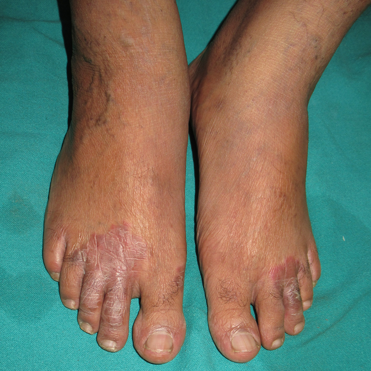
Well-to-ill-defined erythematous plaques on the dorsae of distal feet
Figure 1a:
Well-to-ill-defined erythematous plaques on the dorsae of distal feet
Well to ill-defined erythematous plaque (arrow) on the medial aspect of the right foot
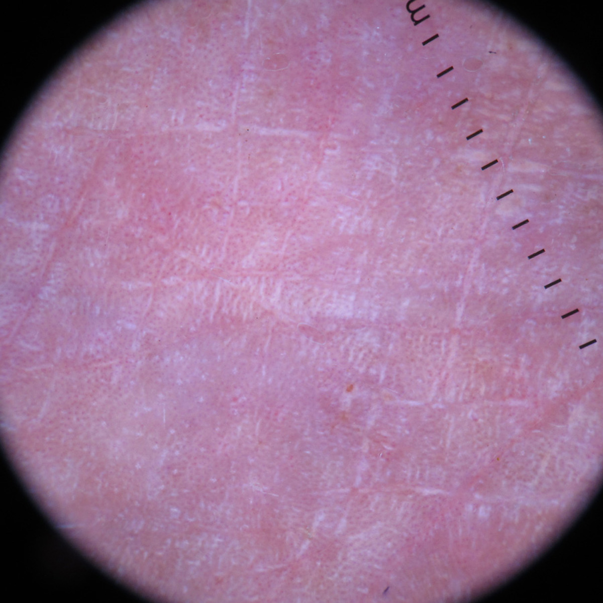
Figure 1b:
Well to ill-defined erythematous plaque (arrow) on the medial aspect of the right foot
Dermoscopy under polarised mode (Dermlite, DL4, ×10 magnification) shows homogenous pink-white areas, diffuse dotted vessels, and shiny white lines
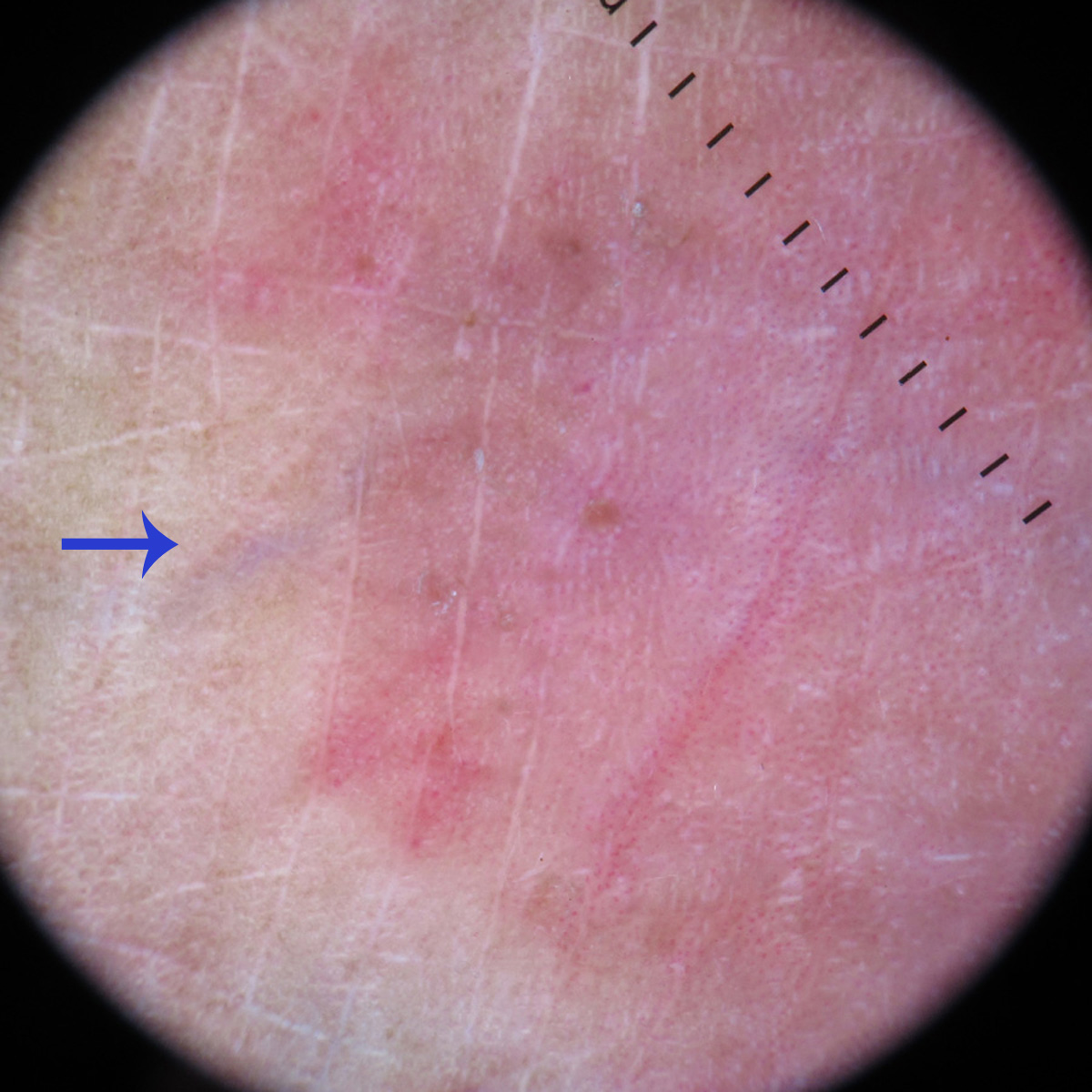
Figure 2a:
Dermoscopy under polarised mode (Dermlite, DL4, ×10 magnification) shows homogenous pink-white areas, diffuse dotted vessels, and shiny white lines
Dermoscopy under polarised mode (Dermlite, DL4, ×10 magnification) shows homogenous pink-white areas, diffuse dotted vessels, and shiny white lines. Arrow points to the normal-looking surrounding skin
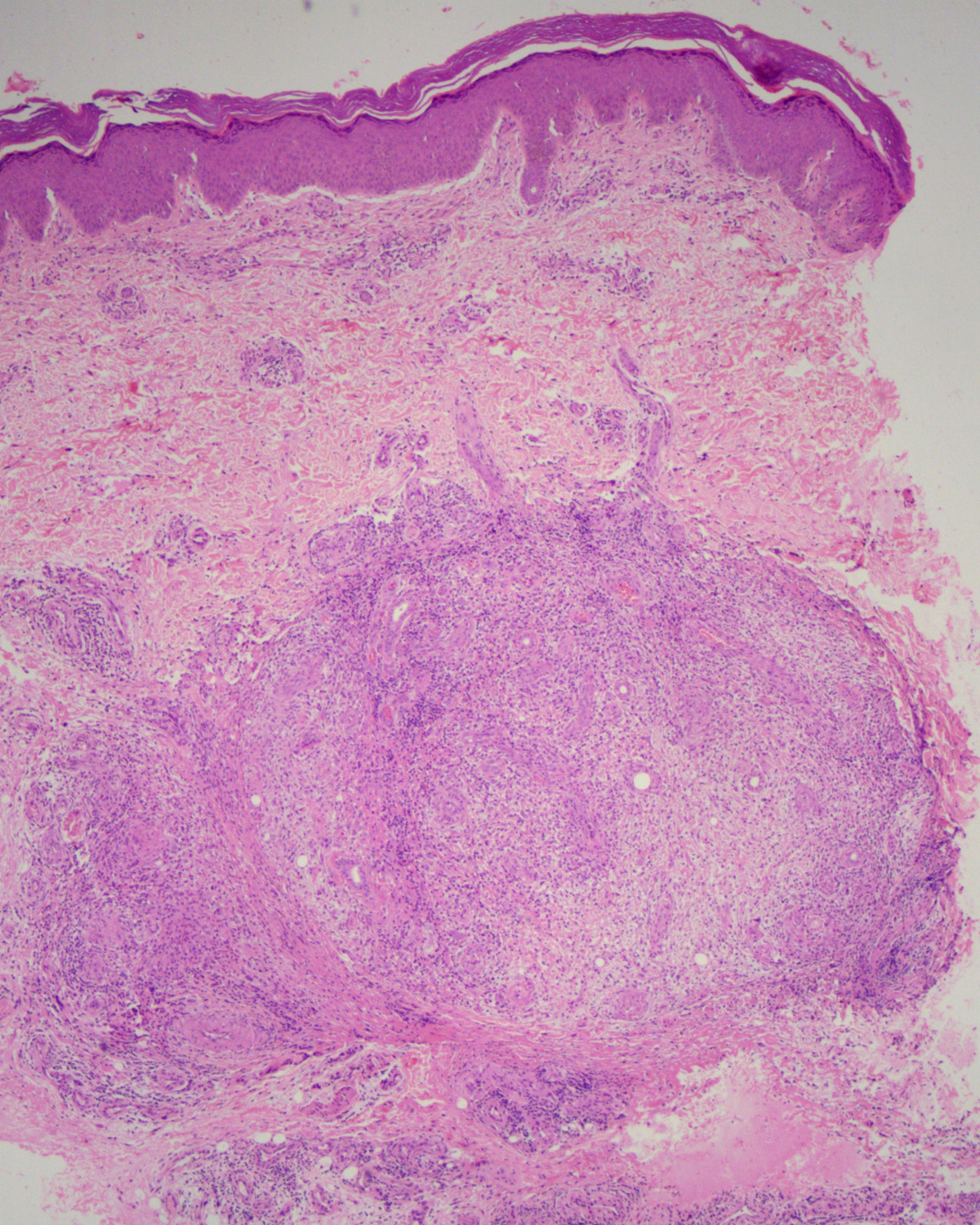
Figure 2b:
Dermoscopy under polarised mode (Dermlite, DL4, ×10 magnification) shows homogenous pink-white areas, diffuse dotted vessels, and shiny white lines. Arrow points to the normal-looking surrounding skin
Histopathology shows an acanthotic epidermis and a well-circumscribed nodular collection of inflammatory infiltrate in the lower dermis (H and E, ×50)
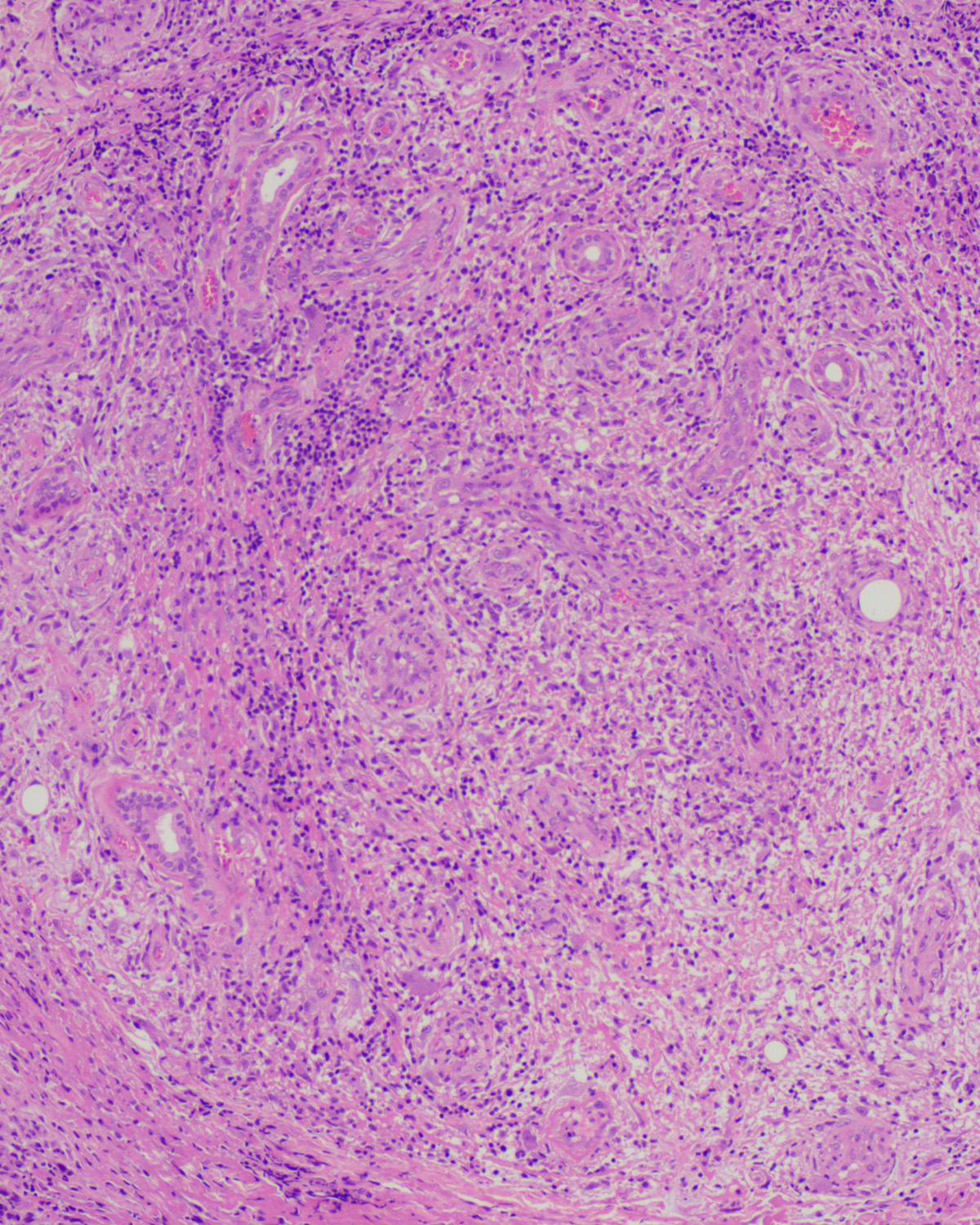
Figure 3a:
Histopathology shows an acanthotic epidermis and a well-circumscribed nodular collection of inflammatory infiltrate in the lower dermis (H and E, ×50)
The nodular collection shows neutrophilic leukocytoclastic vasculitis, fibrin deposition, dermal oedema, and a predominant lymphohistiocytic collection (H and E, ×100)
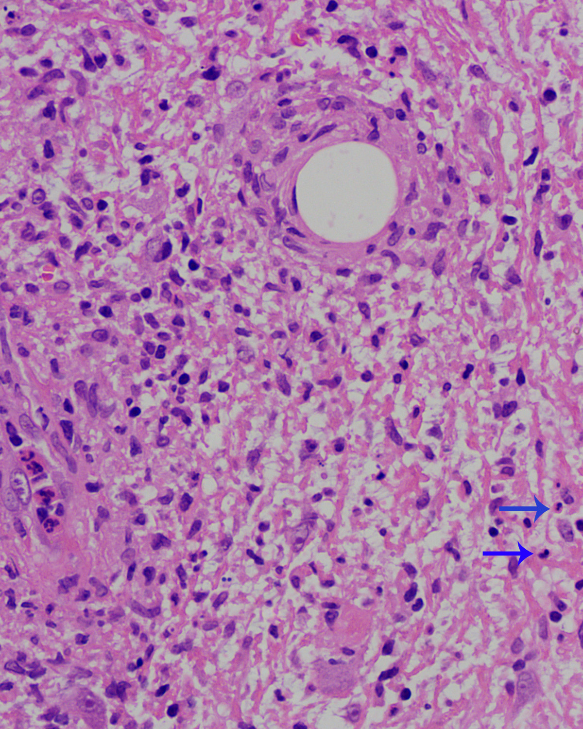
Figure 3b:
The nodular collection shows neutrophilic leukocytoclastic vasculitis, fibrin deposition, dermal oedema, and a predominant lymphohistiocytic collection (H and E, ×100)
Neutrophilic (arrow) leukocytoclastic vasculitis and leukocytoclasia (H and E, ×400)
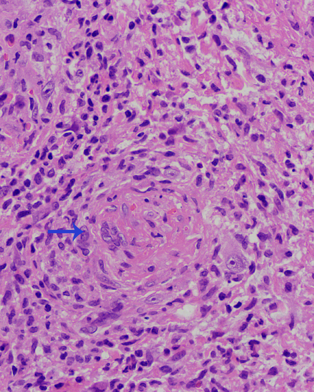
Figure 3c:
Neutrophilic (arrow) leukocytoclastic vasculitis and leukocytoclasia (H and E, ×400)
Endothelial proliferation, fibrin deposition and occlusion of the vascular lumen (arrow) (H and E, ×400)
Figure 3d:
Endothelial proliferation, fibrin deposition and occlusion of the vascular lumen (arrow) (H and E, ×400)

Erythema elevatum diutinum (EED) is a progressive and fibrosing form of immune complex-mediated localised vasculitis. 1 The clinical morphology varies from erythematous, violaceous to brownish papules and nodules in the early stages to yellowish-brown nodules in the advanced stages. Rarely, EED can be vesicular, bullous and ulcerative. 2 The most common sites are extensor aspects of extremities and include skin overlying the joints such as ankles, Achilles tendon, knees, elbow, hands and fingers. Uncommon sites are the retroauricular area, palms, soles, trunk and nipples. 3 The plaque morphology and location over the metatarsophalangeal joints, as in our case, have rarely been reported.

Dermoscopic description of EED is rarely mentioned in the literature. A solitary case of EED had a central white- to-orange area and purple spots on a reddish-purple background. 4 Another case from India showed yellow-red background, arborising telangiectasia and yellow-white streaks. 5 In the index case, the clinicodermoscopic features simulated psoriasis, borderline tuberculoid leprosy with or without reaction, patch-type granuloma annulare and pagetoid reticulosis [Table 1].6,7

Table 1: Dermoscopic features of clinical mimickers of erythema elevatum diutinum in the index case
Clinical diagnosis Dermoscopic feature
Psoriasis vulgaris Regularly distributed red dots or glomerular vessels on a reddish background and white scales
Borderline tuberculoid Hansen with or without type 1 reaction 5 Reddish backgroundWhite structureless areasYellow globulesFine short linear vessels or linear vessels with branching
Granuloma annulare(Patch-type) A pinkish-red backgroundWhitish or yellowish-orange areasDotted, linear-irregular, and branching vesselsShiny white structures (rosettes, crystalline structures)White scales
Pagetoid reticulosis 6 Central homogenous pink areaA whitish network in the marginDotted or glomerular vessels

The diagnosis of EED is solely dependent upon clinicopathological correlation. The pathological features and its differential diagnoses may vary according to the stage of the disease. Neutrophilic infiltration and features of leukocytoclastic vasculitis characterise the early lesion. It can be associated with dermal oedema and may mimic Sweet syndrome. The established lesions show a mixed inflammatory infiltrate, granulation tissue and perivascular onion-skin fibrosis. The late or chronic stage is characterised by increased and thickened collagen bundles, sparse neutrophilic infiltration and variable xanthomatization. The angiocentric and storiform fibroplasia can be mistaken for dermatofibroma, sclerotic fibroma, sclerosing spindle cell perineurioma and sclerotic neurofibroma. 1 As per our search, only a single prior report had luminal occlusion by fibrin and inflammatory cells. 1 In our case, multiple vessels had luminal occlusion within the nodular infiltration.

In conclusion, we report a rare plaque-type of erythema elevatum diutinum in which the clinicodermoscopic features were misleading and the diagnosis was established by pathological examination. In addition, it had an unusual lower dermal nodular inflammatory infiltrate along with features of vasculopathy.

Declaration of patient consent

The authors certify that they have obtained all appropriate patient consent.

Financial support and sponsorship

Nil.

Conflict of interest

There are no conflicts of interest.

References

  1. , , . Erythema elevatum diutinum: Clinical, histopathologic, and immunohistochemical characteristics of six patients. Am J Dermatopathol. 2005;27:397-400.
    [CrossRef] [PubMed] [Google Scholar]
  2. , , . A rare case of erythema elevatum diutinum presenting as diffuse neuropathy. JAAD Case Rep. 2017;3:1-3.
    [CrossRef] [PubMed] [Google Scholar]
  3. , . Atypical presentation of erythema elevatum diutinum. Australas J Dermatol. 2005;46:44-6.
    [CrossRef] [PubMed] [Google Scholar]
  4. , , , . A clinico-dermatoscopic study of acral dermatosis. Int J Dermatol Venereology Leprosy Sci. 2020;3:35-41.
    [Google Scholar]
  5. , , , . Dermatoscopy of a case of erythema elevatum diutinum. J Eur Acad Dermatol Venereol. 2022;36:e316-7.
    [CrossRef] [PubMed] [Google Scholar]
  6. , , , , , . Dermoscopy of type 1 lepra reaction in skin of color. Dermatol Pract Concept. 2020;10:e2020083.
    [CrossRef] [PubMed] [Google Scholar]
  7. , , , , , , et al. Dermoscopy of pagetoid reticulosis, with dermoscopic-pathologic correlation. Photodermatol Photoimmunol Photomed. 2019;35:372-74.
    [CrossRef] [PubMed] [Google Scholar]

Fulltext Views
2,087

PDF downloads
1,094
View/Download PDF
Download Citations
BibTeX
RIS
Show Sections