Translate this page into:
Comment on: “Dermoscopy of Biett's sign and differential diagnosis with annular maculopapular rashes with scaling”
Correspondence Address:
Ellen Maísa Streher
Ambulatório de Dermatologia Sanitária, Av. João Pessoa, 1327, Porto Alegre - RS
Brazil
| How to cite this article: Streher EM, Bonamigo RR. Comment on: “Dermoscopy of Biett's sign and differential diagnosis with annular maculopapular rashes with scaling”. Indian J Dermatol Venereol Leprol 2018;84:441-442 |
Sir,
We read with interest the article “Dermoscopy of Biett's sign and differential diagnosis with annular maculopapular rashes with scaling” by Tognetti et al. The article underlines the importance of dermoscopy in differentiating Biett's sign, a strong indicator of secondary syphilis, from other entities with similar morphologic lesions. The syphilitic lesions demonstrated monomorphic dotted and glomerular vessels on a diffuse, yellowish red background within a outwardly directed circular scaling edge.[1]
Errichetti and Stinco also described a dermoscopic pattern of palmar syphilid consisting of an orangish background and a thin, whitish, annular, scaling edge progressing in an outward direction and often surrounded by an erythematous halo, occasionally associated with peripheral telangiectatic vessels.[2]
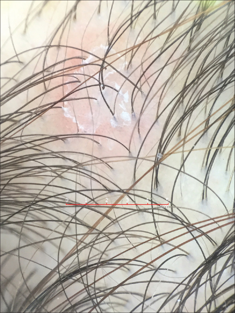
We observed a different finding in a young adult female, with a 4-week history of lesions on forearms, trunk and scalp, which were preceded by a genital ulcer. Physical examination showed slightly erythematous oval macules on the trunk and small oval erythematous plaques with slight peripheral scaling on the occipital region. Dermoscopy of the scaly scalp lesions, interpreted as Biett's sign, revealed monomorphic dotted vessels on a diffuse orangish background with an inner circular scaling edge inwardly oriented, contrasting with the previous observations [Figure - 1]. A venereal disease research laboratory test was positive at a titer of 1:128 and a qualitative test for Treponema pallidum was reactive confirming the suspicion of secondary syphilis. Benzathine Penicillin (2.4 million units) was administered intramuscularly weekly for 2 weeks and the lesions disappeared by the end of the treatment. Syphilis is a well-known sexually transmitted infection and although the primary chancre developing at the site of inoculation usually has typical and well-characterized features, the cutaneous manifestations of secondary syphilis span a wide spectrum and mimic other dermatoses, which may present a diagnostic challenge thereby delaying diagnosis and therapy.[3] Thus, dermoscopy may become a supportive tool to facilitate the recognition and differential diagnosis of this entity.
 |
| Figure 1: Scalp lesion dermoscopy: Monomorphic dotted vessels on a diffuse orangish background with an inner circular scaling edge inwardly oriented. (20× magnification, polarized light, FotoFinder Handyscope®) |
We would like to contribute to the current knowledge emphasizing the role that the orangish background seems to play in the differential diagnosis of secondary syphilis lesions, however, reminding the fact that the scaling may not always be present in an outward direction.
Financial support and sponsorship
Nil.
Conflicts of interest
There are no conflicts of interest.
| 1. |
Tognetti L, Sbano P, Fimiani M, Rubegni P. Dermoscopy of Biett's sign and differential diagnosis with annular maculopapular rashes with scaling. Indian J Dermatol Venereol Leprol 2017;83:270-3.
[Google Scholar]
|
| 2. |
Errichetti E, Stinco G. Dermoscopy in differentiating palmar syphiloderm from palmar papular psoriasis. Int J STD AIDS 2017;28:1461-3.
[Google Scholar]
|
| 3. |
Balagula Y, Mattei PL, Wisco OJ, Erdag G, Chien AL. The great imitator revisited: The spectrum of atypical cutaneous manifestations of secondary syphilis. Int J Dermatol 2014;53:1434-41.
[Google Scholar]
|
Fulltext Views
2,505
PDF downloads
1,011


