Translate this page into:
Condyloma acuminata mimicking cutaneous microcystic lymphatic malformation
Corresponding author: Dr. Pedro Jesús Gómez-Arias, Department of Dermatology and Venerology, Reina Sofía Universitary Hospital, Avenida de Menéndez Pidal s/n 14004, Córdoba, Spain. pjga10@hotmail.com
-
Received: ,
Accepted: ,
How to cite this article: Gómez-Arias PJ, Sanz-Zorrilla A, Salido-Vallejo R. Condyloma acuminata mimicking cutaneous microcystic lymphatic malformation. Indian J Dermatol Venereol Leprol 2022;88:792-3.
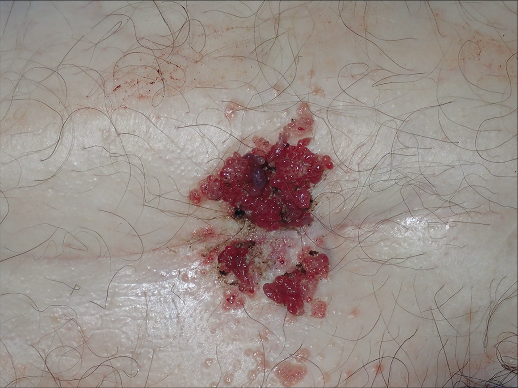
A man in his 80s’ presented with a rapidly growing lesion on his hypogastric region. Agminated small, red and translucent papules (“frog spawn” pattern) were observed [Figure 1] at the site. Koilocytes and a low-risk subtype human papillomavirus virus (HPV) infection were demonstrated in a skin biopsy. A diagnosis of condyloma acuminata was made. The lesion was excised by curettage.

- Grouped multiple, red and translucent small cysts-like lesions (“frog spawn” pattern) in a fold of the pubic region
Immunosenescence and development of the lesion in a moist area might have contributed to this clinical presentation. Unusual clinical forms of anogenital warts do not seem to be related with the presence of high-risk HPV subtypes.
The differential diagnosis of “frog spawn” pattern lesions should not only include microcystic lymphatic malformation, but also condyloma, in keeping with the patient’s age.
Declaration of patient consent
The authors certify that they have obtained all appropriate patient consent.
Financial support and sponsorship
Nil.
Conflicts of interest
There are no conflicts of interest.


