
Translate this page into:
Corymbiform syphilis: A remarkable presentation with several coexistent morphologies

Corresponding author: Dr. Subuhi Kaul, Division of Dermatology, John H Stroger Hospital of Cook County, Chicago, Illinois, United States. dr.subuhi.kaul@gmail.com
-
Received: ,
Accepted: ,
How to cite this article: DeLong BM, Kaul S, Othman D, Mehta S. Corymbiform syphilis: A remarkable presentation with several coexistent morphologies. Indian J Dermatol Venereol Leprol. doi: 10.25259/IJDVL_1874_2024
A 42-year-old man presented with several occasionally itchy and painful lesions on his trunk and extremities for six months. Examination revealed lesions of varying morphologies, including violaceous ovoid macules, depressed erythematous plaques, and crusted ulcers surrounded by multiple discrete and clustered pink, scaly, or oedematous papules [Figure 1]. Histopathology showed superficial and deep mixed inflammatory cell infiltrate, and immunohistochemical staining for Treponema pallidum highlighted numerous spirochetes [Figure 2]. A positive syphilis enzyme immunoassay and reactive plasma reagin test, along with this unique bombshell-like configuration were diagnostic of a rare form of secondary syphilis known as corymbiform syphilis.

- Erythematous to violaceous papules and plaques arranged in a corymbiform configuration on the abdomen.
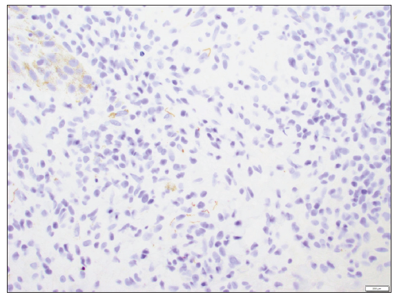
- Immunohistochemistry stained multiple spirochetes, visualised as brown filamentous and coiled figures, at 40x magnification.
Declaration of patient consent
The authors certify that they have obtained all appropriate patient consent.
Financial support and sponsorship
Nil.
Conflicts of interest
There are no conflicts of interest.
Use of artificial intelligence (AI)-assisted technology for manuscript preparation
The authors confirm that there was no use of artificial intelligence (AI)-assisted technology for assisting in the writing or editing of the manuscript and no images were manipulated using AI.


